E-ISSN:2320-8686
P-ISSN:2321-127X
RNI:MPENG/2017/74037

Research Article

Post Pulmonary Tuberculosis

International Journal of Medical Research and Review

2025 Volume 13 Number 1 Jan-Mar
Publisherwww.medresearch.in

Proportion Of Obstructive Airway Disease Among Post Pulmonary Tuberculosis Subjects in a Tertiary Care Setting

Akshaya KT1*, Fathahudeen A2, Kamala R3
DOI:https://doi.org/10.17511/ijmrr.2025.i01.07

1* Akshaya KT, Postgraduate Resident, Department of Respiratory Medicine, Government Medical College, Trivandrum, Kerala, India.

2 Fathahudeen A, Professor and Head of the Department, Department of Respiratory Medicine, Government Medical College, Trivandrum, Kerala, India.

3 Kamala R, Assistant professor, Department of Respiratory Medicine, Government Medical College, Trivandrum, Kerala, India.

Background: Pulmonary tuberculosis can affect lung function and the pulmonary function test of post-patients shows airway obstruction which is not reversible. This group of patients is in the category of post-TB obstruction syndrome with symptoms and signs similar to Chronic Obstructive Pulmonary Disease (COPD). The objectives of our study are: To determine the factors causing the development of obstructive airway disease in post-tuberculous individuals & To assess the severity of obstruction based on GOLD criteria.

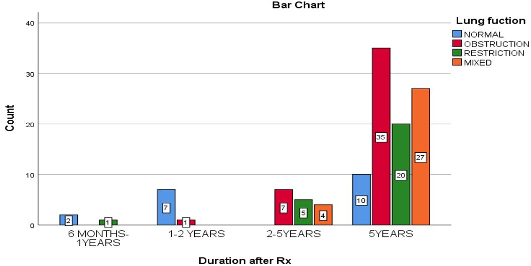
Materials and Methods: The study was conducted in the Department of Pulmonary Medicine, Government Medical College Hospital, Thiruvananthapuram. This was a cross-sectional study that included patients with a history of pulmonary tuberculosis, (≥ 6 months after treatment), the pulmonary function test of each patient was assessed using spirometry as obstruction, restriction, mixed pattern or normal and in obstruction, the severity of obstruction was assessed as mild, moderate, severe, very severe based on GOLD criteria.

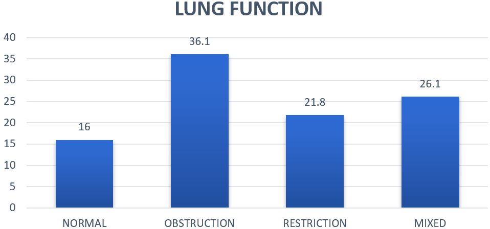
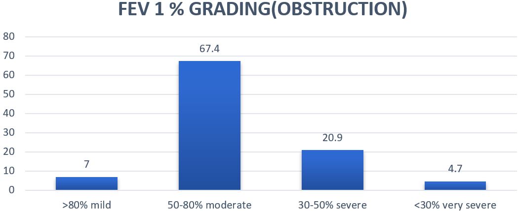
Results: In our study, 84% of patients had abnormal lung function, with obstruction as the predominant abnormality in 36%, 21.8% demonstrated restrictive abnormalities, and 26.1% showed mixed patterns. Among individuals with obstructive lung disease, the severity of obstruction was assessed based on their post-bronchodilator FEV1% according to GOLD grades for COPD. It was observed that 67.4% had moderate obstruction. The mean FEV1 for the moderate obstruction group, the most prevalent among obstructive cases, was 1.41 litres.

Conclusion: Patients with a history of pulmonary tuberculosis in the past, often present with signs of permanent impairment in lung function, which may manifest as obstruction, restriction, or a mixed pattern. Tuberculosis is an independent risk factor for the development of obstructive airway diseases.

Keywords: post tuberculosis, lung function, obstruction, airway diseases

Corresponding Author How to Cite this Article To Browse
Akshaya KT, Postgraduate Resident, Department of Respiratory Medicine, Government Medical College, Trivandrum, Kerala, India.
Email:
Akshaya KT, Fathahudeen A, Kamala R, Proportion Of Obstructive Airway Disease Among Post Pulmonary Tuberculosis Subjects in a Tertiary Care Setting. Int J Med Res Rev. 2025;13(1):42-48.
Available From
https://ijmrr.medresearch.in/index.php/ijmrr/article/view/1520

Manuscript Received Review Round 1 Review Round 2 Review Round 3 Accepted
2024-12-24 2025-01-02 2025-01-10 2025-01-18 2025-01-26
Conflict of Interest Funding Ethical Approval Plagiarism X-checker Note
None Nil Yes 12.43

© 2025by Akshaya KT, Fathahudeen A, Kamala Rand Published by Siddharth Health Research and Social Welfare Society. This is an Open Access article licensed under a Creative Commons Attribution 4.0 International License https://creativecommons.org/licenses/by/4.0/ unported [CC BY 4.0].

Download PDFBack To ArticleIntroductionMethodologyResultsDiscussionConclusionReferences

Introduction

Post-tuberculosis pulmonary sequelae is a problem that frequently arises in the community, especially in countries with a high prevalence of pulmonary tuberculosis. A large number of people recover after full anti-tuberculous treatment, but some patients have clinically symptomatic shortness of breath, especially during exertion. Most often chest radiology shows minimal post-tuberculous changes (fibrosis and calcification) and pulmonary function shows airway obstruction which is not reversible. Patients in this category have post-tuberculous obstructive syndrome with symptoms and signs similar to Chronic Obstructive Pulmonary Disease (COPD).[1] Maximum loss of lung function usually occurs in the first 6 months after diagnosis and stabilizes around 1.8 months after the end of treatment. Structural damage increases with each reactivation and this correlates with an accelerated loss of FEV1. Delayed diagnosis and inappropriate regimens during tuberculosis treatment may play a role in causing post-TB lung disease. There are certain common risk factors for the development of COPD and pulmonary tuberculosis like smoking, biomass fuel exposure, low socioeconomic status, diabetes mellitus, vitamin D deficiency etc. The development of chronic airflow obstruction in post-TB patients may be related to common risk factors or tuberculosis-associated lung damage. [2]

After completion of anti-TB drugs in pulmonary TB patients, these patients need regular follow-up for early identification of post-TB obstructive airway diseases so that proper available treatment can be initiated before further progression of the disease occurs. Efforts to improve long-term lung health should be a part of tuberculosis care. Hence this study aimed to assess the proportion of obstructive airway disease in post-TB patients and thereby help in the early identification of obstructive airway disease and early initiation of treatment, which helps to reduce exacerbations in future and improve quality of life. The objective of the study is to estimate the proportion of obstructive airway disease in patients with a history of pulmonary tuberculosis who have received at least 1 month of anti-tuberculous treatment (≥ 6 months after treatment) and to determine the factors causing the development of obstructive airway disease in post tuberculous individuals and to assess the severity of obstruction based on GOLD criteria.

Methodology

The study was conducted in the Department of Pulmonary Medicine, Government Medical College Hospital, Thiruvananthapuram. This was a cross-sectional study which included patients with a history of pulmonary tuberculosis, (≥ 6 months after treatment), the pulmonary function test of each patient was assessed using spirometry as obstruction, restriction, mixed pattern or normal and in obstruction, the severity of obstruction was assessed as mild, moderate, severe, very severe based on GOLD criteria and those who had bronchodilator reversibility in spirometry were excluded from the study.

Table 1: GOLD Grades and Severity of Airflow Obstruction in COPD

In COPD patients (FEV)/FVC < 0.7):

GOLD 1:MildFEV1 80% predicted
GOLD 2:Moderate50% FEV1 < 80% predicted
GOLD 3:Severe30% FEV1 < 50% predicted
GOLD 4:Very SevereFEV1 < 30% predicted

Inclusion Criteria:

  • All individuals with a history of pulmonary tuberculosis, who have received at least 1 month of anti-tuberculous treatment (≥ 6 months after treatment).
  • Individuals who can do spirometry.
  • Individuals who are willing to participate in the study and give consent.
  • Age >18 years

Exclusion Criteria:

  • Patients with active tuberculosis, underlying malignancy.
  • Diagnosed cases of asthma / COPD.
  • Patients with a smoking index >100, biomass index >60 and occupational exposures.

Study period: One and half years after getting Institutional Ethical Committee clearance.

Sample Size: Taking 44.62% as the prevalence of obstructive airway disease in post-TB patients in a study conducted by Raja Dhar Et Al, a pilot study published in the European Respiratory Journal 2018

Sample size, n =ijmrr-1520-06.JPG


ijmrr-1520-07.JPG

p= prevalence of obstructive airway disease in post-TB patients
q= 100-p 
This sample size, n = 119

Sampling Method: Patients were consecutively recruited till the sample size was reached.

Study Variables

Demographic variables:

  • Age
  • Sex
  • BMI
  • Smoking index
  • Biomass index
  • Occupation

Disease variables:

  • MMRC grading of dyspnea on exertion

1.Grade 0
2. Grade 1
3. Grade 2
4. Grade 3
5. Grade 4

  • Spirometric indices like FVC, FEV1, FEV1/FVC, FEF 25-75
  • Lung function : obstruction , restriction , mixed pattern , normal
  • Duration after completing TB treatment in months
  • The severity of obstruction – mild, moderate, severe, very severe.

Data Collection Tool

Data will be collected in a semi-structured questionnaire after taking informed consent.

Data Collection Technique

After obtaining informed written consent, patients with a history of tuberculosis and who have completed pulmonary TB treatment (≥6 months after completion of treatment) meeting the inclusion criteria attending the OPD of the Department of Pulmonary Medicine and those patients under DTC Trivandrum were enrolled in the study.

These patients were interviewed, and demographic characteristics and clinical history will be noted. Spirometry with a computerized spirometer was done Spirometry with bronchodilator reversibility will be performed as per Joint Indian Chest Society- National College of Chest Physicians guidelines for spirometry. It is a physiological test that measures the maximal volume of air that a person can inspire and expire with maximal effort. FVC, FEV1, FEV1/FVC and FEF25-75 ratio are calculated using spirometry and the values of all indices will be noted. Patient data; including age, sex, weight, height and medication history, were collected before the pulmonary function test. Pulmonary function test was performed by a trained technician. Each patient performs at least three acceptable forced expiratory manoeuvres, which fulfil the criteria of repeatability. The normal pulmonary function should have FEV1 ≥80%, FVC ≥80% and FEV1/FVC ≥70% of predicted values. For abnormal pulmonary function, an obstructive defect is FEV1/FVC <70% and a restrictive defect if FEV1/FVC≥70% and FVC<80% and a mixed defect if FEV1/FVC<70% and FVC<80% of predicted values. Post bronchodilator reversibility is an increase in FEV1 of >12% and >200ml from baseline (greater confidence if the increase is >15% and 400ml) measured 10-15 min after 200-400 mcg salbutamol or equivalent compared with pre-bronchodilator readings and is done to rule out asthma. The pulmonary function test of each patient will be assessed – obstruction, restriction, mixed pattern or normal and in obstruction, the severity of obstruction will be assessed as mild, moderate, severe, or very severe based on GOLD criteria.

Data Analysis

Data was analyzed using the statistical package SPSS 26.0 (SPSS Inc., Chicago, IL) Descriptive statistics were performed to estimate the PROPORTION of the respective groups. The Chi-Square Test was used to check the association between the variables.

Ethical Considerations

  • Informed written consent will be obtained from the patients.
  • Confidentiality of subjects will be maintained throughout the study.
  • No financial burden will be imposed on subjects.

  • The study will commence only after receiving approval from the institute's research committee & institution ethics committee.

Data Analysis

1. Data entry would be done in Excel and analysis using spss27 software.
2. Qualitative variables will be summarised as percentages and quantitative variables as mean and standard deviation.

Ethical Considerations

  • Informed written consent will be obtained from the patients.
  • Confidentiality of subjects will be maintained throughout the study.
  • No financial burden will be imposed on subjects.
  • The study will commence only after receiving approval from the institute's research committee & institution ethics committee.

Results

In this study, 84% of patients exhibited abnormal lung function, with 36% showing obstructive patterns, 21.8% demonstrating restrictive abnormalities, and 26.1% exhibiting mixed patterns. Dyspnoea was the most prevalent symptom observed among post-tuberculosis subjects.

In our study population, a significant majority of patients (44.5%) experienced Grade 2 dyspnoea according to the MMRC dyspnoea grading system, significantly compromising their functional quality. Among individuals with obstructive lung disease, the severity of obstruction was assessed based on their post-bronchodilator FEV1% according to GOLD grades for COPD. It was observed that 67.4% had moderate obstruction (FEV1% between 50-80%), followed by 20.9% with severe obstruction, 7% with mild obstruction, and only 4.7% with very severe obstruction.

The extent of radiological involvement was assessed among our study population, the majority (52.9%) had unilateral or no radiological involvement and 47.1% had bilateral involvement and we could find an association between the extent of radiological involvement and lung function abnormality, with the majority of patients with obstruction had bilateral lung involvement.

Table 2: Association between Duration after Rx vs Lung function

Lung functionTotal
NORMALOBSTRUCTIONRESTRICTIONMIXED
Duration after Rx6 MONTHS-1YEARSCount20103
% within Duration after Rx66.7%0.0%33.3%0.0%100.0%
1-2 YEARSCount71008
% within Duration after Rx87.5%12.5%0.0%0.0%100.0%
2-5YEARSCount075416
% within Duration after Rx0.0%43.8%31.3%25.0%100.0%
5YEARSCount1035202792
% within Duration after Rx10.9%38.0%21.7%29.3%100.0%
TotalCount19432631119
% within Duration after Rx16.0%36.1%21.8%26.1%100.0%
P VALUE0.0001*

Table 2: Association between Duration after Rx vs Lung function

P<0.05 is statistically significant.
The chi-square test reported a significant association between Duration after treatment vs Lung Function (P<0.05).

ijmrr-1520-01.JPG
Figure 1: Pulmonary function assessment of the study population using spirometry

ijmrr-1520-02.JPG
Figure 2: MMRC grading of dyspnoea in the study population


ijmrr-1520-03.JPG
Figure 3: severity of obstruction in my study population based on FEV% grading.

ijmrr-1520-04.JPG
Figure 4: Association between Duration after Rx vs Lung function.

Discussion

Tuberculosis (TB) poses a significant public health challenge worldwide, particularly affecting low and middle-income countries. Despite the successful implementation of standard anti-TB therapy resulting in the cure of the majority of patients, many individuals still suffer from chronic respiratory symptoms such as cough, sputum production, and/or dyspnea. These symptoms significantly impact their quality of life and increase the risk of mortality.

Post-tuberculosis lung disease refers to chronic changes in the lungs that persist following successful treatment of pulmonary tuberculosis. The long-term consequences of post-tuberculosis lung disease (PTLD) manifest as alterations in chest imaging and impairments in pulmonary function. Post-tuberculosis pulmonary function abnormalities include airflow obstruction, restriction, or mixed patterns, with obstruction being the predominant abnormality.

After completing treatment for pulmonary tuberculosis, patients should undergo regular follow-up visits to monitor for any potential relapse and to detect early signs of obstructive or restrictive lung abnormalities. Utilizing tools such as spirometry can facilitate early identification and treatment, thereby preventing adverse outcomes. [3]

The study aimed to determine the proportion of obstructive airway disease in individuals previously treated for pulmonary tuberculosis at a tertiary care centre using spirometry and to evaluate the severity of obstruction based on GOLD criteria. The study was done among 119 individuals with a previous history of pulmonary tuberculosis.

The mean age of the study population is 56.344 years, with ages ranging from 19 to 83 years with 57.1% being males. Similarly, IM Baig et al. found a mean age of 56.4 years for males and 44.2 years for females in their study, and 76.5% were males which aligns closely with our study findings [4]. Additionally, Raja Dhar et al. in their study, reported that the majority of their subjects were above 50 years of age, predominantly males(83%). [5]

In our study, the majority of the participants were nonsmokers (71.4%), while the remaining participants had a very minimal smoking index of less than 100. Almost all participants had no occupational risk for obstructive airway diseases.

Additionally, most participants (67.2%) had no exposure to biomass fuel, and those who did had a biomass index of less than 60. These criteria were intentionally set to minimize common risk factors for the development of obstructive airway diseases, similar to studies by Raja Dhar et al [5] and Tarigan A P et al. [6]

In our study, the majority of patients (77.3%) had completed anti-tuberculous treatment more than 5 years ago. This finding aligns with the studies by Raja Dhar et al. [5] and IM Baig et al. [4], where 65% of patients with obstructive ventilatory defects had been treated over 10 years earlier.

Additionally, P. A. Wilcox et al. [7] reported that obstructive changes become more pronounced after 10 years of follow-up, correlating with residual scarring visible on chest radiographs, regardless of initial findings. Our study also identified a significant association between the duration since completing anti-tuberculous treatment and the decline in lung function, with a p-value of 0.0001 (p < 0.05).

Patients who have recovered from pulmonary tuberculosis are highly susceptible to developing abnormalities in lung function. The decline in pulmonary function following completion of TB treatment correlates with prolonged respiratory symptoms that significantly impact quality of life.


In this study, 84% of patients exhibited some form of abnormal lung function, with 36% showing obstructive patterns, 21.8% demonstrating restrictive abnormalities, and 26.1% exhibiting mixed patterns. These findings emphasize a significant burden of post-treatment pulmonary function impairments among tuberculosis patients, with obstructive patterns being the most frequently observed. Dyspnoea was the most prevalent symptom observed among post-TB subjects. In our study population, a significant majority of patients (44.5%) experienced Grade 2 dyspnoea according to the MMRC dyspnoea grading system, significantly compromising their functional quality.

Many studies have shown similar findings. In the study by IM Baig et al(8), 55.3% had an obstructive ventilatory defect, 29.9% had a restrictive pattern and 14.8% revealed a mixed pattern in spirometry. Similarly in a study by Raja Dhar et al [4] on the prevalence of OAD in patients with a history of Pulmonary Tuberculosis in the absence of risk factors of OAD, the prevalence was 44.62%. These study findings are similar to our study with obstruction as the predominant pulmonary function abnormality. In a study conducted by Pasipanodya et al. (2007) in the USA(49), it was found that 59% of participants had some form of abnormal lung function and the prevalence of obstructive impairment was 15%, restrictive impairment was 31%, and mixed impairment was 13%. In this study the predominant abnormality was restriction. These findings highlight the importance of regular follow-up in post-TB subjects, along with timely interventions as necessary to enhance their quality of life.

In our study population, among individuals with obstructive lung disease, the severity of obstruction was assessed based on their post-bronchodilator FEV1% according to GOLD grades for COPD. It was observed that 67.4% had moderate obstruction (FEV1% between 50-80%), followed by 20.9% with severe obstruction, 7% with mild obstruction, and only 4.7% with very severe obstruction. The mean FEV1 for the moderate obstruction group, the most prevalent among obstructive cases, was 1.41. These findings are consistent with those of Raja Dhar et al. [5] regarding the prevalence of obstructive airway disease in individuals with a history of pulmonary tuberculosis, where the mean FEV1 among those with OAD was 1.27.

According to spirometric criteria (GOLD), none of their patients were classified as having mild COPD, while 62% had moderate disease, 28% had severe disease, and 10% had very severe disease. Similarly, in the study by A P Tarigan et al, [6] 60% of their subject had a moderate obstruction and none had a mild obstruction. These consistent findings indicate the significant impact on lung function in individuals with a history of tuberculosis, highlighting the severity and chronicity of respiratory impairments in this population.

The extent of radiological involvement was assessed among our study population, the majority(62.2%) had unilateral involvement and 37.8% had bilateral involvement and we could find an association between the extent of radiological involvement and lung function abnormality, with the majority of patients with obstruction had bilateral lung involvement This suggests that the severity of radiological findings may correlate with the severity of obstructive airway disease. Comparing these results with other studies, such as those by Raja Dhar et al. [5] and A. P. Tarigan et al. [6], would likely reveal similar patterns of radiological involvement and its impact on lung function among post-tuberculosis individuals. These correlations underline the persistent and clinically significant respiratory sequelae experienced by TB survivors, necessitating comprehensive management strategies.

Conclusion

Our study identified a high prevalence of obstructive airway disease in individuals previously treated for pulmonary tuberculosis, even after excluding other risk factors. Most cases demonstrated moderate obstruction. We observed a significant correlation between radiological findings, especially bilateral lung involvement, and the severity of obstructive lung disease, indicating that radiological abnormalities could predict the extent of lung function impairment following TB treatment. Additionally, our findings suggest that obstructive changes can persist and deteriorate over time, emphasizing the chronic and progressive nature of post-tuberculosis lung sequelae. Our study together with the studies mentioned here collectively highlights the enduring burden of obstructive airway diseases in individuals who have recovered from tuberculosis.


This emphasizes the need for long-term monitoring, early detection, and appropriate management strategies to mitigate respiratory impairments and improve the quality of life for TB survivors.

Limitation: The small study population and the fact that the research was conducted at a single tertiary care centre may limit the generalisability of the findings to other healthcare settings or geographic regions.

Acknowledgement: I would like to express my deepest gratitude to my guide, whose unwavering support and expertise have been invaluable throughout this journey. I also extend my heartfelt thanks to all my teachers for their guidance and encouragement. Lastly, I am grateful to my family and friends for their constant support and belief in me, which kept me motivated to complete this work.

Permission from Institutional research board: Yes

Funding: Nil

Conflict of interest: None Initiated

References

1. Swain S, Pothal S, Behera A, Manjhi R, Dutta P, Pradhan G. Treatment outcome among Post TB obstructive airways diseases and COPD: A prospective cohort study. J Fam Med Prim Care. 2021 Sep;10(9):3411–6. [Crossref][PubMed][Google Scholar]

2. Alvarado González A. Tuberculosis Obstructive Pulmonary Disesase (TOPD). Clin Res Trials [Internet]. 2021 [cited 2024 Jul 22];7(4). [Crossref][PubMed][Google Scholar]

3. Gai X, Allwood B, Sun Y. Post-tuberculosis lung disease and chronic obstructive pulmonary disease. Chin Med J (Engl). 2023 Aug 20;136(16):1923–8. [Crossref][PubMed][Google Scholar]

4. Baig IM, Saeed W, Khalil KF. Post-tuberculous chronic obstructive pulmonary disease. J Coll Physicians Surg--Pak JCPSP. 2010 Aug;20(8):542–4. [Crossref][PubMed][Google Scholar]

5. Dhar R, Biswas B, Kulkarni T, Limaye S, Ghoshal A. Tuberculosis associated Obstructive Lung disease- A Pilot Study. Eur Respir J [Internet]. 2018 Sep 15 [cited 2024 Jul 25];52(suppl 62). Available from: [Article][Crossref][PubMed][Google Scholar]

6. (PDF). Obstructive lung disease as a complication in post pulmonary TB [Internet]. [cited 2024 Jul 25]. Available from. [Crossref][PubMed][Google Scholar]

7. Willcox PA, Ferguson AD. Chronic obstructive airways disease following treated pulmonary tuberculosis. Respir Med. 1989 May;83(3):195–8. [Crossref][PubMed][Google Scholar]

8. Patil S, Patil R, Jadhav A. Pulmonary functions’ assessment in post-tuberculosis cases by spirometry: Obstructive pattern is predominant and needs cautious evaluation in all treated cases irrespective of symptoms. Int J Mycobacteriology. 2018;7(2):128–33. [Crossref][PubMed][Google Scholar]

Disclaimer / Publisher's NoteThe statements, opinions and data contained in all publications are solely those of the individual author(s) and contributor(s) and not of Journals and/or the editor(s). Journals and/or the editor(s) disclaim responsibility for any injury to people or property resulting from any ideas, methods, instructions or products referred to in the content.