E-ISSN:2320-8686
P-ISSN:2321-127X
RNI:MPENG/2017/74037

Research Article

Clinical and Histopathological

International Journal of Medical Research and Review

2025 Volume 13 Number 1 Jan-Mar
Publisherwww.medresearch.in

Clinical & Histopathological Profile Of Patients With Exudative Pleural Effusion Of Unknown Cause Who Undergo Medical Thoracoscopy

Kumar JVS1*, Fathahudeen A2
DOI:DOI:https://doi.org/10.17511/ijmrr.2025.i01.04

1* Jasti Venkata Suneel Kumar, Postgraduate Student, Department of Pulmonary Medicine, Government Medical College, Thiruvananthapuram, Kerala, India.

2 A Fathahudeen, Professor and Head of the Department, Pulmonary Medicine, Government Medical College, Thiruvananthapuram, Kerala, India.

Background: Exudative lymphocytic effusions form a significant proportion of undiagnosed pleural pathologies. This study explores the clinical and histopathological profile of exudative pleural effusion of unknown cause who undergo medical thoracoscopy.

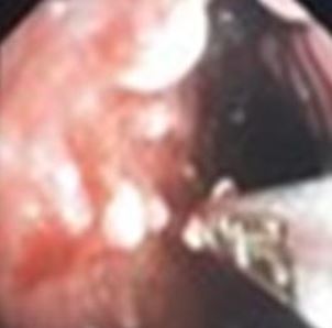
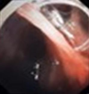
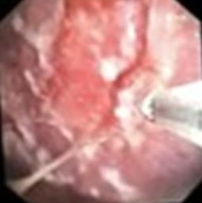
Materials And Methods: This cross-sectional study enrolled 37 patients with exudative pleural effusion. Ultrasound-guided thoracentesis was conducted and samples were sent for pleural fluid cytology. Thoracoscopic pleural biopsy was kept as the gold standard.

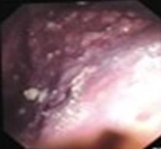
Results: A total of 37 patients were included in the study. 14 (37.83%) were smokers. 19 (51.35%) were exposed to firewood smoke. Only 1 participant (2.7%) reported exposure to passive smoke.5 patients had STEMI/ recent CABG in the last 1 year.1 participant reported a history of Ayurvedic drug intake. There was no asbestos occupational exposure in any of our subjects but 5 of our subjects had mesothelioma. It can be related to an indirect exposure which is absent due to a recall bias. 2 participants reported a history of recent trauma or RTA, but it was unrelated to the final diagnosis. Histopathological profile showed adenocarcinoma in 19(51.35%), tuberculosis in 7(18.9%).Thoracoscopy pleural findings include nodules in 26(70.27%).

Conclusion: Our study concluded that pleural nodules on thoracoscopy were suggestive of malignant aetiology with good cytology yield. Pleural metastasis from lung adenocarcinoma has a good yield of cytology and is the most common cause of exudative pleural effusions that underwent medical thoracoscopy.

Keywords: Medical thoracoscopy, HPE, Pleural Effusion

Corresponding Author How to Cite this Article To Browse
Jasti Venkata Suneel Kumar, Postgraduate Student, Department of Pulmonary Medicine, Government Medical College, Thiruvananthapuram, Kerala, India.
Email:
Kumar JVS, Fathahudeen A, Clinical & Histopathological Profile Of Patients With Exudative Pleural Effusion Of Unknown Cause Who Undergo Medical Thoracoscopy. Int J Med Res Rev. 2025;13(1):19-25.
Available From
https://ijmrr.medresearch.in/index.php/ijmrr/article/view/1517

Manuscript Received Review Round 1 Review Round 2 Review Round 3 Accepted
2024-12-19 2024-12-26 2025-01-02 2025-01-09 2025-01-16
Conflict of Interest Funding Ethical Approval Plagiarism X-checker Note
None Nil Yes 11.32

© 2025by Kumar JVS, Fathahudeen Aand Published by Siddharth Health Research and Social Welfare Society. This is an Open Access article licensed under a Creative Commons Attribution 4.0 International License https://creativecommons.org/licenses/by/4.0/ unported [CC BY 4.0].

Download PDFBack To ArticleIntroductionMaterial and MethodsResultsDiscussionReferences

Introduction

For pulmonologists, undiagnosed pleural effusions continue to be a diagnostic issue [1]. The first thing to determine in a patient with an undiagnosed pleural effusion is whether the fluid is a transudate or an exudate[2]. Stepwise diagnosis should be used when investigating a pleural effusion seen on chest radiographs. [3]. Thoracentesis is performed when necessary after the clinical history, physical examination, and chest radiography have been completed[4].

Most of these exudates in the Western world are cancerous. The pleural space and intrathoracic structures can be seen with a thoracoscopy, a minimally invasive technique. The most common reason for thoracoscopy is the pleural effusion of unknown origin. When TB and malignant pleural effusion are clinical possibilities and initial pleural fluid analysis is inconclusive, individuals with undiagnosed pleural effusions should be evaluated for medical thoracoscopy[5].

With roughly 90% of malignant pleural effusions, its diagnostic yield for patients with malignant pleural disease is 95% & It helps detect 25% of pleural effusions that are missed by other primary diagnostics such as biochemical testing, thoracocentesis, and biopsy. According to the most recent British Guidelines, this technique has the highest diagnostic yield for "aspiration cytology negative exudative effusions," and its efficacy is almost on par with video-assisted thoracoscopic surgery (VATS).In these situations, thoracoscopy is still the gold standard method for diagnosis and treatment. It makes it possible to do pleurodesis, therapeutic drainage of effusions, and pleural biopsies under direct eye, all in one setting.

A minor risk of serious consequences is linked to thoracoscopy(1.95%), and mortality rates are extremely low, comparable to those of mediastinoscopy (0.17%) and transbronchial biopsy (0.22–0.66%). Besides, there is no risk of death or disability from repeated use of thoracoscopy.

Thoracoscopy pleural biopsy under local anesthesia should be aggressively carried out in

patients with undiagnosed exudative pleural effusions since the procedure is simple, safe, and has a high diagnostic rate.

Despite the lower biopsy size, the semirigid thoracoscope achieves a diagnostic yield comparable to that of the traditional rigid instrument. In the assessment and treatment of pleural diseases, both tools are still useful.

In this context, this study aims at assessing the clinical histopathological profile of patients with undiagnosed exudative pleural effusions who qualify for medical thoracoscopy, and its diagnostic yield.

Material and Methods

Study Design: Cross-sectional study

Study Setting: The study was conducted in the Department of Pulmonary Medicine, Government. Medical College, Thiruvananthapuram.

Study Period: One and half years after getting Institutional Ethical Committee clearance.

Study Population: All consecutive patients with exudative pleural effusion of unknown cause at the time of enrollment, aged >18 years who were willing to participate in the study were admitted under the Department of Pulmonary Medicine, Medical College, Thiruvananthapuram.

Inclusion Criteria: Age >18 years
Patients with exudative pleural effusion
Patients who were willing to participate in the study and gave consent.

Exclusion Criteria: Patients not fit for thoracoscopy

Sample Size: As per the study conducted by Laila A. Helala, Gehan M. El-Assal; Diagnostic Yield of Medical Thoracoscopy in cases of undiagnosed pleural effusion in Department of Chest Diseases, Kobri El-Kobba Military Hospital, Egypt.

Sample size was estimated using the formula: n=[(Z1-α/2)2 x p x q]/d2; where Z1-α/2 is 1.96 at 5% level

P=proportion of malignancy=70%, q= 100-70=30% d= relative precision =20% of p

so, n= (1.96)2 x 70 x 30/ 142= 45

The sample size was calculated to be 45.

Sampling Method: Patients were Consecutively Recruited till the sample size was achieved.


Study Variables:

1. Demographic variables: Name, Age, Gender, Education Level, Occupation, Income & Urban/Rural Dwelling
2. Disease variables: Thoracoscopic pleural findings, Malignant Mesothelioma, Small cell carcinoma of the lung, Squamous cell carcinoma of the lung, Adenocarcinoma of lung&Pulmonary Tuberculosis

Study Definitions:

Thoracoscopy: Involves passage of an endoscope through the chest wall for direct visualization of the pleura.

Thoracocentesis: A procedure that involves the use of a needle to remove excess fluid from the pleural space between the lungs and chest wall.

Pleural effusion: Abnormal accumulation of fluid in the pleural space resulting from excess fluid production and/or decreased lymphatic absorption.

Malignant pleural effusion: Accumulation of fluid in the presence of malignant cells or tumor tissue in the pleural space.

Exudative Pleural Effusion of Unknown Cause: Exudative, Lymphocytic pleural effusions with an Adenosine Deaminase level <40 IU/dL were evaluated by a pulmonologist with at least 10 years of experience and found to have no clinically evident cause for the effusion

Thoracoscopic Pleural Appearance: Nodules, Adhesions & Plaques.

Data Collection Tools andTechnique: After obtaining informed written consent, patients with pleural effusion who met the inclusion criteria and attended the Department of Pulmonary Medicine were enrolled in the study. These patients were interviewed, demographic characteristics, clinical history noted and Physical examinations were performed. Ultrasound-guided thoracentesis was conducted, the colour of the fluid was noted and samples were sent for pleural fluid cytology.

As per our institution policy serial thoracentesis on three consecutive days for pleural fluid cytology was done and then the patient was subjected to thoracoscopy. Pleural fluid cytology reporting takes a minimum of 4 days in our institution & as the sensitivity of pleural fluid cytology is only 40-52%, we as a rule send 3 samples on 3 consecutive days to avoid a diagnostic delay.

Thoracoscopic pleural biopsy was kept as the gold standard for diagnosing the cause of exudative pleural effusions. During thoracoscopy, pleural findings were noted, and samples obtained during the procedure were sent for histopathological examination (HPE), mycobacterial tuberculosis culture, fungal culture and the results were analyzed and categorized accordingly. The pleural fluid cytology reports were analysed.

Data Analysis: Data entry was done in Excel, and analysis was conducted using SPSS version 27. Categorical variables were expressed as percentages. Sensitivity, specificity, positive predictive value, and negative predictive value were also determined. All quantitative variables were expressed in mean and standard deviation.

Ethical Considerations: Informed written consent was obtained from the patients. Confidentiality of subjects was maintained throughout the study financial burden was imposed on subjects as all tests were part of the diagnostic process. The study commenced only after receiving approval from the institute's research committee and institute ethics committee.

Results

17 (45.94%) were females and 20 (54.05%) were males.

Among the total sample of 37 individuals, 14 (37.83%) were smokers. 19 (51.35%) were exposed to firewood smoke. Only 1 participant (2.7%) reported exposure to passive smoke. 5 patients had STEMI/ recent CABG in the last 1 year. 2 participants reported a history of recent trauma or RTA, but it was unrelated to the final diagnosis. 1 participant reported a history of hypothyroidism

2 participants reported a history of malignancy in their family. 4 participants reported a history of extrathoracic malignancy. Out of those 4, two had breast carcinoma, one had renal cell carcinoma, and another one had cutaneous squamous cell carcinoma. All 37 participants (100.0%) reported negative sputum AFB. All patients had lymphocytic exudative ADA <40 IU. 24 participants (64.4%) had straw-coloured pleural fluid. 13 participants (35.13%) had hemorrhagic pleural fluid. In our study, all the hemorrhagic pleural effusions were diagnosed as malignancy and all tuberculous effusions were straw-coloured.


16 participants (43.24%) had positive pleural fluid cytology at least once out of three attempts. 21 participants (56.76%) did not have positive pleural fluid cytology at any point. 7 participants (18.91%) had 100% positivity of pleural fluid cytology out of all three attempts and 11 participants (29.72%) had positive pleural fluid cytology in the first attempt. 12 out of 19(63.15%) patients with pleural metastasis from lung adenocarcinoma had pleural fluid cytology positive.

Out of a total of 21 patients with negative cytology on all three attempts,7 had tuberculosis,7 had adenocarcinoma,3 had sarcomatoid mesothelioma,2 poorly differentiated carcinoma,1 mets from renal cell carcinoma and 1 squamous cell carcinoma lung. Out of total 37 patients histopathological profile included adenocarcinoma in 19(51.35%), tuberculosis in 7(18.9%), poorly differentiated carcinoma in 4(10.8%),mesothelioma in 5(13.5%),squamous cell carcinoma in 1(2.7%), metastatic renal cell carcinoma in 1(2.7%) patient.

Table 1: Distribution of the sample according to histopathological profile

FrequencyPercent
Histopathological profileAdenocarcinoma1951.35
Tuberculosis718.9
Mesothelioma513.5
Poorly differentiated410.8
Squamous cell12.7
Metastatic renal12.7
Total37100.0

Thoracoscopy pleural findings include nodules in 26(70.27%), adhesions in 6(16.21%),  plaques in 3(8.1%), and lung covered by a sheath (trapped lung) in 2(5.4%)patients. 24 out of 26 nodules were malignant,5 out of 6 adhesions had tuberculosis, and 2 patients with nodules were tuberculosis.

ijmrr-1517-01.JPG
A. Pleural nodules

ijmrr-1517-02.JPG
B. Pearlywhite nodules

ijmrr-1517-03.JPG
C. Pleural adhesions

ijmrr-1517-04.JPG
D. Pleural plaques

Figure 1: Thoracoscopic Pleural Findings

Discussion

The study was done to look at the clinical& histopathological profile of patients with exudative pleural effusion who undergo medical thoracoscopy. This study was done among 37 patients who presented with exudative pleural effusion and later underwent thoracoscopy.


Aetiological Evaluation To Predict Diagnosis

In our study 17 (45.94%) were females and 20 (54.05%) were males. Among the total sample of 37 individuals, 14 (37.83%) were smokers. In a study conducted by Sophia Magkouta in 2015, smoking was found to be a cause of malignant pleural effusion [2]. In a study conducted by Pavit Tewatia in 2020 with 184 controls, 92 patients with tuberculous pleural effusion were chosen, and a strong correlation between tuberculous pleural effusion and both cigarette and marijuana consumption was found. Tobacco smokers have a dose- and duration-dependent risk for tuberculous pleural effusion [3].

19 (51.35%) were exposed to firewood smoke. Only 1 participant (2.7%) reported exposure to passive smoke. In a study conducted by Renata Báez-Saldaña in 2021, 482 lung cancer patients (cases) and 592 hospital controls participated in the case-control research. Firewood smoke exposure was assessed. In addition to maintaining a dose-response relationship that multiplies with smoking, exposure to firewood smoke was found to be a risk factor for lung cancer[4].

5 patients had STEMI/ recent CABG in the last 1 year. According to an article by R W Light in 2001, about 10% of patients will have a massive unilateral left-sided pleural effusion after cardiac injury or CABG surgery, while the majority of patients will only have a small effusion. These big effusions can be divided into two categories: late effusions, which occur more than 30 days after surgery and are clear yellow lymphocytic exudates, and early effusions, which occur within the first 30 days following surgery and are bloody exudates with a high percentage of eosinophils. Treatments are with NSAIDS and corticosteroids[5]. In our study, these 5 patients did not improve with NSAIDs, and corticosteroids and their final diagnosis was pleural biopsy-proven malignancy. There was no history of previous lung malignancy in any of the participants. 4 participants reported a history of extrathoracic malignancy. Out of those 4, two had breast carcinoma, one had renal cell carcinoma, and another one had cutaneous squamous cell carcinoma. In a study done by A.M. Egan in 2014, quoted that in decreasing order of frequency, lung cancer, breast cancer, lymphoma, ovarian cancer, and gastric cancer are the most common etiologies for malignant pleural effusions, which was previously stated by RW Light[6].

1 participant reported a history of ayurvedic drug intake. In an article published by Veena B. Antony MD in 1998, quoted that drugs like nitrofurantoin can cause pleural effusion and primary treatment is withholding the offending drug which results in partial or complete resolution of pleural effusion[7]. In our study, the drug was stopped long back but there was no reduction in pleural effusion and the final diagnosis was malignancy.

There was no asbestos occupational exposure in any of our subjects but 5 of our subjects had mesothelioma. It can be related to an indirect exposure which is absent due to a recall bias. 2 participants reported a history of recent trauma or RTA, but it was unrelated to the final diagnosis.

1 participant reported a history of hypothyroidism. Our patient’s thyroid function status was maintained within normal limits with thyroxine supplements and it was found to be tuberculosis. None reported a history of abdominal pain or pancreatitis.

2 participants reported a history of malignancy in their family. In a study conducted by Michele L. Coté in 2012, after adjusting for smoking and other confounding factors, the incidence of lung cancer was 1.51 times higher for those with a first-degree relative who had the disease. After adjustment, the link was highest for people who had a sibling with a family history of lung malignancy[8].

All 37 participants (100.0%) reported negative sputum AFB.

Characteristics Of Effusion in The Study Population

All 37 patients had a lymphocytic exudative effusion. In an article published in 2003 which was done by D. Jiménez Castro, G. Díaz Nuevo, E. Pérez-Rodríguez, and R.W. Light, defined that lymphocyte counts greater than 50% of total nucleated cells were considered lymphocytic effusions.

In addition to tuberculous pleuritis, other illnesses like malignancy and collagen vascular disorders can also cause a lymphocytic exudate. R W Light has defined a pleural effusion as exudative if it fulfils one or more of the following: Protein ratio of serum to pleural fluid more than 0.5, LDH/serum LDH ratio of pleural fluid more than 0.6. More than two-thirds of the upper limit of the typical serum LDH value is present in pleural fluid LDH.


Parapneumonic effusions, tuberculosis, malignancy, inflammatory diseases such as pancreatitis, rheumatoid arthritis, postcardiac injury syndrome, chylothorax, hemothorax, postcoronary artery bypass grafting (post-CABG), and benign asbestos pleural effusion are among the common etiologies of exudative pleural effusion.

All 37 participants (100.0%) had low levels(<40 IU) of pleural fluid adenosine deaminase (ADA). An article published in 2003 which was done by D. Jiménez Castro, G. Díaz Nuevo, E. Pérez-Rodríguez, and R.W. Light, concluded that in nontuberculous lymphocytic pleural effusions, increased adenosine deaminase activity is rarely observed. Regarding lymphocytic pleural effusions, an adenosine deaminase level <40 IU almost completely rules out tuberculosis[9].

7 patients in our study had a final diagnosis of tuberculosis despite a Low ADA. This might be due to the higher incidence of tuberculosis in our country.

24 participants (64.4%) had straw-coloured pleural fluid. 13 participants (35.6%) had hemorrhagic pleural fluid. In a study done by Victoria Villena, MD in 2004, 656 non-bloody and 59 bloody pleural fluids were found. Malignancy accounted for 47% of the causes of bloody pleural effusions (BPE). The likelihood of malignancy was marginally elevated by bloody fluid. Transudates and tuberculosis were two unusual causes of BPE. The likelihood of both diseases was lowered by fluid with a bloody appearance.

In our study, all the hemorrhagic pleural effusions were diagnosed as malignancy and all tuberculous effusions were straw-coloured.

16 participants (43.24%) had positive pleural fluid cytology at least once out of three attempts. 21 participants (56.7%) did not have positive pleural fluid cytology at any point. In our study thoracoscopy, pleural findings include nodules in 26(70.27%), adhesions in 6(16.21%), plaques in 3(8.1%), and lung covered by a sheath (trapped lung) in 2(5.4%)patients. 24 out of 26 nodules were malignant,5 out of 6 adhesions had tuberculosis, and 2 patients with nodules had tuberculosis. In a study conducted by Laila A. Helala in 2014, out of 40 patients 92.9% of patients with nodules were malignant, while only 3.6% were non-malignant and this difference was statistically highly significant.

100% of patients with sago grain nodules were non-malignant (all diagnosed as tuberculous pleural effusion), and it was of high statistical significance. Also, 100% of patients with adhesions were non-malignant. It was statistically significant [1].

The recommendations of our study are:

1. Pleural metastasis from lung adenocarcinoma was the most prevalent histopathology and had a significant association with positive pleural fluid cytology.
2. The nodular appearance of the pleura during thoracoscopy was strongly associated with malignancy and adhesions were more suggestive of tuberculosis.

Limitations: An estimated sample size of 45 couldn’t be achieved as the study period has been cut short.

Conclusion: Our study concluded that pleural nodules on thoracoscopy were suggestive of malignant etiology in exudative pleural effusions of unknown cause. All of these findings were more in favour of positive pleural fluid cytology. Pleural metastasis from lung adenocarcinoma has a good yield of positive pleural fluid cytology and is the most common cause of exudative pleural effusions that underwent medical thoracoscopy.

Acknowledgement: I hereby acknowledge the help rendered by my guide, whose unwavering support and expertise have been invaluable throughout this journey.I would like to extend heartfelt thanks to my teachers,seniors&juniors at the Department of Pulmonary Medicine, Government Medical College Trivandrum.

Permission from Institutional research board: Yes

Funding: Nil

Conflict of interest: None Initiated

References

1. Helala LA, El-Assal GM, Farghally AA, El Rady MMA. Diagnostic yield of medical thoracoscopy in cases of undiagnosed pleural effusion in Kobri El-Kobba Military Hospital. Egypt J Chest Dis Tuberc. 2014 Jul 1;63(3):629–34. [Crossref][PubMed][Google Scholar]


2. Magkouta S, Glynos K, Pappas A, Papapetropoulos A, Kalomenidis I. Effect of smoking on experimental malignant pleural effusion. Eur Respir J [Internet]. 2015 Sep 1 [cited 2024 Jul 9];46(suppl 59). Available from: [Article][Crossref][PubMed][Google Scholar]

3. 45. Tewatia P, Kaushik RM, Kaushik R, Kumar S. Tobacco smoking as a risk factor for tuberculous pleural effusion: a case-control study. Glob Health Epidemiol Genomics [Internet]. 2020 [cited 2024 Jul 9];5. Available from: [Article][Crossref][PubMed][Google Scholar]

4. 46. Báez-Saldaña R, Canseco-Raymundo A, Ixcot-Mejía B, Juárez-Verdugo I, Escobar-Rojas A, Rumbo-Nava U, et al. Case-control study about magnitude of exposure to wood smoke and risk of developing lung cancer. Eur J Cancer Prev Off J Eur Cancer Prev Organ ECP. 2021 Nov 1;30(6):462–8 [Crossref][PubMed][Google Scholar]

5. 47. Light RW. Pleural effusions following cardiac injury and coronary artery bypass graft surgery. Semin Respir Crit Care Med. 2001 Dec;22(6):657–64 [Crossref][PubMed][Google Scholar]

6. 48. Egan AM, McPhillips D, Sarkar S, Breen DP. Malignant pleural effusion. QJM Mon J Assoc Physicians. 2014 Mar;107(3):179–84 [Crossref][PubMed][Google Scholar]

7. 49. Antony VB. Drug-Induced Pleural Disease. Clin Chest Med. 1998 Jun 1;19(2):331–40 [Crossref][PubMed][Google Scholar]

8. 50. Coté ML, Liu M, Bonassi S, Neri M, Schwartz AG, Christiani DC, et al. Increased risk of lung cancer in individuals with a family history of the disease: A pooled analysis from the International Lung Cancer Consortium. Eur J Cancer Oxf Engl 1990. 2012 Sep;48(13):1957 [Crossref][PubMed][Google Scholar]

9. 51. Jiménez Castro D, Díaz Nuevo G, Pérez-Rodríguez E, Light RW. Diagnostic value of adenosine deaminase in nontuberculous lymphocytic pleural effusions. Eur Respir J. 2003 Feb;21(2):220–4 [Crossref][PubMed][Google Scholar]

Disclaimer / Publisher's NoteThe statements, opinions and data contained in all publications are solely those of the individual author(s) and contributor(s) and not of Journals and/or the editor(s). Journals and/or the editor(s) disclaim responsibility for any injury to people or property resulting from any ideas, methods, instructions or products referred to in the content.