E-ISSN:2320-8686
P-ISSN:2321-127X
RNI:MPENG/2017/74037

Research Article

Proximal Ureteric Stone

International Journal of Medical Research and Review

2024 Volume 12 Number 6 Nov-Dec
Publisherwww.medresearch.in

The Outcome of Different Endoscopic Modalities in the Management of Large Proximal Ureteric Stone

Ali SMY1*, Islam MS2, Islam MS3, Jahan S4, Islam MR5, Miraj AKA6
DOI:https://doi.org/10.17511/ijmrr.2024.i06.03

1* S M Yunus Ali, Assistant Professor, Department of Urology, Bangabandhu Sheikh Mujib Medical University, Dhaka, Bangladesh.

2 Md Saiful Islam, Professor, Department of Urology, Bangabandhu Sheikh Mujib Medical University, Dhaka, Bangladesh.

3 Md Saiful Islam, Assistant Professor, Department of General Surgery, Bangabandhu Sheikh Mujib Medical University, Dhaka, Bangladesh.

4 Salma Jahan, Associate Professor, Department of Nephrology, Bangabandhu Sheikh Mujib Medical University, Dhaka, Bangladesh.

5 Mohammed Rafiqul Islam, Assistant Professor, Department of Urology, Bangabandhu Sheikh Mujib Medical University, Dhaka, Bangladesh.

6 A K Al Miraj, Research Assistant, Department of Vascular Surgery, Bangabandhu Sheikh Mujib Medical University, Dhaka, Bangladesh.

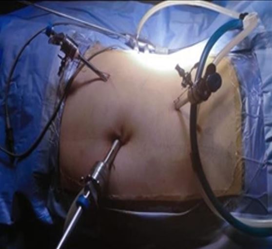
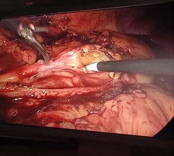
Background: Percutaneous nephrolithotomy. Various techniques have been documented for the management of Large Proximal Ureteric Stones (LPUS), such as laparoscopy (LUL), antegrade approach, retrograde ureteroscopy (RURS), extracorporeal shockwave lithotripsy, and infrequently open surgery. The paper compared the overall results of treating patients with large proximal ureteric stones (15-20 mm) using transperitoneal LUL, RURS, and mini-percutaneous antegrade ureteroscopy.

Methods: This prospective, randomised trial involved 100 individuals of both sexes who were above the age of 18 and had a single, 15-20 mm LPUS. Patients were split into two equal groups at random: Group B received RURS combined with laser fragmentation, and Group A received LUL.

Results: 100 patients who were included in the trial were split evenly between the two groups based on similar demographic information and stone criteria. Regarding patient demographics and stone criteria, both groups were similar. Group A had a substantially greater stone-free percentage (100%) than Group B (72%). There was a significant difference (p<0.001) in the operating duration, hemoglobin deficit, and ultimate stone-free rate between groups A and B. There was little difference in the two groups' conversation rates, mucosal injuries, ureteral perforations, complications, and length of hospital stay.

Conclusions: Compared to RURS, LUL is linked to a greater stone-free rate and fewer complications

Keywords: Proximal Ureteric Stone, Retrograde Ureteroscopy, Laparoscopic Ureter Lithotomy, Stone Free Rate

Corresponding Author How to Cite this Article To Browse
S M Yunus Ali, Assistant Professor, Department of Urology, Bangabandhu Sheikh Mujib Medical University, Dhaka, , Bangladesh.
Email:
Ali SMY, Islam MS, Islam MS, Jahan S, Islam MR, Miraj AKA, The Outcome of Different Endoscopic Modalities in the Management of Large Proximal Ureteric Stone. Int J Med Res Rev. 2024;12(6):176-182.
Available From
https://ijmrr.medresearch.in/index.php/ijmrr/article/view/1515

Manuscript Received Review Round 1 Review Round 2 Review Round 3 Accepted
2024-11-07 2024-11-15 2024-11-23 2024-12-02 2024-12-10
Conflict of Interest Funding Ethical Approval Plagiarism X-checker Note
None Nil Yes 11.77

© 2024by Ali SMY, Islam MS, Islam MS, Jahan S, Islam MR, Miraj AKAand Published by Siddharth Health Research and Social Welfare Society. This is an Open Access article licensed under a Creative Commons Attribution 4.0 International License https://creativecommons.org/licenses/by/4.0/ unported [CC BY 4.0].

Download PDFBack To ArticleIntroductionMethodsResultsDiscussionConclusionReferences

Introduction

Ever since the introduction of shock-wave lithotripsy (SWL), technical advancements in endoscopic approaches, including ureterorenoscopy (URS) and percutaneous nephrolithotomy (PCNL), surgical management of the urinary stone disease has been revolutionized from an open approach. More than 12% of people have urinary calculi, which are frequent [1]. Urolithiasis is the third most common affliction of the urinary tract, exceeded only by UTIs and pathologic conditions of the prostate (benign prostatic hyperplasia and prostate cancer). The treatment of urinary lithiasis has been revolutionized during the last three decades. Minimally invasive therapies in the form of endoscopic surgery in conjunction with the advent of SWL have diminished the role of open stone surgery. While most small distal stones can pass on their own, large proximal ureteral stones (LPUS) with a diameter of 10 mm or more are less likely to do so. In more extreme cases, LPUS can become impacted in the ureter, leading to severe pain, UTIs, hydronephrosis, and renal failure [2]. A variety of techniques, such as antegrade approach, laparoscopic ureter lithotomy (LUL), retrograde ureteroscopy (RURS), extracorporeal shockwave lithotripsy (ESWL), and infrequently open surgery, have been documented for the treatment of LPUS [3]. Furthermore, several criteria, including the size, content, and placement of the stone, clinical considerations, the accessibility of equipment, and the surgeon's skill, determine the best course of action [4]. Since RURS has an overall stone-free rate (SFR) of 81% (range 77-85%) for stones larger than 1 cm, it is now regarded as the first-line technique for treating upper-third ureteric stones [3]. Retrograde stone retropulsion during fragmentation is a prevalent issue with traditional ureteroscopy; it occurs with an incidence of 28-60% lowers the SFR and increases the requirement for supplementary treatments [5]. Additionally, the field of view is restricted by stone impaction and adjacent mucosal edema, which raises the possibility of complications such as perforation and instrument damage [6]. Lately, LUL has shown to be a cutting-edge choice for the management of big proximal ureteral stones [7]. Currently, LUL appears to be most useful for big impacted stones and has approximately a 100% success rate in stone clearing [8].

The study compared the overall results of mini-percutaneous antegrade ureteroscopy, transperitoneal LUL, and RURS in the treatment of patients with large proximal ureteric stones (15-20 mm).

Methods

This prospective, randomized trial involved 100 patients of both sexes who were above the age of 18 and had a single, large, proximal ureteric stone (LPUS) measuring between 15 and 20 mm. The study was conducted from March 2022 to April 2023 in the Department of Urology, BSMMU, Dhaka, Bangladesh with consent from the ethics committee. The patients gave their informed written permission. The following conditions had to be met to be excluded: bleeding diathesis, distal ureteric obstruction, renal insufficiency, pregnancy, patients with congenital kidney anomalies such as horseshoe kidney and ectopic pelvic kidney, multiple ureteric stones or associated renal stones, patients with a history of open abdominal surgery, children, and active urinary tract infections (UTIs). They were split into two equal groups, each with 25 patients, at random. Group B had RURS and holmium laser stone fragmentation treatment, whereas Group A received TPLU treatment. Before surgery, every patient was evaluated through a combination of non-contrast computed tomography (CT) to evaluate stone criteria and a plain abdominal radiograph of the kidneys, ureters, and bladder (PUT), as well as laboratory and radiological investigations. Laboratory investigations included complete blood counts (CBC), kidney and liver function tests, bleeding profiles, and urine tests.

Group A (TPLU): Initially, the patients were put in the lithotomy position while under general anaesthesia. Under the C arm, a retrograde 5Fr or 6Fr ureteral catheter had been placed close below the stone's level. After that, the patients were placed in the lateral decubitus position (kidney), with the side of the stone up and secured to the table with adhesive tapes. All pressure points were properly padded, and the pneumoperitoneum (15 mm Hg) was obtained using an open (Hasson) technique or, in most cases, a Veress needle. Next, a 10-mm camera port, two closed 12-mm and 5-mm ports, a 12-mm trocar (in the operator's right hand), and a 5-mm trocar (in the operator's left hand) were inserted. To form an isosceles triangle,


these trocars were positioned 6-8 cm lateral to the first trocar in the anterior axillary line (Figure 1, A). Following the introduction of the laparoscope into the operating field and colon reflection, the ureter was located anterior to the psoas muscle. An atraumatic grasper was used to confirm the position of the ureteral stone, which is indicated by a bulge in the ureter. If the stone is not perceptible, fluoroscopy may be utilized to aid in its location. Subsequently, a laparoscopic hook or cold knife may be used to perform a longitudinal ureterotomy (Figure 1, B) across the bottom part of the stone.

After antegrade DJ insertion, the ureterotomy was sealed with a 4-0 running Vicryl suture, and a drain was positioned in the peri-ureteral area. The ureterotomy was then prolonged using scissors, and the stone was removed using a nontraumatic grasper and deposited into a bag.

ijmrr-1515-01.JPG
A

ijmrr-1515-02.JPG
B
Figure 1: (A) three ports distribution and (B) longitudinal ureterotomy

Group B (RURS): All patients were placed in the traditional dorsal lithotomy posture while under general anaesthesia. The semi-rigid cystoscope (KARL STORZ 22Fr, Germany) is used for visualizing cystoscopy to locate the ureteric orifice. The next procedure involved inserting a guidewire into the renal pelvis with fluoroscopic assistance (Sensor polytetrafluoroethylene (PTFE)-nitinol guidewire with hydrophilic tip; Boston Scientific, Marlborough, MA, USA).

There are two guidewires inserted into the renal pelvis; the functioning wire is the one that allows the ureteroscopy to pass through. If problems occur, the other line serves as a safety wire and offers ongoing access to the kidney. If the guidewire proved difficult to pass through the stone on the first try, laser fragmentation was used to clear the way for the safety guidewire to pass through. Dilation of the ureter was accomplished using balloons (Uromax Ultra; Boston Scientific). To visualize the ureter, semirigid URS (9.5-11 Fr Karl Sorz, Germany) was employed. To make the repeated withdrawals and reinsertions of the FURS easier, a ureteral access sheath (Boston Scientific 11/13 Fr) was placed over the wire. We attempted to operate without a sheath if the ureter was tight, but if the stone was unreachable or problematic, a DJ stent was implanted for two weeks.

Under fluoroscopic guidance, the FURS (Wiscope Single-Use Digital FURS) was inserted up to the upper ureter. The shaft measured 8.6 Fr with a bullet-like 7.4 Fr tip and 3.6 Fr operating channel. A holmium laser (Lumenis-versa pulse power suit 100-W holmium laser) was used to fracture stone using 365 or 200-μm fibre. The energy was delivered at the following settings: 0.8-1 J/pulse, frequency 6-10 Hz, and extended pulse duration. Larger stone fragments were recovered using a stone basket (Zero Tip Nitinol Stone Basket 3 F., Boston Scientific, USA). Under fluoroscopic supervision, the ureteral access sheath was passed over the guidewire and the FURS was pushed through it if large pieces moved to the kidney.

Under a microscope, the access sheath was taken off to examine any bleeding or holes in the ureteric mucosa. In every patient, an indwelling double J ureteric stent (6 Fr, 26 cm, Percuflex; Boston Scientific) was placed following the surgery. The evaluation of the stone-free rate (SFR) among the three groups was the main result.


Assessing operation time, intraoperative problems, length of hospital stay, visual analogue scale (VAS) [9], auxiliary procedures, and early postoperative issues based on Clavien-Dindo grades were the secondary end goals [10].

Statistical analysis: IBM Inc., Chicago, IL, USA used SPSS v-21 for statistical analysis. The statistical variables were denoted by their mean and standard deviation (SD), and the unpaired Student's t-test was employed to compare them between the two groups. Frequency and percentage (%) were used to represent qualitative variables, and the Chi-square or Fisher's exact test was used for analysis as necessary. A statistically significant result was defined as a two-tailed P value less than 0.05.

Results

Table 1 illustrates how the 100 patients who were included in the trial were split evenly between the two groups based on similar demographic information and stone criteria. Table 2 displays the intraoperative parameters and the postoperative results. Group B's SFR was just 72%, whereas Group A's SFR was substantially greater at 100%. Before the JJ-stent was removed, residual pieces were handled by URS or ESWL. In group A, one subject had an open ureter lithotomy. The Clavien-Dindo Classification was used to grade every complication. One patient in Group B experienced ureteral perforation during laser fragmentation, which was successfully treated by DJ insertion, and four patients (16%) in Group B suffered mucosal damage during guidewire manipulation. Postoperative complications were comparable with two patients in group A who showed post-operative ileus and one patient in group B showed urosepsis which was managed by supportive measures in the ICU. Postoperative pain was comparable between both groups, Table 1.

Data are presented as mean±SD or frequency (%). p: p value for comparing between the studied groups. RURS: Retrograde Ureteroscopy, TPLU: Trans peritoneal Laparoscopic Ureter lithotomy, BMI: Body mass index. Operative time, HB deficit, and final SFR were a significant difference between both groups A and B (p <0.001). Conversation rate, mucosal Injury, ureteral perforation, complications, and hospital stay were insignificantly different between both groups Table 2.

Table 1: Comparison between the three studied groups according to demographic data

TPLU (N=50)RURS (N=50)P
Age (Years)41.37±8.7039.89±8.290.737
SexMale30(60.0%)32(64%)0.756
Female20(40.0%)189(36%)
BMI (kg/m2)26.51±1.4026.91±1.380.144
LateralityRight20(40.0%)22(44.0%)0.888
Left30(60.0%)28(56.0%)
Radio opacityRadio opaque36(72.0%)38(76.0%)0.850
Radiolucent14(28.0%)12(24.0%)
Stone size17.0±1.8317.0±1.710.768
SiteL2-L312(24.0%)20(40.0%)0.768
L320(40.0%)16(32.0%)
L3-L410(20.0%)8(16.0%)
L48(16.0%)6(12.0%)

Table 2: Intraoperative parameters and postoperative outcomes

TPLU (N=50)RURS (N=50)P
Operative time85.0±7.5761.0±8.21< 0.001*
Conversation rate2 (4%)0(0.0%)1.000
Mucosal Injury0(0.0%)8 (16%)0.155
Ureteral perforation0(0.0%)2 (4%)1.000
HB deficit0.50±0.240.33±0.14< 0.001*
P1=0.001*, P2=0.075, p3<0.001*
ComplicationsFever6(12%)8(16%)0.339
Hematuria0(0.0%)6(12%)0.339
Ileus4(8%)0(0.0%)0.339
Sepsis0(0.0%)2(4%)0.339
Hospital stays2.15±0.502.40±0.810.060
Final SFR100%72%0.011*

Data are presented as mean±SD or frequency (%). * Significant p-value < 0.05, the p-value for comparing between the three studied groups, p1: p-value for comparing between TPLU and RURS, p2: p-value for comparing between TPLU L and Antegrade URS, p3: p-value for comparing between RURS and Antegrade URS, RURS: Retrograde Ureteroscopy, TPLU: Transperitoneal Laparoscopic Ureter lithotomy, Hb: haemoglobin, SFR: Stone Free Rate.

Discussion

The most effective therapeutic technique for upper ureteral stones is still up for dispute among endocrinologists, making the decision about LPUS care typically fraught with difficulty.


A variety of therapeutic techniques for LPUS have been documented, including laparoscopy, ESWL, RURS, antegrade approach, and infrequently open surgery [11]. Although, for proximal ureteral stones, SWL is minimally invasive and can be performed as an outpatient procedure, disadvantages include a high retreatment rate, long treatment time, and poor patient compliance. AUA recommends SWL as the first line of management for small (<1 cm) with excellent results but indications were unclear for proximal ureteral stones >1 cm. One significant barrier to minimally invasive surgery is operating time. In terms of operation time, group A had an average operation time of 85.0±7.57 in our study, whereas group B had an average operation time of 61.0±8.21. Comparable to our findings, Elgebaly et al. [12] observed that the RURS group's mean operating time was 64.7 (17.7) minutes, while Kadyan B et al. [13] discovered that the TPLU group's mean operative time was 84.1±16.8 minutes and the RURS group's was 62.8±12.7 minutes. Contrary to our results Güler Y and Erbin A [14] in their study reported that the mean operative time was 147 ±67 in the TPLU group which is longer than the operative time in our corresponding group and this may be due to our good experience in laparoscopy. Rendering the patients stone-free is one of the most crucial outcomes in any stone treatment modality. In our study, SFR was 100% and 72% in the TPLU and RURS groups respectively. According to our results, Güler Y and Erbin A [14] in their study, the SFR was 97.5% and 83.7% in the LUL and Retrograde groups respectively. Also, in Wang, et al. [1] study, the SFR was 72% and 100% in the RURS and RPLU groups, respectively. In Basiri, et al. [15] study, the SFR was 90% and 86% in retrograde and laparoscopy groups, respectively. Additionally, Kumar, et al. [16] contrasted URS with LUL in an RCT. According to our findings, the final SFR was 76% in URS and 100% in LUL (P=0.02). In our study, in group B, 5/25 (20%) patients showed some intraoperative complications, 4/25(16%) cases showed mucosal injury, and 1(4%) case of ureteral perforation while no intraoperative complications were reported in group A. The mucosal injury was mainly 2ry to manipulation of the guidewire and the perforation was due to the disintegration of the impacted stone against the ureteric wall. In Kadyan B, et al. study [13], 3/50 (5%) cases in the RURS group showed mucosal injury and 2/50 (3.3%) cases showed ureteral perforation (nearly as in our study).

Also, In Güler Y and Erbin A's study [14] 2/43 (4.6%) in the RURS group had ureteral perforation. As regards the postoperative complications, in the present study, 6 (12%) patients in group B showed transient hematuria but no cases that needed a blood transfusion. HB deficit was significantly higher in the TPLU group with a mean deficit was 0.50±0.24 while the mean Hb deficit in group B was 0.33±0.14 having significant variations between the A and B groupings (P=0.001). In Güler A and Erbin A's study [14], transfusion was not needed in the two groups either RURS or LUL with HB deficit was 0.5±0.2 and 0.8±0.3 in RURS and LUL groups respectively. In the current study, fever (grade 2) was reported in 6 (12%) cases in group A and 8 (16%) cases in group B and managed by antipyretics and proper antibiotics, In Güler A and Erbin A study, 1/43 (2.3%) and 2/41 (2.4%) cases in RURS and LUL groups respectively showed fever. Dissection might raise the risk of urine leak, paralytic ileus, and postoperative discomfort due to bigger impacted stones, which are especially detrimental in LUL [15]. In our study, two (8%) cases in the laparoscopy group showed ileus, a lower incidence of ileus was reported in Abdel Raheem A et al. [17] study, as postoperative ileus, after TPLU occurred only in one patient (2.3%). In our study, a more prolonged hospital stay was seen in group B; nevertheless, there was no statistically significant difference between groups A and B. The mean hospital stay (days) was 2.14±0.49, 2.40±0.81, and 2.89±1.02, respectively. In Güler Y and Erbin A study [14], the mean hospital stay in the retrograde group was nearly similar to our results (2.0 ±1.3) while it was longer in the laparoscopic group Wang et al. [1] also found similar results, reporting that the mean hospital stay for the RURS and laparoscopic groups was 2.5 and 4.3 days, respectively. Nevertheless, there are several limits to our research. Firstly, the relatively small sample size may not attain adequate power to generate accurate results. Secondly, large-scale randomized trials should be established to allow for the verification of strong conclusions with a higher level of statistical power.

Conclusion

In the hands of a skilled laparoscopic practitioner, LUL may be the best option for managing LPUS since it has been linked to the greatest rate of stone removal and the fewest problems.


Furthermore, in situations where many centres lack flexible endoscopes, equipment, and laser machines for budgetary reasons, LUL may be a useful substitute for other solutions.

Permission from Institutional research board: Yes

Funding: Nil

Conflict of interest: None Initiated

References

1. Wang Y, Zhong B, Yang X, Wang G, Hou P, Meng J. Comparison of the efficacy and safety of URSL, RPLU, and MPCNL for treatment of large upper impacted ureteral stones: a randomized controlled trial. BMC Urol. 2017 Jun 29;17(1):50. doi: 10.1186/s12894-017-0236-0. PMID: 28662708; PMCID: PMC5492714 [Crossref][PubMed][Google Scholar]

2. Mousavi Bahar SH, Amirhassani S, Nouralizadeh A, ZerafatJou N, Rasiuli J. Percutaneous Nephrolithotomy Versus Laparoscopy in the Management of Large Proximal Ureteral Stones: The Experience of Two Different Settings. Urol J. 2019 Oct 21;16(5):448-452. doi: 10.22037/uj.v0i0.4538. PMID: 30882178 [Crossref][PubMed][Google Scholar]

3. Preminger GM, Tiselius HG, Assimos DG, Alken P, Buck C, Gallucci M, Knoll T, Lingeman JE, Nakada SY, Pearle MS, Sarica K, Türk C, Wolf JS Jr; EAU/AUA Nephrolithiasis Guideline Panel. 2007 guideline for the management of ureteral calculi. J Urol. 2007 Dec;178(6):2418-34. doi: 10.1016/j.juro.2007.09.107. PMID: 17993340 [Crossref][PubMed][Google Scholar]

4. Kijvikai K, Haleblian GE, Preminger GM, de la Rosette J. Shock wave lithotripsy or ureteroscopy for the management of proximal ureteral calculi: an old discussion revisited. J Urol. 2007 Oct;178(4 Pt 1):1157-63. doi: 10.1016/j.juro.2007.05.132. Epub 2007 Aug 14. PMID: 17698126 [Crossref][PubMed][Google Scholar]

5. Elashry OM, Tawfik AM. Preventing stone retropulsion during intracorporeal lithotripsy. Nat Rev Urol. 2012 Dec;9(12):691-8. doi: 10.1038/nrurol.2012.204. Epub 2012 Nov 20. PMID: 23165399 [Crossref][PubMed][Google Scholar]

6. Chow GK, Patterson DE, Blute ML, Segura JW. Ureteroscopy: effect of technology and technique on clinical practice. J Urol. 2003 Jul;170(1):99-102. doi: 10.1097/01.ju.0000070883.44091.24. PMID: 12796655 [Crossref][PubMed][Google Scholar]

7. Cohen J, Cohen S, Grasso M. Ureteropyeloscopic treatment of large, complex intrarenal and proximal ureteral calculi. BJU Int. 2013 Mar;111(3 Pt B):E127-31. doi: 10.1111/j.1464-410X.2012.11352.x. Epub 2012 Jul 3. PMID: 22757752 [Crossref][PubMed][Google Scholar]

8. Kaygısız O, Coşkun B, Kılıçarslan H, Kordan Y, Vuruşkan H, Özmerdiven G, Yavaşcaoğlu İ. Comparison of ureteroscopic laser lithotripsy with laparoscopic ureterolithotomy for large proximal and mid-ureter stones. Urol Int. 2015;94(2):205-9. doi: 10.1159/000368374. Epub 2015 Jan 29. PMID: 25633596 [Crossref][PubMed][Google Scholar]

9. Boonstra AM, Schiphorst Preuper HR, Balk GA, Stewart RE. Cut-off points for mild, moderate, and severe pain on the visual analogue scale for pain in patients with chronic musculoskeletal pain. Pain. 2014 Dec;155(12):2545-2550. doi: 10.1016/j.pain.2014.09.014. Epub 2014 Sep 17. PMID: 25 [Crossref][PubMed][Google Scholar]

10. Dindo D, Demartines N, Clavien PA. Classification of surgical complications: a new proposal with evaluation in a cohort of 6336 patients and results of a survey. Ann Surg. 2004 Aug;240(2):205-13. doi: 10.1097/01.sla.0000133083.54934.ae. PMID: 15273542; PMCID: PMC1360123 [Crossref][PubMed][Google Scholar]

11. Preminger GM, Tiselius HG, Assimos DG, Alken P, Buck C, Gallucci M, Knoll T, Lingeman JE, Nakada SY, Pearle MS, Sarica K, Türk C, Wolf JS Jr; EAU/AUA Nephrolithiasis Guideline Panel. 2007 guideline for the management of ureteral calculi. J Urol. 2007 Dec;178(6):2418-34. doi: 10.1016/j.juro.2007.09.107. PMID: 17993340 [Crossref][PubMed][Google Scholar]

12. Elgebaly O, Abdeldayem H, Idris F, Elrifai A, Fahmy A. Antegrade mini-percutaneous flexible ureteroscopy versus retrograde ureteroscopy for treating impacted proximal ureteric stones of 1-2 cm: A prospective randomised study. Arab J Urol. 2020 Aug 23;18(3):176-180. doi: 10.1080/2090598X.2020.1769385. PMID: 33029428; PMCID: PMC7473252 [Crossref][PubMed][Google Scholar]


13. Kadyan B, Sabale V, Mane D, Satav V, Mulay A, Thakur N, Kankalia SP. Large proximal ureteral stones: Ideal treatment modality? Urol Ann. 2016 Apr-Jun;8(2):189-92. doi: 10. 4103/0974-7796.157963. PMID: 27141190; PMCID: PMC4839237 [Crossref][PubMed][Google Scholar]

14. Güler Y, Erbin A. Comparative evaluation of retrograde intrarenal surgery, antegrade ureterorenoscopy and laparoscopic ureterolithotomy in the treatment of impacted proximal ureteral stones larger than 1. 5 cm. Cent European J Urol. 2021;74(1):57-63. doi: 10.5173/ceju.2021.0174.R1. Epub 2021 Jan 23. PMID: 33976917; PMCID: PMC8097644 [Crossref][PubMed][Google Scholar]

15. Basiri A, Simforoosh N, Ziaee A, Shayaninasab H, Moghaddam SM, Zare S. Retrograde, antegrade, and laparoscopic approaches for the management of large, proximal ureteral stones: a randomized clinical trial. J Endourol. 2008 Dec;22(12):2677-80. doi: 10.1089/end.2008.0095. PMID: 19025388 [Crossref][PubMed][Google Scholar]

16. Kumar A, Vasudeva P, Nanda B, Kumar N, Jha SK, Singh H. A Prospective Randomized Comparison Between Laparoscopic Ureterolithotomy and Semirigid Ureteroscopy for Upper Ureteral Stones >2 cm: A Single-Center Experience. J Endourol. 2015 Nov;29(11):1248-52. doi: 10.1089/end.2013.0791. Epub 2014 Oct 30. PMID: 25177768 [Crossref][PubMed][Google Scholar]

17. Abdel Raheem A, Alowidah I, Hagras A, Gameel T, Ghaith A, Elghiaty A, Althakafi S, Al-Mousa M, Alturki M. Laparoscopic ureterolithotomy for large proximal ureteric stones: Surgical technique, outcomes and literature review. Asian J Endosc Surg. 2021 Apr;14(2):241-249. doi: 10.1111/ases.12861. Epub 2020 Sep 1. PMID: 32875735 [Crossref][PubMed][Google Scholar]