E-ISSN:2320-8686
P-ISSN:2321-127X

Research Article

Bone marrow

International Journal of Medical Research and Review

2013 Volume 1 Number 5 Nov-Dec
Publisherwww.medresearch.in

Blue round cell tumors – Bone marrow metastasis

Namrata NR1*
DOI:10.17511/ijmrr.2013.i05.12

1* Namrata N Rajakumar, Assistant Professor, Pathology, Kidwai Memorial Institute of Oncology, Bangalore, Karnataka, India.

Objective: Bone marrow involvement by solid tumor implicates advanced disease and bad prognosis. Bone marrow aspiration and biopsy, are performed routinely for staging for small round cell tumors and unexplained cytopenia in other solid tumors. It is important to rule out bone marrow involvement before planning for any definitive, curative treatment.

Materials and Methods: This was a retrospective observational study of bone marrow involvement by, Small round cell tumors / solid tumors and their hematological manifestation, especially the neuroblastomas in children.

Results: Evaluation of Bone marrow evaluation during the past 5 years, in solid malignancies, revealed Out of 574, 240 were pediatric cases and 334 were adult cases. Bone marrow was involved in 65 patients. In children, bone marrow involvement was present in 34 cases, and in adults, bone marrow involvement was diagnosed in 31 cases. Neuroblastoma was the most common malignancy, which involved the bone marrow in pediatric cases, followed by Ewing’s sarcoma& retinoblastoma. Among adult patients, neuroendocrine carcinoma involving bone marrow, a primary malignancy in the breast is common in women and prostatic carcinoma in men.

Conclusion: small round blue cell tumors are the major cause of bone marrow involvement in pediatric, Use of immunohistochemistry markers on bone marrow biopsies results in a higher detection rate.

Keywords: blue cell tumors.metastasis, Bone marrow Immunohistochemistry

Corresponding Author How to Cite this Article To Browse
Namrata N Rajakumar, Assistant Professor, Pathology, Kidwai Memorial Institute of Oncology, Bangalore, Karnataka, India.
Email:
Namrata NR, Blue round cell tumors – Bone marrow metastasis. Int J Med Res Rev. 2013;1(5):258-260.
Available From
https://ijmrr.medresearch.in/index.php/ijmrr/article/view/1509

Manuscript Received Review Round 1 Review Round 2 Review Round 3 Accepted
2013-02-01 2013-02-04 2013-02-11 2013-02-18 2013-02-25
Conflict of Interest Funding Ethical Approval Plagiarism X-checker Note
None Nil Yes 12.62

© 2013by Namrata NRand Published by Siddharth Health Research and Social Welfare Society. This is an Open Access article licensed under a Creative Commons Attribution 4.0 International License https://creativecommons.org/licenses/by/4.0/ unported [CC BY 4.0].

Introduction

Neoplasms that exhibit sharp peaks in incidence in children younger than age 10 years, ALL neuroblastoma, Wilms tumor, hepatoblastoma, retinoblastoma, rhabdomyosarcoma, Ewing sarcoma etc Histologically, many of the malignant non-hematopoietic pediatric neoplasms are unique. In general, they tend to have a more primitive (embryonal) undifferentiated appearance and are often characterized by sheets of cells with small, round nuclei, childhood tumors have been collectively referred to as their malignant small round blue cell tumors, due to their primitive histologic appearance [1] The most frequent childhood cancers arise in the hematopoietic system, nervous tissue, soft tissues, bone, and kidney. This is in sharp contrast to adults, in whom the skin, lung, breast, prostate, and colon are the most common sites. This study analyzed blue cell tumour, and metastasis to bone marrow, over 5 years (2006-2011), from a regional cancer centre in India. The majority of SRCT were Neuroblastoma which metastasised to bone marrow in the current study.

Materials and Methods

In Kidwai Cancer Hospital, Bengaluru, India, between January 2006 to January 2011, it was a retrospective observational study. All suspected nonhematological malignancies involving marrow were included in the study.. lymphoma was not included in this study. Bone marrow was obtained from the posterior iliac crest by Jamshidi needle. Bone marrow smears and peripheral smears and stained by Romanowsky stains, Bone marrow biopsies were stained with hematoxylin and eosin. Immunohistochemistry followed in a few cases. Patient’s name, age, gender, diagnosis, and clinical, radiological findings were recorded. Children age<14 years. The bone marrow was considered to be ‘involved by the tumour’ if tumour cells were detected in bone marrow aspirate, biopsy, or both.

Results

The bone marrow examinations, of suspected SRCT /solid malignancies patients were evaluated. Out of 574 total cases, 240 were pediatric cases and 334 were adult cases.(Table 1)

Table 1: Patient Information

Number of casesNumber of positive cases
Pediatric cases24034
Adult cases33431
Total57465

Bone marrow was involved in 65 patients(fig1).
ijmrr_1509_01.JPG
Figure 1:
Bone marrow smear showing small cells with hyperchromatic nucleus and increased N/C ratio.
ijmrr_1509_02.JPG
Figure 2:
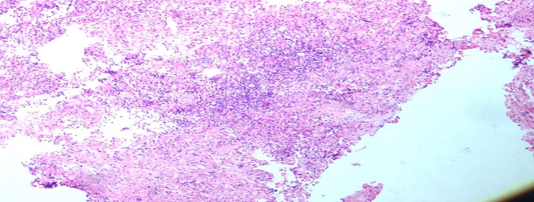
H&E 20X Tissue biopsy showing small cells in fibrillary matrix

Tissue confirmation from the primary site was available in 40 cases. (fig 2) Bone marrow involvement was present in 34 cases, in children, and 31 adult cases, showed metastasis, primarily from the breast and prostate. Neuroblastoma was the most common malignancy, which involved the bone marrow among pediatric cases( Table 2), followed by Ewing’s sarcoma& retinoblastoma.

Table 2: Distribution of bone marrow involvement among pediatric patients

DiagnosisNumber of cases
Neuroblastoma14
Ewing’s sarcoma12
Retinoblastoma6
Rhabdomyosarcoma2
Total34

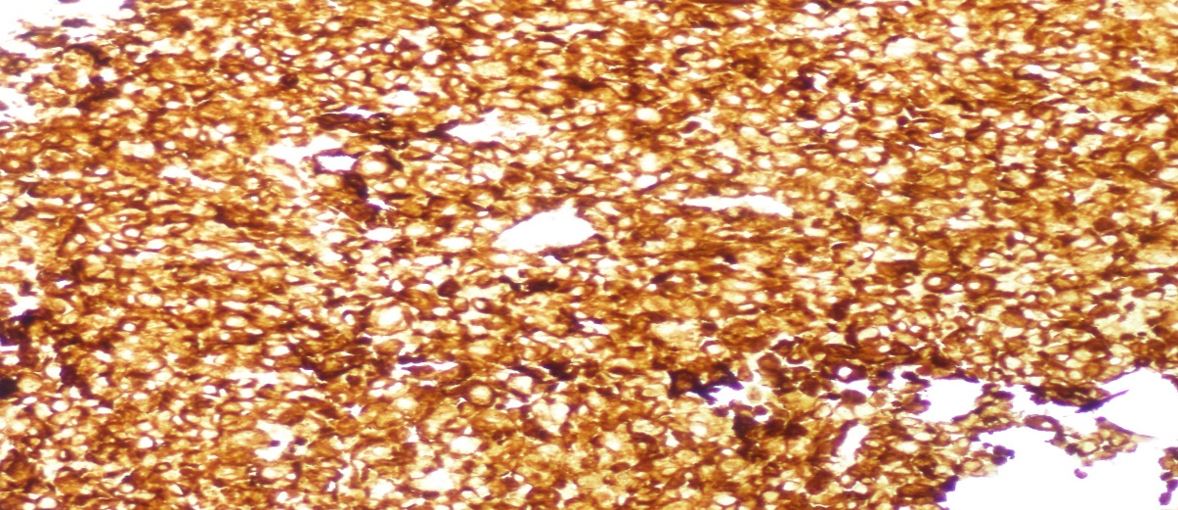
Commonest was the Neuroblastoma which metastasised to bone marrow in the current study. IHC confirmation was available in most cases. (fig 3). Anaemia was the common abnormality due to bone marrow involvement, the leucopenia/neutropenia and thrombocytopenia followed respectively. Pancytopenia was found in 8 children. Normal blood pictures, in the majority of patients, confirmed haematological abnormality may not be always present in bone marrow metastasis. IHC confirmation was available in most cases.(fig3)
ijmrr_1509_03.JPG
Figure 3:
IHC positive for synaptophysin

Discussion

Bone marrow involvement in non-haematological solid tumors is rare. Disseminated malignant cells from the primary tumor, are the cause of metastases to bone marrow. Detection of metastatic tumors in the bone marrow is mandatory, for staging and has prognostic value. Rolf et al showed NB frequently metastatic [1] [2]. Molenar et al. studied sequencing of neuroblastomas [3]. Diligent Morphological examination of the bone marrow aspiration with ancillary tests such as immunohistochemistry aids in detecting the bone marrow involvement early, also still, remains the easiest, cheapest, and least time-consuming procedure for the diagnosis of clinically suspected bone marrow involvement.[3&4]. Excess secretion of catecholamines may be noted.


Though radiological suggestion might help, morphological examination is confirmatory. Reich et al showed as early as 1935, metastatic neoplastic cells appeared in bone marrow. [5] Curtis studied solid cancer after bone marrow transplantation [6]. Valdes et al studied bilateral bone marrow biopsy having more detection rate. [7] Blue cell tumors metastasis common in children Bilateral bone marrow biopsies improves the metastatic detection rate.

The bone marrow metastasis in adults primarily were from, the breast, prostate, and lung.[8], [9], [10], [11] In a retrospective study, Mohanty and Dash reported that, in children, neuroblastoma was the common primary tumour involving bone marrow. In this study, we also found the neuroblastoma as the most common malignancy metastasizing to bone marrow among pediatric cases, comprising around 40 % of all patients. Neuroblastoma was followed by Ewing’s sarcoma and retinoblastoma rhabdomyosarcoma. In childhood, about 40% of neuroblastomas arise in the adrenal medulla. Tumours may arise in numerous other sites, including the pelvis, the neck, and within the brain (cerebral neuroblastomas). Neuroblastomas range in size from minute nodules (so-called in situ lesions) to large masses of more than 1 kg in weight. In adult women, the commonest metastasis is from primary breast carcinomas[8] [9]

Histologically, classic neuroblastomas are composed of small, primitive-appearing cells with dark nuclei, scant cytoplasm, and poorly defined cell borders growing in solid sheets. Such tumours may be difficult to differentiate morphologically from other small round blue cell tumours, Mitotic activity, nuclear breakdown ("karyorrhexis"), and pleomorphism may be prominent. Eosinophilic fibrillary material neuropil corresponds to neuritic processes of the primitive neuroblasts, rosettes (Homer-Wright pseudorosettes) can be found, tumor cells are concentrically arranged about a central space filled with neuropil .immunohistochemical reactions for neuron-specific enolase will confirm the diagnosis[10]. Ultrastructural seen are dense core granules. Signs of maturation are seen in some, that can be spontaneous or therapy-induced. After the availability of immunohistochemistry markers, these tumors involving marrow are increasingly diagnosed [10] [11] [12] Mohanty and Dash reported that, in children, neuroblastoma was the common primary tumor with metastasis [12].

Conclusion

Neuroblastoma involves bone marrow frequently among all blue cell tumors. Use of immunohistochemistry markers on bone marrow biopsies may result in a higher detection rate. NB cells within the BM are the most powerful negative prognostic factor for NB patients. Future Studies are needed to detect proteins differentially expressed by metastatic NB cells, which may be new prognostic markers and novel targets for therapy.

Abbreviations: SRCT small round cell tumors, NB-Neuroblastoma

Permission from Institutional Research Board (IRB): Yes

Funding: Nil

Conflicts of interest: none

References

1. Park JR, Eggert A, and Caron H: Neuroblastoma: Biology, prognosis, and treatment. Hematol Oncol Clin North Am. 24:65-86. 2010. View Article: Google Scholar : [Crossref][PubMed][Google Scholar]

2. Rolf et al. Frequency and significance of bone marrow involvement by metastatic solid tumors. Cancer39;1337. 1977. [Crossref][PubMed][Google Scholar]

3. Molenaar JJ, Koster J, Zwinenburg DA. et al: Sequencing of neuroblastoma identifies chromothripsis and defects in Neuri-togenesis genes. Nature 483:589-93. 2012. [Crossref][PubMed][Google Scholar]

4. Olshan AF Bunin GR. Epidemiology of Neuroblastomain BORDEU GM pa eds Neuroblastoma Amsterdam Elsevier Science. BV;2000;33-39. . [Crossref][PubMed][Google Scholar]

5. Rcich C. A study of the diagnostic value of stenal puncture in clinical hematology. Am J Med 1935:189:515-20. . [Crossref][PubMed][Google Scholar]

6. Curtis re. et al solid cancer after bone marrow transplantation. N Engl J Med 336:897. 1997. [Crossref][PubMed][Google Scholar]

7. Valdes-Sanchez M. Nava-Ocampo AA. Palacios-Gonzalez RV. Perales-Arroyo A, Medina-Sansion A. Martinez-Avalos A.er al Diagonosis of bone marrow metastases in children with solid tumors and lymphomas. Aspiration, or unilateral or bilateral biopsy? Arch Med Res 2000:31:58-61 [Crossref][PubMed][Google Scholar]

8. Landys K. Prognostic value of bone marrow biopsy in breast cancer. Cancer 1982:49:513-8. . [Crossref][PubMed][Google Scholar]

9. Anner RM. Drewinko B. Frequency and significance of bone marrow involvement by metastatic solid tumors. Cancer 1977:39:1337-14. [Crossref][PubMed][Google Scholar]

10. Reid MM, Wallis JP, McGuckin AG, Pearson AD. Malcolm AJ, Routine histological compared with immunohistological examination of bone marrow trephine biopsy specimens in disseminated neuroblastoma. J Clin Pathol 1991:44:493-6. . [Crossref][PubMed][Google Scholar]

11. Kilickap S. Erman M, Dincer M, Aksoy S. Hakan H. Yalcin Y. Bone marrow metastasis of solid tumors: Clincopathological evaluation of 73 cases. Turk J cancer 2007:37:85-8 [Crossref][PubMed][Google Scholar]

12. Mohanty SK, Dash S. Bone marrow metastasis in solid tumors. Indian J Pathol Microbiol 2003:46:613-6. . [Crossref][PubMed][Google Scholar]