E-ISSN:2320-8686
P-ISSN:2321-127X

Case Report

Appendicitis Revealed

International Journal of Medical Research and Review

2024 Volume 12 Number 4 Jul-Aug
Publisherwww.medresearch.in

A Suspected Case of Acute Appendicitis Revealed to be Isolated Submucosal Lipomatosis Of Appendix on Computed Tomography: A Case Report

Biswas P1*, Supriya M2
DOI:https://doi.org/10.17511/ijmrr .2024.i04.05

1* Pintu Biswas, Senior Resident, Agartala Government Medical College and Gb Pant Hospital, Agartala, Tripura, India.

2 Matam Supriya, Post Graduate Trainee, Agartala Government Medical College and Gb Pant Hospital, Agartala, Tripura, India.

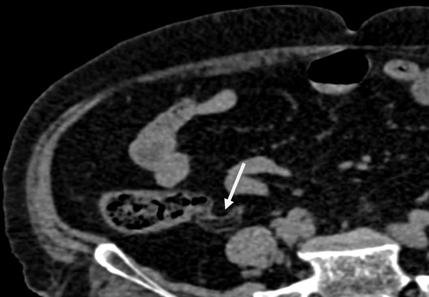
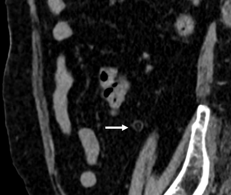
Symptomatic isolated submucosal intestinal lipomatosis is rare. Also, few cases have been reported in the literature. Here, we are presenting computed tomography findings of a rare case of isolated submucosal lipomatosis of the appendix presumptively diagnosed as acute appendicitis in a 72-year-old female. This case highlights the importance of considering isolated submucosal lipomatosis as a differential diagnosis in instances of suspected acute appendicitis, especially when clinical findings are inconclusive.

Keywords: Acute appendicitis, Computed tomography, isolated submucosal lipomatosis, intestinal lipomatosis

Corresponding Author How to Cite this Article To Browse
Pintu Biswas, Senior Resident, , Agartala Government Medical College and Gb Pant Hospital, Agartala, Tripura, India.
Email:
Biswas P, Supriya M, A Suspected Case of Acute Appendicitis Revealed to be Isolated Submucosal Lipomatosis Of Appendix on Computed Tomography: A Case Report. Int J Med Res Rev. 2024;12(4):123-125.
Available From
https://ijmrr.medresearch.in/index.php/ijmrr/article/view/1495

Manuscript Received Review Round 1 Review Round 2 Review Round 3 Accepted
2024-07-01 2024-07-04 2024-07-10 2024-07-16 2024-07-22
Conflict of Interest Funding Ethical Approval Plagiarism X-checker Note
Nil None Yes 12.32

© 2024by Biswas P, Supriya Mand Published by Siddharth Health Research and Social Welfare Society. This is an Open Access article licensed under a Creative Commons Attribution 4.0 International License https://creativecommons.org/licenses/by/4.0/ unported [CC BY 4.0].

Introduction

Symptomatic isolated submucosal intestinal lipomatosis is a rare entity [1,2]. Lipomatosis is characterized by an increased infiltration of well-differentiated fat within the submucosal layer of the bowel. Unlike lipomas, lipomatosis lacks a defining capsule [3]. It is commonly observed in the cecum, ascending colon, sigmoid colon, transverse colon, rectum, and descending colon with decreasing frequency [3]. The presence of lipomatosis in the appendix is quite rare. The exact etiology of this condition remains unknown.

Case Report

A 72-year-old female presented to the emergency department with symptoms indicative of an acute abdomen. She was conscious, coherent, and cooperative, with stable vital signs. Physical examination revealed tenderness in the right iliac fossa. Suspecting acute appendicitis, a computed tomography (CT) scan was performed for further evaluation. The computed tomography (CT) scan of the abdomen and pelvis revealed the appendix in a pelvic position, exhibiting a uniformly hypodense thickened wall.

The maximum wall-to-wall diameter measures approximately 89 mm, with the submucosa showing fat density ranging from -30 to -90 HU. No other signs of inflammation like peri appendiceal fat stranding, fluid accumulation, or lymphadenopathy were noted. A diagnosis of isolated submucosal lipomatosis of the appendix was made. Consequently, the patient was discharged the following day after her symptoms had resolved.

ijmrr_1495_01.JPG
Figure 1:
Non-contrast Enhanced Computed Tomography Axial plane

ijmrr_1495_02.JPG
Figure 2:
Non-contrast Enhanced Computed Tomography coronal plane

ijmrr_1495_03.JPG
Figure 3:
Non-contrast Enhanced Computed Tomography sagittal plane

Images show the fatty infiltration in the submucosa of the appendix resulting in the thickening of the wall. (Siemens CT Definition AS 128 slice CT (2017). Protocol: 50 mAs, 120 kV, 1 mm Slice thickness.

Discussion

Gastrointestinal lipomatosis is characterized by the infiltration of mature adipose tissue into the submucosal layer of the bowel, distinguishing it from lipomas due to its lack of a capsule. The cecum is the most common site for this condition, followed by the ascending colon, sigmoid colon, transverse colon, rectum, and descending colon [3]. Isolated lipomatosis of the appendix is rare and can present as diffuse, asymmetric, or focal [2]. Initially described by Hellstrom in 1906, intestinal lipomatosis typically involves the submucosa in 90% of cases, though it may extend into the muscularis propria or subserosa in up to 10% of cases.


The pathogenesis of appendiceal lipomatosis remains unclear, with potential etiological factors including embryonic displacement of adipose tissue, degenerative changes affecting fat metabolism, post-chemotherapy fat deposition, chronic irritation, low-grade infection, and hamartomata’s syndromes [4].

Lipomatosis can present with a range of non-specific symptoms, including abdominal pain, diarrhoea, constipation, sub ileus or ileus, and bleeding. In some cases, it may manifest as acute appendicitis. The presumed cause is a mechanical obstruction of stool discharge from the appendix due to lipomatosis [5].

On radiological imaging techniques, thickening of the appendiceal wall which is in favour of fatty tissue without peri appendiceal inflammation, fluid accumulation, or lymphadenopathy is suspicious for the diagnosis. Hypodense homogeneous thickening between -80 to -120 HU on CT, high intensity in T1 and T2 weighted sequences on MRI and decreasing intensity in fat suppression sequence support fatty tissue infiltration. Despite its rarity, appendiceal lipomatosis should be considered in the differential diagnosis of acute appendicitis when clinical findings are uncertain.

Conclusion

Gastrointestinal lipomatosis is a rare occurrence, with isolated submucosal lipomatosis being exceptionally uncommon. Symptoms are only present in a small number of patients. Physicians need to be mindful of this condition to avoid unnecessary surgical procedures, especially when both clinical and laboratory results appear stable.

Despite their rarity, this condition should be considered in the differential diagnosis of suspected cases of acute appendicitis where clinical uncertainty exists, a CT scan is a reliable diagnostic tool. A definitive diagnosis necessitates a histopathological examination.

References

1. Anatolia AM. Isolated submucosal lipomatosis of appendix mimicking acute appendicitis: computed tomography findings. Acta Medica Anatolia 2014;3 (2):76-77. doi :10. 15824/actamedica.35025 [Crossref][PubMed][Google Scholar]

2. Chandanwale SS, Patel P, Verma A, Patel N. A Focal Submucosal Lipomatosis: An Extremely Rare Cause of Appendicitis. Indian Journal of Medical Specialities. 2023 Jan 1;14(1):45-6. doi: 10.4103/injms.injms_107_223 [Crossref][PubMed][Google Scholar]

3. Dajti G, Vagliasindi A, Bosi S, Guerra E, Zanzi F, Raulli GD. An unusual clinical presentation of colonic lipomatosis: A case report. International journal of surgery case reports. 2020 Jan 1;75:273-5. doi: 10.1016/j.ijscr.2020.08.036 [Crossref][PubMed][Google Scholar]

4. Thakur B, Kishore S, Bhardwaj A, Kudesia S. Diffuse intestinal submucosal lipomatosis with incidental epidermal inclusion cyst of caecum clinically masquerading as carcinoma caecum. Rare Tumors. 2014 Aug 26;6(3):114-5. doi: 10.4081/rt.2014.5380 [Crossref][PubMed][Google Scholar]

5. Shanmugarajah I, Sundrehagen BH, Warberg EA, Kazaryan AM. Lipomatosis of appendix in a teenager. Clinical Case Reports. 2021 Jul;9(7):e04595. [Article][Crossref][PubMed][Google Scholar]