E-ISSN:2320-8686
P-ISSN:2321-127X

Case Series

Congenital pulmonary

International Journal of Medical Research and Review

2024 Volume 12 Number 4 Jul-Aug
Publisherwww.medresearch.in

A Case Series Report on Congenital pulmonary airway malformation – antenatal diagnosis and post-natal follow up

Sahu S1, Khokan M2, Guha Roy A3*
DOI:https://doi.org/10.17511/ijmrr .2024.i04.06

1 Suparna Sahu, Senior Resident, Department of Radiodiagnosis, Nil Ratan Sircar Medical College, Kolkata, West Bengal, India.

2 Mandi Khokan, Demonstrator, Department of Radiology, Deben Mahato Government Medical College and Hospital, Purulia, West Bengal, India.

3* Anik Guha Roy, Post Graduate Resident, Md Radiodiagnosis, Nil Ratan Sircar Medical College, Wbuhs, West Bengal, India.

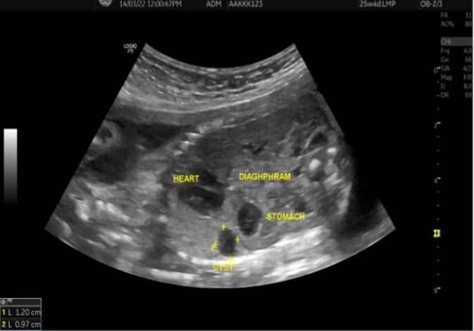
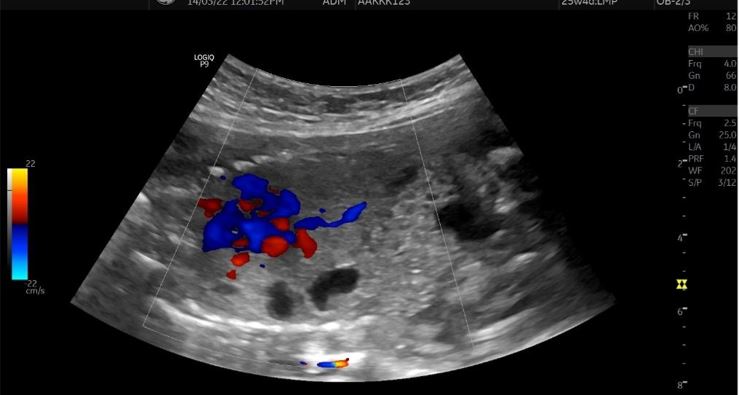
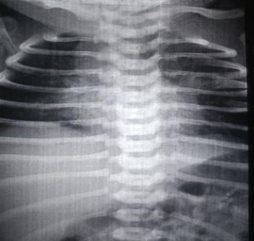
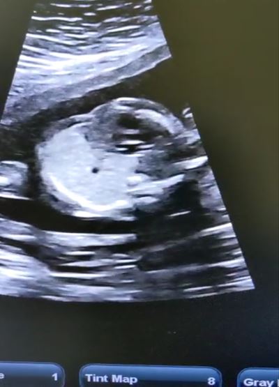
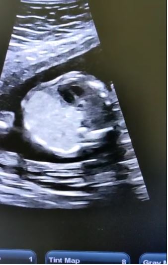
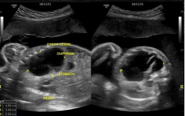
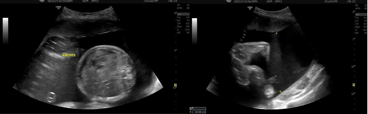
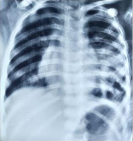
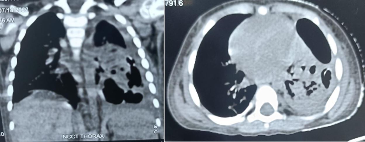
Congenital pulmonary airway malformation is the most commonly detected congenital lung defect in the ante-natal period. CPAM is a rare developmental, non-hereditary dysplastic lung lesion. No obvious association with maternal age, race or exposure to any given factor could be ascertained. It has an incidence of about 1:1500-4000 of all live births and accounts for approximately 25% of all congenital lung lesions. Abnormal proliferation of the fetal tracheobronchial tree in the early weeks of gestation can lead to the development of this condition. Frequent associated congenital defects include congenital heart defects, tracheoesophageal fistula, congenital diaphragmatic hernia, and renal agenesis. Post-natal prognosis of CPAM depends on the size of the lesion, degree of pulmonary hypoplasia, presence of other anomalies and development of fetal hydrops. We present four cases of congenital pulmonary airway malformation: antenatally detected by ultrasound with their follow-up in the post-natal period. Three of them have normal uneventful post-natal periods. One child was normal for six months of age then developed frequent respiratory tract infections.

Keywords: CPAM, USG, cystic lung lesion, Types of CPAM, respiratory tract infection

Corresponding Author How to Cite this Article To Browse
Anik Guha Roy, Post Graduate Resident, Md Radiodiagnosis, Nil Ratan Sircar Medical College, Wbuhs, West Bengal, India.
Email:
Sahu S, Khokan M, Guha Roy A, A Case Series Report on Congenital pulmonary airway malformation – antenatal diagnosis and post-natal follow up. Int J Med Res Rev. 2024;12(4):126-131.
Available From
https://ijmrr.medresearch.in/index.php/ijmrr/article/view/1485

Manuscript Received Review Round 1 Review Round 2 Review Round 3 Accepted
2024-07-01 2024-07-04 2024-07-10 2024-07-16 2024-07-22
Conflict of Interest Funding Ethical Approval Plagiarism X-checker Note
Nil None Yes 13.32

© 2024by Sahu S, Khokan M, Guha Roy Aand Published by Siddharth Health Research and Social Welfare Society. This is an Open Access article licensed under a Creative Commons Attribution 4.0 International License https://creativecommons.org/licenses/by/4.0/ unported [CC BY 4.0].

Introduction

Congenital pulmonary airway malformation is a rare developmental dysplastic lesion of the lung [1] [2]. It is also known as congenital adenomatoid malformation. It has been found that it occurs due to arrested development of the foetal bronchial tree during the 6th and 7th week of foetal development [3] [4].

It is believed that congenital pulmonary airway malformation is a part of the spectrum of bronchopulmonary foregut malformation [5]. About ~25% of a congenital lung lesion is CPAM [6] [7]. It has an incidence of about 1:1500-4000 of total live births [8].

It is usually diagnosed antenatally by routine ultrasound examination. The lesion is usually unilateral and involves a single lobe [9] [10]. It appears as an intra-thoracic cystic or solid-appearing echogenic lesion on ultrasound [11]. Differential diagnosis like broncho-pulmonary sequestration, and congenital diaphragmatic hernia can be excluded by doing B mode & doppler USG [12].

Depending on the size of the cysts we can also detect the type of CPAM [13] [14]. The mortality rate of CPAM is low and has a favourable prognosis: hence with proper care & counselling pregnancy can be continued with postnatal follow-up [15]. Here, we are presenting 4 cases of ante-natally detected CPAM with their sonographic appearance and post-natal follow-up.

Case 01

A 21-year-old female patient, primigravida attended our department at 23 weeks 4 days of gestational age for a routine anomaly scan. The ultrasound examination revealed a small cystic lesion (1.2x 0.9 cm) in the left lung without significant mass effect or vascularity on colour Doppler study.

The lesion was seen above the diaphragm and separated from the fundic bubble. The lesion is classified as type 2 according to Stoker’s classification. Amniotic fluid volume was normal. No other abnormality was noted.

The patient was counselled and the pregnancy was continued. At term, LUCS was performed: baby cried immediately after birth and the birth weight was 2950 grams.

Post-natal chest x-ray was done and it showed a small cystic lesion with an internal air-fluid level in the left hemithorax. No significant mediastinal shift. Both dome diaphragms are delineated.

ijmrr_1485_01.JPG
Figure 1: Ante-natal USG showed a small cystic lesion in the left lung region with a normal heart

ijmrr_1485_02.JPG
Figure 2: Usg demonstrates no significant vascularity within the cystic lesion

ijmrr_1485_03.JPG
Figure 3: Post-natal chest X-ray of the baby shows a small radiolucent lesion in the left hemithorax with internal air-fluid level and no significant mass effect


Case 02

28 years female primigravida came to our department at 34 weeks of gestational age for a routine ultrasound. The ultrasound examination showed increased echogenicity in unilateral lung parenchyma with ipsilateral hyperechogenic lung also appearing larger than normal, predominantly in the left hemi thorax. No significant blood supply was seen.

ijmrr_1485_04.JPG
Figure 4: Ante-natal USG showing increased echogenicity of unilateral lung parenchyma

ijmrr_1485_05.JPG
Figure 5: USG also demonstrates the mass effect of the echogenic enlarged lung on the heart which is pushed to the right side

A significant mass effect was observed as the heart was pushed to the right side of the thoracic cavity. These lesions were classified as type III according to Stoker’s classification. No associated abnormalities were noted.

At term, LUCS was done, the baby cried immediately after birth and the birth weight was 1800 grams. There was no respiratory distress.

Chest x-ray was done on the 7th post-natal day and showed multiple lucent areas in both lower lung zones, predominantly in the left lower zone. The baby was discharged on the 10th post-natal day.

Case 03

27-year-old second gravida came to our department at 30 weeks of gestation for routine USG. Ultrasound examination revealed a large (5x3.4x5.5 cm) thin-walled cystic lesion in the left hemithorax with the contralateral mediastinal shift.

The patient also had polyhydramnios with minimal fetal ascites. No other abnormality was detected. At term, LUCS was performed: baby cried immediately after birth and the birth weight was 270 0 grams.

The post-natal period was uneventful for 6 months, after that the child presented with repeated attacks of lower respiratory tract infection & few attacks of respiratory distress. Chest x-ray and HRCT thorax were performed.

X-ray showed an area of consolidation in the left lower zone. HRCT showed a thick-walled cystic lesion in the left lower lobe with surrounding consolidation. The child was managed symptomatically and discharged. Operative intervention is not done yet.

ijmrr_1485_06.JPG
Figure 6: Ante-natal USG showing large thin-walled cystic lesion in left hemithorax with contralateral mediastinal shift


ijmrr_1485_07.JPG
Figure 7: USG also demonstrates the presence of fetal ascites and Polyhydramnios(SDVP - 10.6 cm)

ijmrr_1485_08.JPG
Figure 8:
Chest X-ray at 6 months age shows consolidation in left lower lung zone

ijmrr_1485_09.JPG
Figure 9: NCCT thorax showing thick-walled cystic lesion in left lower lobe with surrounding consolidation


Case 04

32-year-old 2nd gravida attended our department at 19 weeks of gestational age for routine examination. A routine ultrasound revealed a few cystic lesions with more than 2cm lesion in the left lung. No significant mass effect or internal vascularity was seen. Diagnosis of type 1 CPAM was made. On serial follow-up, these cystic lesions gradually became smaller in size.

The baby was born by normal vaginal delivery and cried immediately after birth. The post-natal period was uneventful. Chest X-ray was done after a few months and showed near homogeneous opacity in the left upper region in the paramedical location. As the child did not develop any symptoms of chest infection or respiratory distress, no further investigations were done.

Discussion

Congenital pulmonary airway malformations were first identified in 1949 by Ch’in and Tang as distinct and rare lesions that occurred in stillborn or premature newborns with anasarca. These are the most common congenital lung malformations with an estimated incidence of about 1:1500-4000 live births constituting about ~ 25% of congenital lung lesions.

It is a benign nonhereditary congenital lung lesion which can present as a cystic or solid mass. The pathophysiology of CPAM development is controversial. Some author believes it is due to the arrested development of the localized portion of the bronchial tree during the 6th and 7th weeks of foetal development. Others believe these lesions are hamartomata lesions of the bronchial tree. TTF1 play an important role in the development of CPAMS. Disruption of TTF-1 either by mutation or deletion can lead to the development of CPAMs. CPAMs are usually diagnosed antenatally by routine ultrasound and present as cystic [9] or solid-appearing lesions.

Lesions can remain stable in size, enlarge in size or be associated with polyhydramnios or foetal hydrops fetalis, which carries a poor prognosis. On some occasions, they present with respiratory distress or recurrent chest infection. Ultrasound is the imaging modality of choice for diagnosis, and determination of size. Echogenicity, vascularity, and any other associated abnormality.

CPAM is now classified into five different types that vary depending on the origin of the mass as well as the presence of the cyst, dimension and sonographic appearance. It is usually unilateral and mostly seen in the lower lobe. Bilateral involvement is very rare and carries a lethal prognosis.

Type 0: it arises from the trachea or bronchus of the lung. It is the least common accounting for approximately 1% to 3% of cases. Sonologically, it appears as a solid echogenic mass and carries a poor prognosis.

Type 1: this is the most common form. It arises from the distal bronchus or proximal bronchus. It is the most common and accounts for 50% to 70% of cases. Sonologically, it appears as one or a few large cystic lesions present in the lung.

Type 2: it is the second most common accounting for 15% to 30% of cases. This type of lesion contains cysts that are smaller in size and also have a solid area. This type of lesion is associated with other foetal anomalies.

Type 3: it arises from the alveolus and it is a rare form. It accounts for about 5% to 10% of cases. Sonographically, this lesion shows an echogenic solid mass.

Type 4: it arises from the alveolar and contains multiple cysts. This type accounts for about 5% to 15% of CPAM cases.

CPAMs have to be differentiated from bronchopulmonary sequestration, bronchogenic cyst, and congenital diaphragmatic hernia. Bronchopulmonary sequestration appears as a solid, echogenic, homogenous, well-circumscribed mass that can be round or wedge-shaped. The most difficult type of CPAM to differentiate from pulmonary sequestration is type 3. It is very important to identify the vascular origin of the mass to determine if the mass is fed by systemic vasculature or if it is by pulmonary vasculature. Pulmonary sequestration is supplied by the thoracic or abdominal aorta, so colour Doppler plays a major role in differentiation. In cases of congenital diaphragmatic hernia, abdominal contents are seen in the thoracic cavity with mass effect. An important clue for the differentiation is careful observation of the diaphragm. In cases of CPAM, no evidence of any herniation of abdominal contents is noted. In bronchogenic cysts, abnormal budding occurs during the development of the foetal tracheobronchial tree.


To our knowledge, CPAM carries a favourable prognosis with a low mortality rate. Many of the lesions gradually reduce in size with no definite symptoms in the post-natal period. Type 1 CPAM is associated with neoplastic transformation. Bronchoalveolar cell carcinoma is the most common form of malignancy associated with CPAM in adults. The vast majority of authors recommend surgical excision of the mass for definite lesions.

Conclusion

Congenital pulmonary airway malformation is a rare disease. Ultrasound is the imaging modality of choice for ante-natal detection of the lesion. USG also plays a major role in characterising the type of lesion along with differentiating it from others. As it carries a favourable prognosis pregnancy can be continued in the absence of other major congenital abnormalities. The post-natal period can be asymptomatic or the child may be presented with recurrent chest infection or respiratory distress, hence further investigation and management depend upon the clinical scenario.

References

1. McDonough RJ, Niven AS, Havenstrite KA. Congenital pulmonary airway malformation: a case report and review of the literature. Respiratory care. 2012;57(2):302–306. ] [Crossref][PubMed][Google Scholar]

2. Chan I-C, Lee Y-S, Tsao P-C, Jeng M-J, Soong W-J. Congenital pulmonary airway malformation type 4: a case report. Journal compilation. 2013;9(2):48–52. [Crossref][PubMed][Google Scholar]

3. Laberge JM, Flageole H, Pugash D, Khalife S, Blaie S, Filiatraut D, et al. Outcome of the prenatally diagnosed congenital cystic adenomatoid lung malformation: A Canadian experience. Fetal Diagn Ther 2001; 16:178-86. . [Crossref][PubMed][Google Scholar]

4. Henningsen C, Kuntz K, Youngs D. Clinical Guide to Sonography Exercises for Critical Thinking. 2nd ed. Amsterdam Elsevier Mosby 2014. [Crossref][PubMed][Google Scholar]

5. Ch'In K. Congenital adenomatoid malformation of one lobe of a lung with general anasarca. Arch pathol. 1949;48:221–229. [Crossref][PubMed][Google Scholar]

6. Sfakianaki AK, Copel JA. Congenital cystic lesions of the lung. congenital cystic adenomatoid malformation and bronchopulmonary sequestration. Rev Obstet Gynecol 2012;5:85–93. [Crossref][PubMed][Google Scholar]

7. Laberge J, Flageole H, Pugash D, Khalife S, Blair G, Filiatrault D, et al. Outcome of the prenatally diagnosed congenital cystic adenomatoid lung malformation: a Canadian experience. Fetal diagnosis and therapy. 2001;16(3):178–186. [Crossref][PubMed][Google Scholar]

8. Seear M, Townsend J, Hoepker A, Jamieson D, McFadden D, Daigneault P, et al. A review of congenital lung malformations with a simplified classification system for clinical and research use. Pediatr Surg Int 2017;33:657-64. . [Crossref][PubMed][Google Scholar]

9. Laberge J, Flageole H, Pugash D, Khalife S, Blair G, Filiatrault D, et al. Outcome of the prenatally diagnosed congenital cystic adenomatoid lung malformation: a Canadian experience. Fetal diagnosis and therapy. 2001;16(3):178–186. [Crossref][PubMed][Google Scholar]

10. Avitabile AM, Greco MA, Hulnick DH. Congenital cystic adenomatoid malformation of the lung in adults. Am J Surg Pathol 1984;8:193–202. 10. 1097/00000478-198403000-00005 [Crossref][PubMed][Google Scholar]

11. Riedlinger WF, Vargas SO, Jennings RW et al. . Bronchial atresia is common to extralobar sequestration, intralobar sequestration, congenital cystic adenomatoid malformation, and lobar emphysema. Pediatr Dev Pathol 2006;9:361–73. 10.2350/06-01-0023.1 [Crossref][PubMed][Google Scholar]

12. Feng A, Cai H, Sun Q et al. . Congenital cystic adenomatoid malformation of lung in adults: 2 rare cases report and review of the literature. Diagn Pathol 2012;7:37 10. 1186/1746-1596-7-37 [Crossref][PubMed][Google Scholar]

13. Kumar A, Aggarwal S, Halder S, Kumar S, Khilnani GC. Thoracoscopic excision of mediastinal bronchogenic cyst: A case report and review of literature. Indian J Chest Dis Allied Sci. 2003; 45: 199-201. [Crossref][PubMed][Google Scholar]

14. Chaparro A, María. Resultado postnatal de fetos con malformación adenomatoidea quística pulmonar en la unidad maternofetal clínica Colombia. [Colombia]. Universidad del Rosario. 2013 [Crossref][PubMed][Google Scholar]