E-ISSN:2320-8686
P-ISSN:2321-127X

Research Article

Lymphocytic colitis

International Journal of Medical Research and Review

2024 Volume 12 Number 3 May-Jun
Publisherwww.medresearch.in

Lymphocytic colitis: an Important but Underdiagnosed Cause of Chronic Diarrhoea in Adult Nigerians

Okonkwo K1*, Ojo O2, Betiku O3
DOI:https://doi.org/10.17511/ijmrr.2024.i03.03

1* Kenechukwu Okonkwo, Lecturer, Department of Medicine, University of Medical Sciences, Ondo, Nigeria.

2 Olusegun Ojo, Professor, Department of Morbid Anatomy and Forensic Medicine, Obafemi Awolowo University, Ileife, Nigeria.

3 Omolade Betiku, Lecturer, Department of Morbid Anatomy and Forensic Medicine, Obafemi Awolowo University, Ileife, Nigeria.

Introduction: Lymphocytic colitis is an important cause of chronic diarrhoea which is amenable to appropriate treatment which helps to reduce the morbidity and mortality associated with chronic diarrhoea. In Africa unfortunately, there is poor awareness of this treatable cause of chronic diarrhoea on account of a paucity of skilled experts necessary to make a diagnosis and implement effective therapy.

Aim: We present the clinical aspects, endoscopic findings and pathological features of twenty-one Nigerian patients with lymphocytic colitis. A short literature review of the epidemiological, clinical, endoscopic, and pathological features of this important condition is also presented.

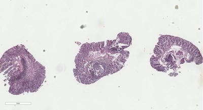
Materials and Methods: This was a retrospective analysis of the clinical, endoscopic and pathological findings of twenty-one patients who were diagnosed with lymphocytic colitis following colonoscopy in an open-access endoscopy setting in Akure, Ondo State, Nigeria over eighteen months.

Results: Twenty-one diagnoses of lymphocytic colitis were made from a pool of one hundred and twenty-two colonoscopies done between December 2021 and May 2023. There were slightly more females diagnosed with an M: F ratio of 3:4 with a mean (SD) age of 52.2 (± 14.3) years. All of the patients had a history of passage of watery diarrhoea while normal colonoscopy findings were present in 87.5%. The mean (SD) time to diagnosis in five of these study subjects was 18.4 ± 9.1 months. These patients had standard medical treatment with good outcomes.

Conclusion: In sub-Saharan Africa, lymphocytic diarrhoea is a treatable cause of chronic diarrhoea. It is important to refer patients with chronic diarrhoea to facilities where they can benefit from the expertise of endoscopists and pathologists.

Keywords: Lymphocytic colitis, chronic diarrhoea, Nigeria

Corresponding Author How to Cite this Article To Browse
Kenechukwu Okonkwo, Lecturer, Department of Medicine, University of Medical Sciences, , Ondo, Nigeria.
Email:
Okonkwo K, Ojo O, Betiku O, Lymphocytic colitis: an Important but Underdiagnosed Cause of Chronic Diarrhoea in Adult Nigerians. Int J Med Res Rev. 2024;12(3):84-92.
Available From
https://ijmrr.medresearch.in/index.php/ijmrr/article/view/1471

Manuscript Received Review Round 1 Review Round 2 Review Round 3 Accepted
2024-04-06 2024-04-12 2024-04-18 2024-04-24 2024-04-30
Conflict of Interest Funding Ethical Approval Plagiarism X-checker Note
None Nil Yes 13.25

© 2024by Okonkwo K, Ojo O, Betiku Oand Published by Siddharth Health Research and Social Welfare Society. This is an Open Access article licensed under a Creative Commons Attribution 4.0 International License https://creativecommons.org/licenses/by/4.0/ unported [CC BY 4.0].

Introduction

Chronic diarrhoea is one of the most common presentations in the Gastroenterology clinic. [1] Aside from infectious diarrhoea, it is increasingly becoming more apparent that lymphocytic colitis is an important cause of diarrhoea. [1] Lymphocytic colitis was described by Read and Lazenby as part of the entity called microscopic colitis, which also includes collagenous colitis. [1] Though lymphocytic colitis is an established cause of chronic diarrhoea, there are only sparse reports of it in Africa. [2,3] This is largely due to the shortage of skilled personnel required to make the diagnosis. [4,5] It is important to make the diagnosis of lymphocytic colitis in patients with chronic diarrhoea as treatment options are available to reduce the significant morbidity associated with the disease.

Materials And Methods

A retrospective analysis of prospectively collected data of patients with a diagnosis of lymphocytic colitis retrieved from the Endoscopy Register of an open access endoscopy setting in Akure, Ondo State, Nigeria as well as the outcomes of some of the patients who were consecutively afterwards seen in a Specialty Gastroenterology Clinic in Akure, Ondo State was carried out. The information obtained from the register between December 2021 and May 2023 (eighteen months) included the age and sex of the patients, indication and diagnosis. The study complied with all ethical protocols as contained in the Helsinki Declaration.

All the patients underwent a 2-day bowel preparation before the procedure. The bowel preparation consisted of a liquid diet, 30 mg of bisacodyl tablets, and Epsom salt solution. All patients signed an informed written consent before undergoing colonoscopy. Pre-medications were given to all the patients, which consisted of intravenous midazolam 2.5–5 mg and pentazocine 30 mg. The patient’s vital signs were monitored before, during, and after the procedure. A digital rectal examination was carried out on all the patients before the insertion of the colonoscope. The Pentax EC-3890 LX Video colonoscope was used for the duration of the study. The procedures were carried out with the patient initially in the left lateral position. A change in posture and abdominal pressure were applied where necessary. Caecal intubation was defined as deep intubation into the caecum with visualization of the appendiceal orifice.

Biopsies were taken and sent for histological examination.

Histological examination of the biopsies was conducted and reviewed by seasoned histopathologists with expertise in this field. The diagnostic criteria used were as follows: lymphocytic colitis was recognized by an increased intra-epithelial lymphocyte content (greater than 20 per 100 epithelial cells) and an associated increase in inflammatory cells in the lamina propria but no thickened collagen band. Data Analysis All data were collated and analyzed using the statistical package for social sciences (SPSS) version 20.0 (IBM, Armonk, NY, USA). The ages of the patients were summarized using mean and standard deviation (SD). Variables such as ages, sex, colonoscopy indications, endoscopy findings, time to diagnosis and outcomes were presented in frequency tables.

Results

The mean age (SD) of the studied population was 52.2 (± 14.3) years. There were 12 (57.1%) females and 9 (42.9%) males, with a male: female ratio of 3:4. The major complaint was the passage of loose stools (100%) while the commonest colonoscopy finding was normal-looking colonic mucosa (85.7%). Five of these patients were managed in the Gastroenterology Specialty Clinics and had good outcomes. The mean (SD) time to diagnosis in these five patients was 18.4 ± 9.1 years.

Summaries of patients seen in the Gastroenterology Specialty Clinic
Case 1: 52-year-old woman who presented on account of recurrent passage of loose stools which had been on and off for eighteen months. The stools did not resolve with over-the-counter medications. There was associated upper abdominal pain for which she used PPIs namely Rabeprazole. Endoscopy revealed an erythematous patch in the descending colon. There was oedema of the mucosa of the transverse colon. She was commenced on Prednisolone and Cholestyramine with improvement in clinical symptoms.
ijmrr_1471_01_02.JPG


Case 2: 59-year-old woman who presented with a history of recurrent passage of loose stools of a year duration. She had been to several hospitals with improvement in symptoms. A colonoscopy revealed oedematous colonic mucosa with widespread patchy erythema. She was commenced on Prednisolone with some improvement in symptoms.

Case 3: 67-year-old male who presented on account of passage of loose stools of a year’s duration. He is a known diabetic. The colonic mucosa was normal apart from healed ulcers in the rectal mucosa. He was commenced on Budesonide with significant improvement in clinical symptoms.

Case 4: 66-year-old woman with complaints of recurrent passage of loose stools of 14 months duration. The colonic mucosa was normal throughout on colonoscopy. She was commenced on Prednisolone with a good response to treatment.

Case 5: 21-year-old woman who had been passing loose stools for three years. On endoscopy, the colonic mucosa appeared normal. She was absent from school for a significant amount of time on account of frequent passage of loose stools.

She made remarkable improvements on steroids and is now able to pay more attention to her education.

Informed consent: informed consent was sought and obtained from the patients.

Table 1: Socio-demographic and clinical characteristics of patients

Sr.Age
(years)
SexIndicationEndoscopy
findings
Time to
diagnosis
(months)
Outcome
168FPassage of loose watery stoolsNormal looking mucosa______Was not referred to the gastroenterology speciality clinic
275FPassage of loose watery stools. There was also a history of abdominal painNormal looking mucosa______Was not referred to the gastroenterology speciality clinic
364FPassage of loose watery stoolsNormal looking mucosa______Was not referred to the gastroenterology speciality clinic
450MPassage of loose watery stools. There was also a history of abdominal painNormal looking mucosa______Was not referred to the gastroenterology speciality clinic
544MPassage of loose watery stools and faecal incontinenceNormal looking mucosa______Was not referred to the gastroenterology speciality clinic
621FPassage of loose watery stoolsNormal looking mucosa36 monthsGood
766FPassage of loose watery stoolsNormal looking mucosa14 monthsGood
847FPassage of loose watery stoolsOedematous and erythematous colon______Was not referred to the gastroenterology speciality clinic
957FPassage of loose watery stools. There was also a history of haematocheziaNormal-looking mucosa. Internal haemorrhoids______Was not referred to the gastroenterology speciality clinic
1040MPassage of loose watery stools. There was also a history of haematocheziaOedematous and erythematous colon. Internal haemorrhoids______Was not referred to the gastroenterology speciality clinic
1167MPassage of loose watery stoolsNormal looking mucosa______Was not referred to the gastroenterology speciality clinic
1235FPassage of loose watery stoolsNormal looking mucosa______Was not referred to the gastroenterology speciality clinic

1362MPassage of loose watery stools. Recurrent haematocheziaNormal-looking mucosa. Internal haemorrhoids______Was not referred to the gastroenterology speciality clinic
1467MPassage of loose watery stools.Normal looking mucosa12 monthsGood
1579MPassage of loose watery stools. Recurrent haematocheziaNormal-looking mucosa. Internal haemorrhoids______Was not referred to the gastroenterology speciality clinic
1646MPassage of loose watery stools. There was also a history of weight loss.Normal looking mucosa______Was not referred to the gastroenterology speciality clinic
1738FPassage of loose watery stoolsNormal looking mucosa______Was not referred to the gastroenterology speciality clinic
1859FPassage of loose watery stoolsOedematous and erythematous colon12 monthsGood
1952FPassage of loose watery stoolsNormal looking mucosa______Was not referred to the gastroenterology speciality clinic
2070MPassage of loose watery stools. History of chemotherapy for colorectal cancerNormal looking mucosa______Was not referred to the gastroenterology speciality clinic
2152FPassage of loose watery stoolsNormal looking mucosa18 monthsWas not referred to the gastroenterology speciality clinic

Mean ±SD
Age: 52.2 ± 14.3
Time to diagnosis : 18.4 ± 9.1
Range
Age: 21-75
Time to diagnosis: 12-36

Figures 1-6 highlight and describe some of the characteristic microscopic features found in lymphocytic colitis in one of the cases in this study


Figure 1: showing three bits of colonic tissue.


Figure 2: showing evidence of loss of gland and architectural distortion and oedema


Figure 3: showing evidence of mild to moderate loss of gland and oedema (green circle)


Figure 4: showing lymphoid aggregate with germinal centre (green arrow 1) and mucosal erosion (green arrow 2)


Figure 5: Showing loss of apical mucin (green arrow)


Figure 6: showing intraepithelial lymphocytes (green arrows)

Discussion

Chronic diarrhoea causes significant morbidity and mortality [6] The role lymphocytic colitis plays in the aetiology of chronic diarrhoea is increasingly being recognized and said to account for 10-20% of chronic diarrhoea.[7] The prevalence and incidence rates in developed economies are 0.5-12.9/100 000(8) and 2.3 to 16/100 000 respectively[9] [10] However, the incidence has been noted to plateau in the above-developed economies [11][12] The landscape in Africa has not been well described due to the paucity of skilled endoscopists, diagnostic facilities and histopathologists [4][5]. Nonetheless, case reports are beginning to describe it [2][3] The role of histopathologists in its diagnosis cannot be overemphasized as the diagnostic criteria for lymphocytic colitis which is intraepithelial lymphocyte ≥ 20 per 100 inter cryptal epithelial cells, change in surface epithelium (flattening and mucin depletion), mononuclear infiltration of the lamina propria as well as subepithelial collagen layer less than 10µm is within the realm of histopathology.[13] [14] It is important to define the role lymphocytic colitis plays in the aetiology of chronic diarrhoea in Africa as there is effective treatment for it.

In this study, there was a female preponderance as observed in previous studies [8][12] The average age was 55 years which was lower than the peak age of 65 years as has been previously described [7]. Indeed, 25% of patients with lymphocytic colitis are under 45 years old [15].All of them had loose, watery diarrhoea which is the classic presenting complaint [13][16]. Other symptoms found in some of the patients include weight loss, faecal incontinence and abdominal pain. It is important to note that other studies have recognized that the presentation can be myriad

including symptoms like nocturnal diarrhoea, weight loss, urgency, faecal incontinence and abdominal pain [15][17] Four of the patients had a history of haematochezia in addition to the primary complaint of watery diarrhoea. In these patients, the additional finding on colonoscopy was internal haemorrhoids which could have been responsible for the bleeding per rectum. Oluyemi et al noted similar findings of co-existing haemorrhoids and haematochezia in patients who were diagnosed with lymphocytic colitis [3]. One of the patients who had presented for colonoscopy on account of chronic diarrhoea had a history of treatment of colorectal cancer. Studies have reported that patients treated with chemotherapeutic agents may develop lymphocytic colitis though the exact therapeutic agent(s) the patient in this index study received was not ascertained [18]. Conditions that have been documented as being associated with lymphocytic colitis include coeliac disease, uveitis, idiopathic pulmonary fibrosis, juvenile diabetes mellitus, pernicious anaemia, autoimmune thyroiditis and oligoarthritis [19][20][21] These associations are however more linked with collagenous colitis which is within the spectrum of microscopic colitis.(19) Genetic susceptibility in the HLA-DR3-DQ2 haplotype, altered epithelial barrier function and mucosal inflammation have been suggested as possible pathophysiologic mechanisms for lymphocytic colitis [22][23]

One of the patients was documented to have a history of chronic use of PPIs namely Rabeprazole for upper abdominal discomfort. It is interesting to note that PPIs in particular, lansoprazole have been particularly implicated in the causation of lymphocytic colitis.[1] Other drugs thought to play a role in the pathology of lymphocytic colitis include NSAIDs, statins, SSRIs, pembrolizumab and flutamide.[24] However, there is conflicting evidence as most of these drugs are also known to cause chronic diarrhoea.[25]

As has been noted previously, many patients are missed on initial presentation if proper evaluation is not done. [25] An earlier study showed how lymphocytic colitis can be mistaken for irritable bowel syndrome [2][25]. Indeed, one of the patients in this study had intermittent diarrhoea which can simulate irritable bowel syndrome. Chronic diarrhoea is however mostly first treated as infectious colitis in sub-Saharan Africa and expectedly, all of the patients in this study


had a history of prior treatment with antibiotics before colonoscopy [26]. In this case series presented, the patients had a mean lag time of eighteen months before definitive diagnosis. This is in contrast to Europe where the median time to diagnosis is eight weeks [27]. This in no small regard contributes to the diminished quality of life that patients with lymphocytic colitis suffer as a result of their symptoms particularly unremitting diarrhoea [28]. This was seen in one of the patients who missed school on account of diarrhoea. Other debilitating features documented in patients with lymphocytic colitis include worry about losing control of bowel function, feelings of guilt and embarrassment, lack of social participation as well as impaired sexual functioning. [6]

The classical picture on endoscopy is normal colonic mucosa which was noted in the majority of the patients discussed. Colonic mucosal oedema and patchy erythema were noted on colonoscopy in three patients. Other endoscopic lesions that may be present include increased mucosal vascularity, ulcerations, erosions, mucosa friability and scars [21] We did not find any of these in this cohort of patients. The growing consensus is that samples should be taken from both sides of the colon, particularly the ascending and descending colon. [29][30] Part of the rationale behind this is that patients may sometimes present with patchy colitis. It is a diagnosis of exclusion as other causes of chronic diarrhoea such as ulcerative colitis and colorectal cancer have to be ruled out. It is important to note that these entities may also cause intraepithelial lymphocytosis [31].

Fortunately, the disease runs a benign course. As noted above, the patients who received treatment showed significant improvement in symptoms of steroids. Unfortunately, most of the patients could not be followed up as they were not referred to the gastroenterology speciality clinic by their primary managing physician. Most of the patients who received treatment were placed on Prednisolone as Budesonide which is the gold standard is not readily available. TNF alpha therapy has been suggested for patients with severe disease who do not respond to steroids or immunomodulators. [32]

Conclusion

This study provides more information on the clinicopathological presentations of lymphocytic

colitis in Nigeria and hopefully will increase awareness of this treatable cause of chronic diarrhoea. A limitation of this study is its retrospective nature such that we could not probe fully into the social, medical and drug histories of the patients. We hope that in the future additional studies which will be large-scale, longitudinal, and preferably, prospective will be able to further elaborate on the risk factors, including genetics, associated with lymphocytic colitis in this environment.

What does this study add to existing knowledge? : This study highlights the fact that lymphocytic colitis may not be as uncommon in sub-Saharan Africa as previously thought. It also shows that lymphocytic colitis in the environment usually have good outcomes when treated with steroids.

Permission from Institutional research board: Yes
Conflict of interest: None
Funding: Nil

References

1. Guidelines for the investigation of chronic diarrhoea in adults: British Society of Gastroenterology, 3rd edition. Gut. 2018 Aug;67(8):1380–99. doi: 10. 1136/gutjnl-2017-315909 [Crossref][PubMed][Google Scholar]

2. Mohamed N. Microscopic colitis as a missed cause of chronic diarrhea. World J Gastroenterol. 2011;17(15):1996. doi: 10.3748/wjg.v17.i15.1996 [Crossref][PubMed][Google Scholar]

3. Read NW, Krejs GJ, Read MG, Santa Ana CA, Morawski SG, Fordtran JS. Chronic diarrhea of unknown origin. Gastroenterology. 1980;78(2):264–71. doi: 10.1016/0016-5085(80)90575-2 [Crossref][PubMed][Google Scholar]

4. Lazenby AJ, Yardley JH, Giardiello FM, Jessurun J, Bayless TM. Lymphocytic (“microscopic”) colitis: A comparative histopathologic study with particular reference to collagenous colitis. Hum Pathol. 1989 Jan;20(1):18–28. doi: 10.1016/0046-8177(89)90198-6 [Crossref][PubMed][Google Scholar]


5. Ekrikpo UE, Otegbayo JA, Oluwasola AO. Lymphocytic colitis presenting as difficult diarrhoea in an African woman: a case report and review of the literature. J Med Case Rep. 2010 Dec 29;4(1):31. doi: 10.1186/1752-1947-4-31 [Crossref][PubMed][Google Scholar]

6. Oluyemi A, Momoh M, Odeghe E, Keshinro S. Lymphocytic Colitis in Nigeria: A Case Series. Annals of African Surgery. 2021 Jul 20;18(3):180–4. doi: 10.4314/aas.v18i3.10 [Crossref][PubMed][Google Scholar]

7. Mudenda V, Malyangu E, Sayed S, Fleming K. Addressing the shortage of pathologists in Africa: Creation of a MMed Programme in Pathology in Zambia. Afr J Lab Med. 2020 Jun 3;9(1). doi: 10.4102/ajlm.v9i1.974 [Crossref][PubMed][Google Scholar]

8. Perl D, Leddin D, Bizos D, Veitch A, N’Dow J, Bush-Goddard S, et al. Endoscopic capacity in West Africa. Afr Health Sci. 2016 May 9;16(1):329. doi: 10.4314/ahs.v16i1.44 [Crossref][PubMed][Google Scholar]

9. Pihl Lesnovska K, Münch A, Hjortswang H. Microscopic colitis: Struggling with an invisible, disabling disease. J Clin Nurs. 2019 Oct 29;28(19–20):3408–15. doi: 10.1111/jocn.14916 [Crossref][PubMed][Google Scholar]

10. Olesen M. Microscopic colitis: a common diarrhoeal disease. An epidemiological study in Orebro, Sweden, 1993-1998. Gut. 2004 Mar 1;53(3):346–50. doi: 10.1136/gut.2003.014431 [Crossref][PubMed][Google Scholar]

11. Tysk C, Bohr J, Nyhlin N, Wickbom A, Eriksson S. Diagnosis and management of microscopic colitis. World J Gastroenterol. 2008;14(48):7280. doi: 10.3748/wjg.14.7280 [Crossref][PubMed][Google Scholar]

12. Williams JJ, Kaplan GG, Makhija S, Urbanski SJ, Dupre M, Panaccione R, et al. Microscopic Colitis–Defining Incidence Rates and Risk Factors: A Population-Based Study. Clinical Gastroenterology and Hepatology. 2008 Jan;6(1):35–40. doi: 10.1016/j.cgh.2007.10.031 [Crossref][PubMed][Google Scholar]

13. Wickbom A, Bohr J, Eriksson S, Udumyan R, Nyhlin N, Tysk C. Stable Incidence of Collagenous Colitis and Lymphocytic Colitis in Örebro, Sweden, 1999–2008. Inflamm Bowel Dis. 2013 Oct;19(11):2387–93. doi: 10.1097/MIB.0b013e31829ed8cd [Crossref][PubMed][Google Scholar]

14. Tong J, Zheng Q, Zhang C, Lo R, Shen J, Ran Z. Incidence, prevalence, and temporal trends of microscopic colitis: a systematic review and meta-analysis. Am J Gastroenterol [Internet]. 2015 Feb 5 [cited 2023 Feb 4];110(2):265–76. doi: 10.1038/AJG.2014.431 [Crossref][PubMed][Google Scholar]

15. Tong J, Zheng Q, Zhang C, Lo R, Shen J, Ran Z. Erratum: Incidence, Prevalence, and Temporal Trends of Microscopic Colitis: A Systematic Review and Meta-Analysis. American Journal of Gastroenterology. 2015 Jul;110(7):1121. doi: 10.1038/ajg.2015.46 [Crossref][PubMed][Google Scholar]

16. Veress B, Lofberg R, Bergman L. Microscopic colitis syndrome. Gut. 1995 Jun 1;36(6):880–6. doi: 10.1136/gut.36.6.880 [Crossref][PubMed][Google Scholar]

17. Olesen M. Lymphocytic colitis: a retrospective clinical study of 199 Swedish patients. Gut. 2004 Apr 1;53(4):536–41. doi: 10.1136/gut.2003.023440 [Crossref][PubMed][Google Scholar]

18. Münch A, Langner C. Microscopic Colitis: Clinical and Pathologic Perspectives. Clinical Gastroenterology and Hepatology. 2015 Feb;13(2):228–36. doi: 10.1016/j.cgh.2013.12.026 [Crossref][PubMed][Google Scholar]

19. Pardi DS, Ramnath VR, Loftus E V. , Tremaine WJ, Sandborn WJ. Lymphocytic colitis: clinical features, treatment, and outcomes. Am J Gastroenterol. 2002 Nov;97(11):2829–33. doi: 10.1111/j.1572-0241.2002.07030.x [Crossref][PubMed][Google Scholar]

20. Storr MA. Microscopic Colitis: Epidemiology, Pathophysiology, Diagnosis and Current Management—An Update 2013. ISRN Gastroenterol. 2013 Apr 18;2013:1–12. doi: 10.1155/2013/352718 [Crossref][PubMed][Google Scholar]


21. Choi K, Samdani R, Shuttlesworth G, Wang Y. P016 case series: immunotherapy-induced lymphocytic colitis. Gastroenterology. 2018 Jan;154(1):S9. doi: 10.1053/j.gastro.2017.11.048 [Crossref][PubMed][Google Scholar]

22. Koskela RM, Niemelä SE, Karttunen TJ, Lehtola JK. Clinical characteristics of collagenous and lymphocytic colitis. Scand J Gastroenterol. 2004 Jan 8;39(9):837–45. doi: 10.1080/00365520410006468 [Crossref][PubMed][Google Scholar]

23. Kao KT, Pedraza BA, McClune AC, Rios DA, Mao YQ, Zuch RH, et al. Microscopic colitis: A large retrospective analysis from a health maintenance organization experience. World J Gastroenterol. 2009;15(25):3122. doi: 10.3748/wjg.15.3122 [Crossref][PubMed][Google Scholar]

24. Mellander MR, Ekbom A, Hultcrantz R, Löfberg R, Öst Å, Björk J. Microscopic colitis: a descriptive clinical cohort study of 795 patients with collagenous and lymphocytic colitis. Scand J Gastroenterol. 2016 May 3;51(5):556–62. doi: 10.3109/00365521.2015.1124283 [Crossref][PubMed][Google Scholar]

25. Koskela RM, Karttunen TJ, Niemelä SE, Lehtola JK, Ilonen J, Karttunen RA. Human leucocyte antigen and TNFα polymorphism association in microscopic colitis. Eur J Gastroenterol Hepatol. 2008 Apr;20(4):276–82. doi: 10.1097/MEG.0b013e3282f2468d [Crossref][PubMed][Google Scholar]

26. Jackson K. Are Collagenous Colitis and Lymphocytic Colitis Distinct Syndromes? Digestive Diseases. 1995;13(5):301–11. doi: 10. 1159/000171510 [Crossref][PubMed][Google Scholar]

27. Bonderup OK, Nielsen GL, Dall M, Pottegård A, Hallas J. Significant association between the use of different proton pump inhibitors and microscopic colitis: a nationwide Danish case-control study. Aliment Pharmacol Ther. 2018 Sep;48(6):618–25. doi: 10.1111/apt.14916 [Crossref][PubMed][Google Scholar]

28. Keszthelyi D, Jansen S V. , Schouten GA, De Kort S, Scholtes B, Engels LGJB, et al. Proton pump inhibitor use is associated with an increased risk for microscopic colitis: a case-control study. Aliment Pharmacol Ther. 2010 Nov;32(9):1124–8. doi: 10.1111/j.1365-2036.2010.04453.x [Crossref][PubMed][Google Scholar]

29. Baert F, Wouters K, D’Haens G, Hoang P, Naegels S, D’Heygere F, et al. Lymphocytic colitis: a distinct clinical entity? A clinicopathological confrontation of lymphocytic and collagenous colitis. Gut. 1999 Sep 1;45(3):375–81. doi: 10.1136/gut.45.3.375 [Crossref][PubMed][Google Scholar]

30. Gueye MN, Diop SN, Diallo S, Diouf G, Cissé CAB, Fall MP, et al. Ulcerative Colitis in Sub-Saharan Africa: Analysis of 24 Cases in Dakar (Senegal). Open J Gastroenterol. 2020;10(06):128–36. doi: 10.1136/gut.45.3.375 [Crossref][PubMed][Google Scholar]

31. Loreau J, Duricova D, Gower-Rousseau C, Savoye G, Ganry O, Ben Khadhra H, et al. Long-Term Natural History of Microscopic Colitis: A Population-Based Cohort. Clin Transl Gastroenterol. 2019 Sep;10(9):e00071. doi: 10.14309/ctg.0000000000000071 [Crossref][PubMed][Google Scholar]

32. Nielsen OH, Fernandez-Banares F, Sato T, Pardi DS. Microscopic colitis: Etiopathology, diagnosis, and rational management. Elife. 2022 Aug 1;11. doi: 10.7554/eLife.79397 [Crossref][PubMed][Google Scholar]

33. Malik A, Nadeem M, Javaid S, Malik MI, Enofe I, Abegunde AT. Estimating the optimum number of colon biopsies for diagnosing microscopic colitis: a systematic review. Eur J Gastroenterol Hepatol. 2022 Jul 14;34(7):733–8. doi: 10.1097/MEG.0000000000002355 [Crossref][PubMed][Google Scholar]

34. Virine B, Chande N, Driman DK. Biopsies From Ascending and Descending Colon Are Sufficient for Diagnosis of Microscopic Colitis. Clinical Gastroenterology and Hepatology. 2020 Aug;18(9):2003–9. doi: 10.1016/j.cgh.2020.02.036 [Crossref][PubMed][Google Scholar]


35. Topel G, Cakir E, Aydin I, Dilek FH, Sari AA. Analysis of clinical and histopathological findings in microscopic colitis. Turkish Journal of Pathology. 2022; 38(8):275-283 doi: 10. 5146/tjpath.2022.01574 [Crossref][PubMed][Google Scholar]

36. Fernández-Bañares F, Casanova MJ, Arguedas Y, Beltrán B, Busquets D, Fernández JM, et al. Current concepts on microscopic colitis: evidence-based statements and recommendations of the Spanish Microscopic Colitis Group. Aliment Pharmacol Ther. 2016 Feb;43(3):400–26. doi: 10.1111/apt.13477 [Crossref][PubMed][Google Scholar]