E-ISSN:2320-8686
P-ISSN:2321-127X

Case Series

Caudal Duplication Syndrome

International Journal of Medical Research and Review

2024 Volume 12 Number 2 Mar-Apr
Publisherwww.medresearch.in

The Double Trouble: Caudal Duplication Syndrome-A rare case series with a review of literature

Datta D1, Saha S2, Dakshit D3, Maity A4*, Bansal R5, Basu N6
DOI:https://doi.org/10.17511/ijmrr.2024.i02.05

1 Debarpita Datta, Senior Resident, Department of Radiodiagnosis, Medical College and Hospital , Kolkata, West Bengal, India.

2 Sudipta Saha, Associate Professor, Department of Radiodiagnosis, Medical College and Hospital, Kolkata, West Bengal, India.

3 Debashis Dakshit, Professor, Department of Radiodiagnosis, Medical College and Hospital , Kolkata, West Bengal, India.

4* Arup Maity, Senior Resident, Department of Radiodiagnosis, Medical College and Hospital, Kolkata, West Bengal, India.

5 Ruchi Bansal, Assistant Professor, Department of Radiodiagnosis, Medical College and Hospital, Kolkata, West Bengal, India.

6 Nupur Basu, Associate Professor, Department of Radiodiagnosis, Medical College and Hospital, Kolkata, West Bengal, India.

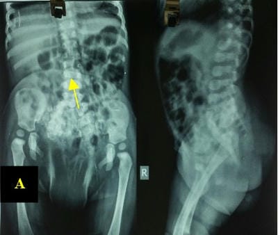
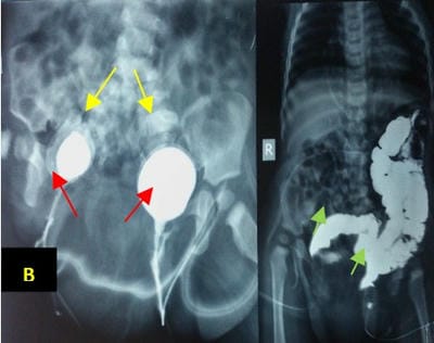
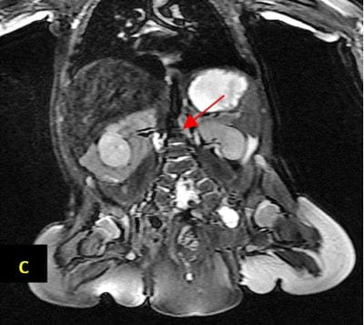
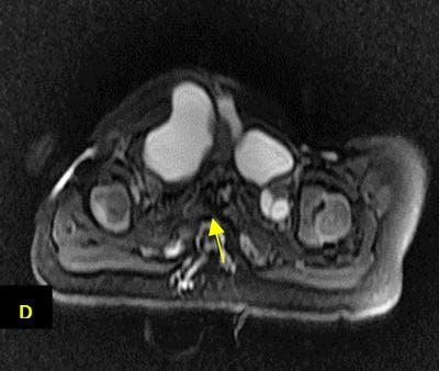
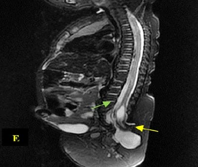
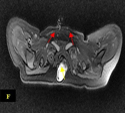
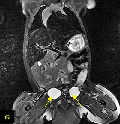
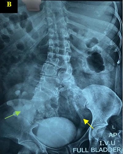
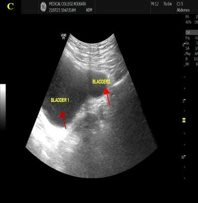
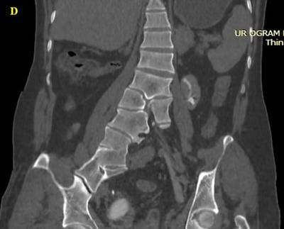
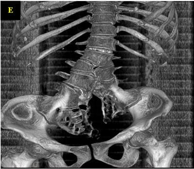
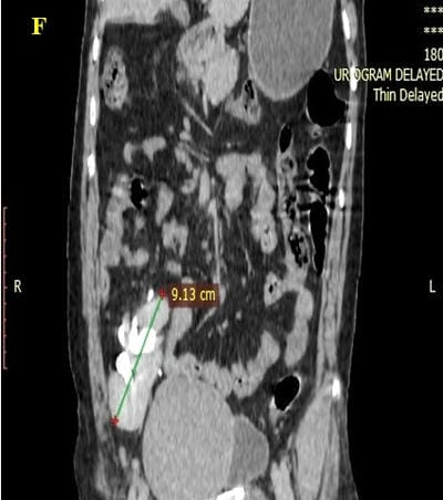
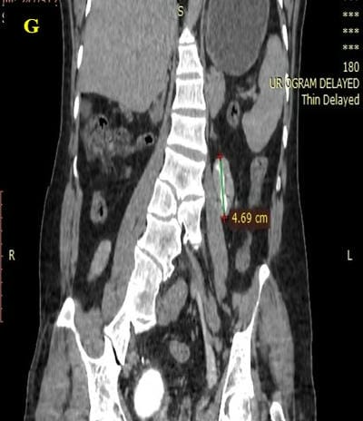
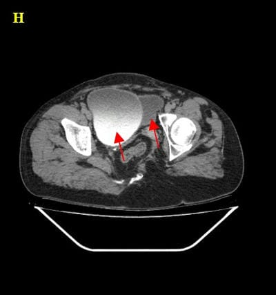
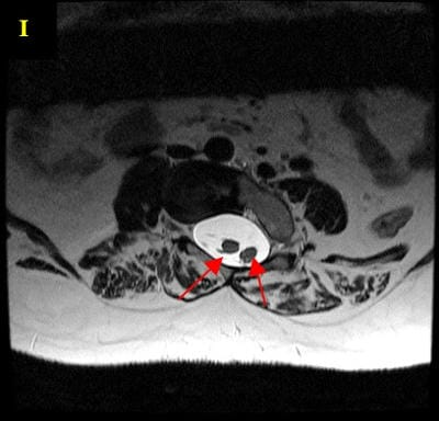
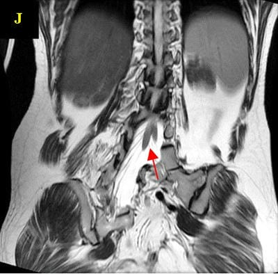
Caudal duplication syndrome (CDS) is a very rare disease entity with prevalence of less than 1 per 100,000 at birth. It includes a broad range of abnormalities and clinical symptoms, from single or partial duplication of the gastrointestinal, genitourinary and distal neural tube system organs to total duplication. There have been several ideas proposed to explain the complicated yet symmetrical abnormalities and the wide range of clinical manifestations of caudal duplication syndrome. In this case series, we present two confirmed cases of Caudal Duplication Syndrome in our institute. The management for this syndrome is individualized and may include surgical intervention to fuse or excise the duplicated organs.

Keywords: Caudal Duplication Syndrome, Duplication, Rare

Corresponding Author How to Cite this Article To Browse
Arup Maity, Senior Resident, Department of Radiodiagnosis, Medical College and Hospital, Kolkata, West Bengal, India.
Email:
Datta D, Saha S, Dakshit D, Maity A, Bansal R, Basu N, The Double Trouble: Caudal Duplication Syndrome-A rare case series with a review of literature. Int J Med Res Rev. 2024;12(2):57-64.
Available From
https://ijmrr.medresearch.in/index.php/ijmrr/article/view/1463

Manuscript Received Review Round 1 Review Round 2 Review Round 3 Accepted
2024-02-27 2024-02-29 2024-03-07 2024-03-14 2024-03-21
Conflict of Interest Funding Ethical Approval Plagiarism X-checker Note
Nil Nil Yes 17%

© 2024by Datta D, Saha S, Dakshit D, Maity A, Bansal R, Basu Nand Published by Siddharth Health Research and Social Welfare Society. This is an Open Access article licensed under a Creative Commons Attribution 4.0 International License https://creativecommons.org/licenses/by/4.0/ unported [CC BY 4.0].

Introduction

Caudal duplication syndrome, also known as split notochord syndrome, is a very uncommon congenital condition in which several caudal tissues, embryonic cloaca, and neural tubes show a range of abnormalities, including duplication and deformities. Less than one in 100,000 newborns are born with this rare congenital disease, and there are only about 100 cases known globally [1].

The condition's precise cause is not known [1]. Even though several hypotheses point to incomplete monozygotic twin separation as the etiological cause [2], structural anomalies in the caudal region have also been associated with damage to the caudal cell mass and the posterior gut, abnormal ectoderm-endoderm adhesion during gastrulation [3], polytopic primary developmental field defects [8], somatic and germline mutations in developmental genes [2], and polytopic primary developmental field defects. The HOX gene, specifically HOX10 and HOX11, is thought to be connected to the condition [2]. Misexpression of the genetic components, which normally code for the mammalian appendicular and axial skeleton, may cause the caudal mesenchyme to grow abnormally [2]. The wide range of gastrointestinal (GI), genitourinary (GU), spinal, and limb malformations causes a wide range of symptoms for the disorder.

Gastrointestinal tract anomalies[1]

  • Duplication of colon, cecum, ani, appendix, and ileum
  • Agenesis, atresia, cyst, herniation, omphalocele, imperforate anus, and rectal fistula
  • Meckel’s diverticulum, intestinal malrotation, and situs inversus

Genitourinary tract anomalies[1]

  • Duplication of external and internal genitalia, vagina, cervix, bladder, urethra, uterus, ureter, scrotum, and testis
  • Bilateral exstrophy of the bladder, cryptorchidism, malrotation, agenesis of the kidney, rectourethral fistula, urinary disturbances including incontinence and retention

Spinal anomalies[1]

  • Duplication of lumbar and thoracic vertebrae, sacrum, coccyx, ilium, and spinal cord
  • Hemivertebra, sacral agenesis, myelomeningocele, diplomyelia, spina bifida, and diastematomyelia

Anomaly scans during the second trimester of pregnancy or soon after birth are frequently used to make the diagnosis [2]. Before prognosis, a comprehensive medical evaluation is necessary to comprehend the anatomy of the patient and choose the most appropriate treatment plan. Anomalies can be thoroughly examined using imaging techniques [4] such as conventional X-rays, magnetic resonance imaging (MRI), ultrasonography, barium enema, computed tomography (CT) scans, and micturating cystourethrography (MCU).

Case Report 1

The patient is a 1-year-old infant female child diagnosed with imperforate anus and presented to our department for imaging studies. No foetal screening had been carried out, and there was no family history of congenital abnormalities. Physical examination revealed imperforate anus and abdominal distension.

Radiographs of the lumbar spine revealed splitting of posterior elements and duplication of lumbar and sacral vertebrae. The urethrogram showed the presence of two bladder with two urethras. Barium meal in anteroposterior view showed malrotation of the bowel to the left side with two rectums. MRI showed diastematomyelia (split cord) Pang type 2 from the level of L4-L5. There was also the presence of rachischisis with myelomeningocele and tethered cord from the same level.

ijmrr_1463_01.jpg
(A): Radiographs of lumbar spine showing complex malformation with duplication of lower lumbar and sacral segments [yellow arrow].


ijmrr_1463_02.jpg
(B): Urethrogram and barium follow-through studies showing double bladder [yellow arrows] with separate urethra [red arrows] and double rectum [green arrows].

ijmrr_1463_03.jpg
(C): Coronal T2-weighted MR sequence demonstrates splitting of lower lumbar and sacral segments [red arrow].

ijmrr_1463_04.jpg
(D): Diastematomyelia (Pang type II) and rachischisis [yellow arrow] is also evident in axial T2-weighted FSE MR sequence.

ijmrr_1463_05.jpg
(E): Sagittal T2-weighted MR sequence showing tethered cord [green arrow] and myelomeningocele [yellow arrow].

ijmrr_1463_06.jpg
(F): Double ureter and myelomeningocele are both observed in this axial T2-weighted FSE MR sequence [red arrows, yellow arrow].

ijmrr_1463_07.jpg
(G): Coronal T2- weighted MR sequence showing double bladder [yellow arrows].


ijmrr_1463_08.jpg
(H): Bowel loops protruding from the abdominal cavity on an axial T2-weighted FSE MR sequence are indicative of an umbilical hernia [yellow arrow].

An ostomy was made as a loop on the skin of the left side of the abdomen since the patient had an imperforate anus. In a vaginoscopy, there were two distinct vaginas, and each hemi-vagina had a distinct cervix. Each urethra had its own bladder and ureteral opening during a cystoscopy or ureteroscopy

A transverse laparotomy was carried out. Two unique columns with discrete mesocolon joined above the peritoneal reflection at the terminal ileum site shared a proximal aperture. The second colon was removed from the ileocecal to the rectum since the first colon had undergone a colostomy at a previous operation site

Case Report 2

A 27-year-old adult male presented to our department with complaints of haematuria and occasional dull lower abdominal pain. He had a limping gait on his left side since childhood. There was no family history of such complaints. He supported three similar incidents in the previous ten years, some of which were connected to UTIs (UTIs).On physical examination, his abdomen was soft with diffuse rebound tenderness but no guarding or rigidity. Hyperactive bowel sounds were present diffusely across the abdomen.

Intravenous pyelography was done. The control film incidentally revealed malformation in the vertebral column and showed duplication

of the lower lumbar vertebrae with a defect extending to the left ala of the sacrum. The excretory projection in the full bladder showed an ectopic mal-rotated pelvic kidney on right side and dilated left ureter. Ureters appeared normal. A pelvic ultrasound was conducted, followed by contrasted computed tomography urography and MRI spine with and without contrast. The identical findings and presence of two bladders were visualised with a pelvic ultrasound.

Computed tomography urography demonstrated the ectopic mal-rotated right kidney which drained into the right bladder. A small dysplastic kidney was noted on left side draining into the left bladder. The left ureter was dilated on its distal end. There was duplication of bladders with separate single ureter draining into it. The rectum was also duplicated, with one colon containing normal well-formed stool. The other colon appeared stenosed. Midline fusion defects of L4, L5 and the sacrum were also noted.

MR lumbosacral spine sequences showed splitting of the lower lumbar spinal cord from L4-L5 level and caudal spinal nerve roots with two hemicords in a single dural sac-diastematomyelia Pang type 2. Two rectums were also noted.

The gastro surgery and urosurgery departmentsrecommended that the patient undergo surgery to remove the duplicate colon and bladder, but he opted to be released from the hospital and receive follow-up care as an outpatient.

ijmrr_1463_09.jpg
(A): Intravenous Pyelography in control projection showing the split malformation in lower lumbar and sacral vertebral segments [yellow arrow].


ijmrr_1463_10.jpg
(B): Same study in excretory phase showing malrotated pelvic right kidney [green arrow] and dilated left ureter [yellow arrows].

ijmrr_1463_11.jpg
(C): Pelvic ultrasound showing two urinary bladders [red arrows].

ijmrr_1463_12.jpg
(D): Coronal reconstructed computed tomographic image demonstrates duplication of lower lumbar and sacral segments.

ijmrr_1463_13.jpg
(E): Three-dimensional reconstruction of pelvic computed tomographic image showing the midline fusion defects of the L4, L5 vertebral body and the sacrum.

ijmrr_1463_14.jpgijmrr_1463_15.jpg

(F), (G): Coronal reconstructed computed tomographic image demonstrates the ectopic


mal-rotated right kidney[annotated] and a small dysplastic left kidney[annotated].

ijmrr_1463_16.jpg
(H): Axial computed tomographic image in delayed phase showing double urinary bladder [red arrows].

ijmrr_1463_17.jpgijmrr_1463_18.jpg
(I), (J): Axial and coronal T2-weighted MR sequences demonstrates the splitting of spinal cord from the level of L4-L5[red arrows].

ijmrr_1463_19.jpg
(K): Axial T2-weighted MR sequence showing double rectum [red arrows].

Discussion

Caudal Duplication Syndrome is a rare entity. As of 2014, there were just 2 patients diagnosed as adults and less than 100 cases of Caudal Duplication Syndrome (CDS) had been reported in the literature globally [6]. There is no evidence of a genetic or racial predisposition, while there are about an equal proportion of male and female cases [10]. In 1993, the term "caudal duplication syndrome" was initially proposed and the first comprehensive study of caudal duplication symptoms was conducted [1].

An adult female with duplication of the colon, rectum, anus, urinary bladder, urethra, uterus, cervix, vagina, and external genitalia was the first example to be reported in India [11]. Less than 100 CDS cases have been documented globally, with the majority of cases being identified before birth. Only four adult examples have been documented to date, one of which involved a patient who had a cloacal abnormality corrected as a baby [11,13]. Except for a CDS case published in 2013 [14], there hasn't been any information about a prior adolescent presentation.

Even though patients frequently display a wide range of symptoms, no adverse consequences were seen in a female adult with duplication of the colon, rectum, anus, urinary bladder, urethra, uterus, cervix, vagina, and external genitalia [4]. This shows that the gastrointestinal tract and urogenital duplication, which are extremely unusual, are frequently asymptomatic [7].


Embryology is suggested to have an intimate association with the development of caudal duplication syndrome.[1] The cloaca is the common embryological source of origin of both the genitourinary and gastrointestinal tracts which is the reason behind the frequent association of anomalies involving these systems[1,2]. The notochord develops from the primitive knot on day 15 following fertilisation, where it invaginates and creates the notochord canal. [1] On day 20, the notochord's ventral wall gradually disintegrates while connections are generated between the amniotic and yolk sacs. [1] The Kovalevsky Canal is one such link. The spinal cord develops from the 23rd to the 25th day of gestation, except for its distal-most portion, where the notochord and neural tube are fused to form the caudal cell mass [1].Many insults directed at the cell mass and hindgut during the stage of development may result in the development of caudal anomalies, one of which is caudal duplication syndrome. The canal of Kovalevsky crosses the caudal cell mass, while the endoderm located anterior to the cell mass develops into the hindgut [1].

Incomplete Kovalevsky's canal regression may also result in the creation of fibrous bands connecting the spinal canal and the hindgut,[5] which may eventually cause diastematomyelia. These bands have the potential to split the notochord, leading to duplicates of the spinal cord and lower spine [4]. Additionally, the adjacent mesoderm is split, resulting in duplicated genitourinary and gastrointestinal tracts. [4] The presence of a variety of malformations, such as spina bifida, dorsal enteric fistulas, enteric cysts, malformed or duplicated colons, bladders, sacrums, and lower spinal cord, can then result from the duplications [1, 9].

Furthermore, a midline pelvic mass defect during pregnancy may prevent the Müllerianduct structuresfrom migrating caudally, which could result in the duplication of the genital tract [1]. It is uncommon to have intestinal duplications that extend into the rectum or anus [1, 6].

The recommended practice of treatment for CDS entails the correction of duplication abnormalities in stages, with an emphasis on anatomical, functional, and aestheticreconstructions [16]. In UB, a septal excision is performed to create a single chamber.

It is possible to do mucosal stripping or resection of the duplicated colon and rectum because the duplicated colons are typically not joined and have distinct blood supplies [15]. If left untreated, hindgut duplication may signal the onset of intestinal blockage or potentially malignant change. Otherwise, as most female patients can be expected to experience normal menstruation, coitus, and pregnancy, reconstructive surgery is primarily of an aesthetic nature [17].

Conclusion

Caudal Duplication Syndrome (CDS) is a rare congenital condition characterized by the duplication of structures within the caudal region. Through the presentation of these case studies, we effectively demonstrate the complexity of diagnosing and managing this anomaly, highlighting the importance of advanced imaging techniques like MRI, CT scans, and ultrasonography in identifying key structural duplications such as in the lumbar and sacral vertebrae, bladders, and ureters. The cases discussed illustrate the need for a tailored, multidisciplinary approach to treatment, depending on the specific anatomical abnormalities and associated symptoms of each patient. Surgical interventions often play a critical role in managing the duplicated organs and correcting anatomical malformations to improve the quality of life for affected individuals.

References

1. Dominguez R, Rott J, Castillo M, Pittaluga RR, Corriere JN. Caudal Duplication Syndrome. Arch Paediatr Adolesc Med. 1993 Oct;147(10):1048-52. [Crossref][PubMed][Google Scholar]

2. Sur A, Sardar SK, Paria A. Caudal duplication syndrome. J Clin Neonatol. 2013 Apr;2(2):101-2. [Crossref][PubMed][Google Scholar]

3. Swaika S, Basu S, Bhadra RC, Sarkar R, Maitra SK. Caudal duplication syndrome-report of a case and review of literature. Indian J Surg. 2013 Jun;75(Suppl 1):484-7. [Crossref][PubMed][Google Scholar]


4. Samuk I, Levitt M, Dlugy E, Kravarusic D, Ben-Meir D, Rajz G, et al. Caudal Duplication Syndrome: The Vital Role of a Multidisciplinary Approach and Staged Correction. Eur J Pediatr Surg Rep. 2016 Dec;4(1):001-005. [Crossref][PubMed][Google Scholar]

5. Molinaro F, Cerchia E, Bulotta AL, Angotti R, Di Maggio G, Bianchi A, Messina M. Caudal 'duplication' or 'split' syndrome: Is there a misnomer? J Pediatr Surg Case Rep. 2013 Oct;1(10):351-356. . [Crossref][PubMed][Google Scholar]

6. Goil P, Banerjee A. A Rare Presentation of Caudal Duplication Syndrome in an Adult with No Functional Impairment. J Case Rep. 2015 Apr 10;4(2):324-327. [Crossref][PubMed][Google Scholar]

7. Shah KR, Joshi A. Complete genitourinary and colonic duplication: a rare presentation in an adult patient. J Ultrasound Med. 2006 Mar;25(3):407-11. [Crossref][PubMed][Google Scholar]

8. Kroes HY, Takahashi M, Zijlstra RJ, Baert JA, Kooi KA, Hofstra RM, van Essen AJ. Two cases of the caudal duplication anomaly including a discordant monozygotic twin. Am J Med Genet. 2002 Nov;112(4):390-3. [Crossref][PubMed][Google Scholar]

9. Pang D, Dias MS, Ahab-Barmada M. Split cord malformation: Part I: A unified theory of embryogenesis for double spinal cord malformations. Neurosurgery. 1992 Sep;31(3):451-80. [Crossref][PubMed][Google Scholar]

10. Ramzan M, Ahmed S, Ali S. Caudal duplication syndrome. J Coll Physicians Surg Pak. 2014 Jan;24(1):64-6. [Crossref][PubMed][Google Scholar]

11. Shah KR, Joshi A. Complete genitourinary and colonic duplication: a rare presentation in an adult patient. J Ultrasound Med. 2006 Mar;25:407-411. [Crossref][PubMed][Google Scholar]

12. Perez-Brayfield MR, Clarke HS, Pattaras JG. Complete bladder, urethral, and vaginal duplication in a 50-year-old woman. Urology. 2002;60:514. [Crossref][PubMed][Google Scholar]

13. Greenberg JA, Hendren WH. Vaginal delivery after cloacal malformation repair. Obstet Gynecol. 1997 Oct;90(4, Pt 2):666-667. [Crossref][PubMed][Google Scholar]

14. Liu H, Che X, Wang S, et al. Multiple-stage correction of caudal duplication syndrome: a case report. J Pediatr Surg. 2009;44:2410-2413. [Crossref][PubMed][Google Scholar]

15. Dunetz GN, Bauer SB. Complete duplication of the bladder and urethra. Urology. 1985;25:179-182. [Crossref][PubMed][Google Scholar]

16. Kajbafzadeh A-M, Aghdas FS, Tajik P. Complete covered duplication of the bladder, urethra, vagina, uterus, and visceral sequestration. Int J Urol. 2006;13:1129-1131. [Crossref][PubMed][Google Scholar]

17. Ravitch MM. Hind gut duplication; doubling of colon and genital urinary tracts. Ann Surg. 1953;137:588-601. [Crossref][PubMed][Google Scholar]