Orthodontic Marvel: 2x4 Appliance for Impacted Incisor Traction - A Case Series
Isha S1*, Gupta S2, Gumro M3
DOI: https://doi.org/10.17511/ijmrr.2023.i05.04
1* Simran Isha, Junior Resident, Department of Pediatric And Preventive Dentistry, Kd Dental College And Hospital, Mathura, Uttar Pradesh, India.
2 Sonal Gupta, Professor and Head, Department of Pediatric and Preventive Dentistry, KD Dental College and Hospital, Mathura, Uttar Pradesh, India.
3 Menia Gumro, Junior Resident, Department of Pediatric and Preventive Dentistry, KD Dental College and Hospital, Mathura, Uttar Pradesh, India.
Impacted incisors can pose significant esthetic and functional challenges for patients. Various treatment approaches have been proposed. The 2x4 orthodontic appliance is one of the most effective treatment plans for aligning the teeth.7 This article presents two cases showing surgical traction's effectiveness using the 2x4 orthodontic appliance in managing impacted incisors. The results of this case series demonstrated successful outcomes in terms of impacted incisor alignment and overall occlusion. Patients exhibited improved esthetics and function. Therefore, Surgical traction of impacted incisors using the 2x4 orthodontic appliance can be a viable and effective treatment approach.
Keywords: Impacted incisors, Orthodontic Marvel, Unerupted teeth
| Corresponding Author | How to Cite this Article | To Browse |
|---|---|---|
| , Junior Resident, Department of Pediatric And Preventive Dentistry, Kd Dental College And Hospital, Mathura, Uttar Pradesh, India. Email:  |
Isha S, Gupta S, Gumro M, Orthodontic Marvel: 2x4 Appliance for Impacted Incisor Traction - A Case Series. Int J Med Res Rev. 2023;11(5):123-129. Available From https://ijmrr.medresearch.in/index.php/ijmrr/article/view/1442 |
 |
 ©
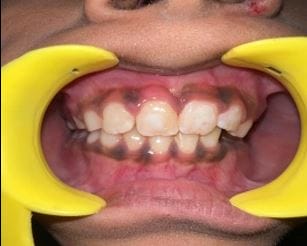
©  PRE-OPERATIVE
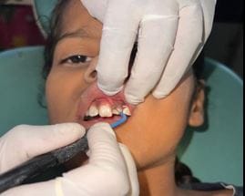
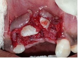 SURGICAL EXPOSURE OF IMPACTED INCISORS
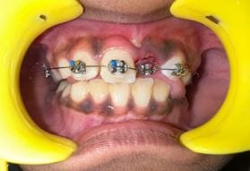
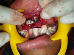 BRACKETS PLACED ON IMPACTED CENTRAL INCISOR
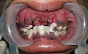 NITI 0.12 WIRE PLACED
 NITI 0.14 WIRE PLACED
 NITI 0.16 WIRE PLACED
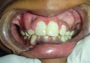 POST OPERATIVE
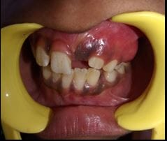


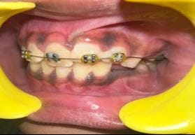
 Post-Operative After 3 months