E-ISSN:2320-8686
P-ISSN:2321-127X

Research Article

T -prolymphocytic leukemia

International Journal of Medical Research and Review

2023 Volume 11 Number 5 September-October
Publisherwww.medresearch.in

True-Tales Of Ten T- prolymphocytic leukemia (T-PLL) Over A Decade

N Rajakumar N1*, H Vijay R2, Balikai G3, B Horatti L4, Shamnur M5
DOI: https://doi.org/10.17511/ijmrr.2023.i05.03

1* Namrata N N Rajakumar, Associate Professor, Pathology Molecular biology research, Kidwai Memorial Institute Of Oncology, Bangalore, Karnataka, India.

2 Raghavendra H Vijay, Professor, Department of Pathology, Kidwai Memorial Institute Of Oncology, Bangalore, Karnataka, India.

3 Girish Balikai, Professor, Department of Haematology, SDM College Of Medical Sciences And Hospital, Dharwad, Karnataka, India.

4 Laxmi B Horatti, Assistant Professor, Department Of Dermatology, Shri Atal Bihari Vajpayee Medical College And Research Center, Bangalore, Karnataka, India.

5 Murgesh Shamnur, Professor, Department Of Dermatology, JJM Medical College, Davangere, Karnataka, India.

Introduction: T-PLL is a mature T-cell leukemia typically presenting at stages of exponentially rising lymphocyte counts in peripheral blood, accompanied by splenomegaly and bone marrow involvement. They are rare and inherently aggressive and notoriously refractory to therapeutics. To our knowledge, this is the largest series of T-PLLs from India. Objectives; We studied Immunophenotypic characteristics, prognostic factors, outcomes, and treatments of 10 patients with T-PLL. Methods: Out of 4500 clinically suspected chronic leukemias, during 10 years, at Kidwai Memorial Institute of Oncology, which is a state cancer institute, diagnostic flow cytometric analysis was done and leukemias were classified based on WHO 2008 criteria, along with, morphology, cytogenetics, clinical, immunophenotyping and molecular findings. Results: out of 4500 cases of Chronic lymphoproliferative disorders sent for flow cytometric immunophenotyping, only 10 cases were diagnosed as T-PLL, accounting for 0.4 % mature leukemias of the lymphoid lineage. multiorgan involvement was common but effusion as a presenting feature was seen in only 10% of patients. Surprisingly skin involvement was evident in more number 70% of cases. single case showed cytogenetic abnormalities, later confirmed by FISH. Conclusions: Evaluation of the immunophenotype of this entity by flow cytometry is a critical part of diagnosis and is an indispensable tool in distinguishing T-PLL from other mature T-cell lymphoid neoplasms.

Keywords: T -prolymphocytic leukemia, immunophenotyping; T-leukemias. flow cytometry, Next generation sequencing

Corresponding Author How to Cite this Article To Browse
Namrata N N Rajakumar, Associate Professor, Pathology Molecular biology research, Kidwai Memorial Institute Of Oncology, Bangalore, Karnataka, India.
Email:
N Rajakumar N, H Vijay R, Balikai G, B Horatti L, Shamnur M, True-Tales Of Ten T- prolymphocytic leukemia (T-PLL) Over A Decade. Int J Med Res Rev. 2023;11(5):116-122.
Available From
https://ijmrr.medresearch.in/index.php/ijmrr/article/view/1432

Manuscript Received Review Round 1 Review Round 2 Review Round 3 Accepted
2023-07-15 2023-07-18 2023-07-25 2023-08-01 2023-08-08
Conflict of Interest Funding Ethical Approval Plagiarism X-checker Note
Nil Nil Yes 17%

© 2023by N Rajakumar N, H Vijay R, Balikai G, B Horatti L, Shamnur Mand Published by Siddharth Health Research and Social Welfare Society. This is an Open Access article licensed under a Creative Commons Attribution 4.0 International License https://creativecommons.org/licenses/by/4.0/ unported [CC BY 4.0].

Introduction

T-LPD are heterogenous groups, resulting in clonal expansion of T cells [1,2]. Because of the rarity of T-PLL (T -T-prolymphocytic leukemia), there are very few studies reported, and many reported series are multi-centre studies. The present study includes Ten cases of T-PLL, which are extremely rare, the study period was from January 2012 till January 2022.

 At the time of diagnosis, generally, patients with T-PLL have widely disseminated disease and primary clinical features include a striking leukocytosis with atypical lymphocytosis, hepatosplenomegaly, and lymphadenopathy. serous effusions chiefly pleural in a minority. ,20% of T-PLL patients show skin involvement, Surprisingly skin involvement was evident in more patients, accounting for 70% of cases in our study [1,2,3].

By flow cytometry, T-PLL has post-thymic (Tdt-, CD1a-) mature T-cell immunophenotype ( CD5+, CD7+, CD16-, CD56-) with variable CD4 and CD8 expression with a variety of phenotypic abnormalities, including aberrant expression of pan T-cell antigens. Flow cytometry, plays an indispensable role in diagnosing mature T-cell leukemias, especially in T-PLL

Methods

Study at Kidwai Memorial Institute of Oncology, 4500 clinically suspected chronic leukemias, during a period of 10 years, from January 2012 to January 2022.

Sample were received for diagnosis, no surgical procedure was performed scoring system was involved. immunophenotypic panel included CD3, CD4, CD5, CD7, CD34, CD1a, TDT, CD19, CD20, CD200, CD10, CD8, CD79a, CD38,cd117,MPO,CD13 ,CD33, CD14,CD16,CD56,CD117, Kappa and Lambda.

Sample size calculation; formula

n’= n/1+Z(square)*p^(1-p^)/E(Square)*N. 

Inclusion criteria: all clinically suspected cases of leukemias, received in the unit of hematology for diagnosis.

Exclusion criteria: other hematologic neoplasms, myelomas, and lymphomas were not included in the study.

Giemsa stain was used on air-dried smears, and morphologic examination of Bone marrow aspiration, biopsy, and peripheral blood was done.

Flow cytometry analysis was performed on Bone marrow aspirate by Becton Dickinson’s FACS calibre and Beckman coulter, multi-colour flow cytometers. The strategy followed for gating was CD45 vs side scattering. And CD19 negative population. Immunophenotype features were analysed with morphology.

G banding was done on overnight-unstimulated cultures of bone marrow & were analysed.

A single case also had a Skin biopsy. Yet another case had a lymph node biopsy.

Haematoxylin and eosin staining with immunohistochemistry was also done on skin and lymph node biopsy.

Results

Out of 4500 cases of Chronic lymphoproliferative disorders sent for flow cytometric immunophenotyping, only 10 cases were diagnosed as T-PLL, accounting for 0.4 % of mature lymphocytic leukaemias. The majority were men 80%, median age was 60 years.100% of cases showed increased total count of more than 1lakh /cu mm. Although multiorgan involvement was common, effusion as a presenting feature was seen in only 20% of patients. Surprisingly skin involvement was evident in more number 70% of cases. Lymphadenopathy, splenomegaly and skin lesions were seen in the majority of cases(7 cases). Routine biochemical analysis revealed increased LDH in 7 cases, the highest LDH value noted was 480 U/L( normal range 120-240U/L. Total calcium, LFT, RFT and Electrolytes were within normal range. Serology for HIV, HBsAg, and HCV were non-reactive by Elisa, Only a single case showed cytogenetic abnormalities, later confirmed by FISH. Median haemoglobin was 12g/dl, Total count >1 Lakh/Cumm, raised absolute lymphocyte count with lymphocytosis (>90%) in all cases, median platelet was 90,000/cumm. Peripheral smear revealed >90% lymphocytes.

Giemsa stain was used on air-dried smears, morphologic examination of Bone marrow aspiration, biopsy and peripheral blood revealed medium-sized lymphocytes. ( fig 1 and 2) majority


of cases where diagnostic flow cytometric analysis was done and leukaemias were classified based on WHO 2008 criteria, along with morphology, cytogenetics, clinical immunophenotyping and molecular findings. however single case was reported on peripheral blood as blasts, suggestive of Acute leukemia. Immunophenotype, the flow cytometric findings of T-PLL cases are presented in ( fig3 and 4)

Skin biopsy was done in a single case which was reported as T cell Lymphoma/leukemia. Lymphnode biopsy in a single case was done and reported as peripheral T cell lymphoma.

The majority of cases had normal karyotypes except for a single case that revealed 45 XY,t (3:21)(q11:q22) dup (6) (p21 p26), del (7)(q22) -10, dup(12)(q13q24) add (13)(q340,-14,-15,+2 marker chromosomes as chromosomal abnormalities. Single case BCR-Abl was requested by the clinician, and it was negative by RT-PCR. 

ijmrr_1432_01.JPG
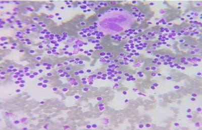
Figure 1: Bone marrow aspiration slides showing medium-sized prolymphocytes.

ijmrr_1432_02.JPG
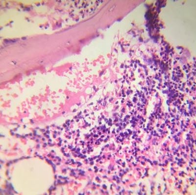
Figure 2: Bone marrow core biopsy revealing increase in prolymphocytes.

ijmrr_1432_03.JPG
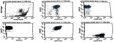
Figure 3: Flow cytometric histogram showing Neoplastic cells( prolymphocytes) positive for CD markers of T cell type and negative for CD1a and Tdt.

ijmrr_1432_04.JPG
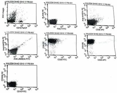
Figure 4: Flow cytometric histogram showing Neoplastic cells (Prolymphocytes)Negative for CD markers of B cell type.

Statistical methods: Event-free survival was estimated using the Kaplan –Meier method. Peto et al, Method was used to determine SEs. R software was used for survival comparison

Discussion

The World Health Organization (WHO) classification of hematopoietic malignancies defines many types of mature T-cell leukemia including T-cell pro-lymphocytic leukemia (T-PLL), T-cell large granular lymphocytic (T-LGL) leukemia and Sezary syndrome (SS). These neoplasms frequently show overlapping features with each other and as these are not commonly encountered, also these T-cell lymphomas involve peripheral blood. Despite the recent advances in our understanding of the molecular biology of T-PLL, survival is poor1,2

This study, to our knowledge the largest series of T-PLL and immunophenotypic characterization from India. Disease characteristics, responses to treatment and factors were included in this study, that may be useful in determining prognosis.


T-PLL accounts for up to one-third of mature T-cell malignancies with a leukemic presentation. Even in the present study, the majority of T leukemias were T-PLL[1,2]. The minority of patients (15%) are asymptomatic at diagnosis and this can persist as an indolent phase for a variable length of time. [3,4,5]. In the present study, all the cases had aggressive clinical course, presentation was in days to months, and precedent indolent phase was not present.

Clinical data was available in all cases, leukocytosis,(>1lakh/cumm ) and lymphocytosis (>90%) were seen in all cases at presentation. None of our cases presented with cytopenia nor eosinophilia was noted. [2]

Similar to earlier reported series male predominance (80%) was noted with a median age of 60 years. Initial presentation with B-symptoms, lymphadenopathy, organomegaly, and pleural and pericardial effusions are suggestive of a cytokine-mediated syndrome. And the data is very sparse. [3]

The hepato splenomegaly. Lymphadenopathy, skin lesions, pleural effusion and were seen in 60 %,10 %,70 %, and 10 %, of patients, respectively. similar to the other reported series, enlarged lymph nodes and uncommon features. The serous effusions chiefly pleural are noted in a minority, of patients.(3,4,5 )We came across a single patient with Serous effusion, ascites and pericardial effusion comprising (10%), fluid cytology was reported as lymphocytic rich effusion, followed by ADA analysis which was normal range.LDH reflects the disease burden in lymphoproliferative disorder, as well as in T-PLL.(3) Routine biochemical analysis revealed increased LDH in 7 cases, accounting for 70% of the total cases, highest LDH value noted was 480 U/L( normal range 120-240U/L). Total calcium, LFT, RFT and Electrolytes were within normal range. Serology for HIV, HBsAg, and HCV were non-reactive by Elisa.

Morphologic features were variable, as reported in the literature. Peripheral smear and bone marrow revealed pro lymphocytes, medium-sized cells, with a high nuclear-cytoplasmic ratio, and an irregular convoluted nucleus with a single prominent nucleolus. the cytoplasm was basophilic and agranular, cytoplasmic blebs and protrusions were commonly seen in most of the cases fig (1)

Skin lesions are evident in 20% of patients [3,5]. Surprisingly skin involvement was noted in 70 % of cases Skin biopsy was done in a single case which was reported as T cell Lymphoma/leukemia. Lymph node biopsy in a single case was done and was reported as Peripheral T-cell Lymphoma. central nervous system involvement feature of T-PLL was not observed in our cohort. Single case BCR-Abl was requested, and it was negative by RT-PCR.

flow cytometry with a large combination of antibodies, gives the immunophenotypic clues that help in distinguishing T-PLL from other malignancies, especially T-cell leukemias . It also allows the recognition of unusual immune phenotypic features.[8] The flow cytometric findings of T-PLL cases are as follows. All cases revealed a monogeneous abnormal population while a single case showed two distinct abnormal subsets on CD3. CD3 was positive in all cases. As expected for a mature T-cell neoplasm CD45 was typically strongly positive. (fig 2). Abnormal gated populations, all expressed pan-T-cell markers CD2, CD3, CD5, and CD7. (fig 3,4) Aberrant expression of antigen (dimmer or brighter than normal T cells) was observed in a few cases .aberrant CD5 (50%) (fig 2). Among those populations showing immune phenotypic abnormalities, 3 cases revealed two aberrancies and one case presented three aberrancies bright cCD3, CD5&weak CD7 .bright CD7 is the hallmark of T-PLL in many reported series ( fig 3,4), however, CD7 was weak in single T-PLL case in present study.

All 100% of cases were negative for CD34 & Tdt. CD1a was done in 2 cases and was negative. (fig 3,4)- 100% of cases of T-PLL were CD4+ in the present study, and 80% of patients cells are CD4+CD8-.Coexpressing CD8 was seen in a single case (CD4+/CD8+ co-expression accounting for 10% ), and a small-cell variant of T –PLL shows co-expression of CD4 and CD8.( fig 3,4) However it was not small cell variant. Nucleoli were seen.

CD4-/CD8+ & CD4-/CD8- immune phenotype populations were not seen in the present study. Aberrantly decreased CD4 expression was noted in a single case ( 10 %).Given the reported rare cases of T-PLL showing decreased surface CD3 and CD45 as well as the more common phenomenon of double positivity of CD4 and CD8, the differential diagnosis for T-PLL may include a T-cell lymphoblastic leukaemia (T-ALL). All cases were evaluated for


markers of immaturity, including CD1a, CD34, and TdT, which were negative, which aided in differentiating T-PLL from immature T-cell neoplasms.

B-cell markers ( CD19, CD10 and CD20) were negative in all cases ( fig 1), and natural killer cell markers (CD16 and CD56) were also negative. CD25 was analysed in a case & was positive. A single case was reported as blasts suggestive of acute leukemia, CD13, CD33, MPO, CD11C, CD64, and CD14, along with immature markers, were found negative.CD200 and CD23 Were also negative. The majority of cases consisted of a single homogeneous population and also showed bright CD7 in the present study.

The distinction of T-PLL from other mature T-lymphoid leukemias and circulating T-cell lymphomas may be more difficult at times.

Adult T-cell leukemia (ATLL) and Sezary syndrome (SS) are often primary considerations in the differential diagnosis of T-PLL. These T-cell neoplasms could be indistinguishable from T-PLL based on morphology and are usually CD4+, similar to most cases of T-PLL. T-PLL usually presents with a bright CD7, decreased in SS and ATLL [1,5,6]. Patients with dim CD7 did not reveal any morphologic features( cerebriform cells) of ATLL/SS, & total calcium levels were within normal range and the CT scan did not reveal any bone lesions. The presence of PB eosinophilia was a highly characteristic and diagnostically useful feature seen in both primary and secondary SS but not other disease categories. None of our cases had eosinophilia. Furthermore, CD25 was negative in our cohort with T-PLL, further distinguishing these cases from ATLL, in which CD25 is strongly positive.

peripheral blood involvement of Peripheral T-cell lymphoma (PTCL) is rare and expresses a wide spectrum of morphologic and immune phenotypic markers. In comparison to T-PLL, PTCL often shows aberrant expression of pan-T-cell antigens, with CD7 and CD3 the most common, followed, by CD2, and CD5. PTCL are most often CD4+, co expression of CD4 and CD8 is not common.

T-cell large granular lymphocyte (LGL) leukemia is frequently indolent and expresses CD16, CD56 and CD57. The majority of LGL leukemia is CD8+, with co-expression of CD4 and CD8 rare. reduced expression of CD5 is noted,

which in combination with a distinct morphology, allows for differentiation from T-PLL. low numbers of circulating tumor T cells and cytopenias characterize -LGL leukaemia. None of our cases were indolent, also none of our cases had cytopenia. The CD4-CD8+ immune phenotype seen in T-LGL leukemia.1 tumour cells in all our cases showed CD5 expression, and all cases expressed CD4.

Pan-t-cell antigens generally have an abnormal expression in a variety of mature T-cell leukemias, preservation of pan-T-cell antigen expression at normal levels and co-expression of CD4 and CD8 helpful findings to distinguish T-PLL from other circulating T-cell leukemias. Also, the presence of multiple subsets appears more common in T-PLL than in other T-cell neoplasms that typically present as an abnormal population with a relatively uniform phenotype,( 5)however this was not the observation in the current study.

There are case reports of patients who initially presented with CD4+/CD8- T-PLL but progressed to the more aggressive course with high CD8 expression, which may be a harbinger of the worst prognosis. we did not come across any such change in immune phenotype during follow-up in our study. some studies indicate an improved response with CD4+/CD8- phenotype. CD4 in the absence of CD8 was the most frequent phenotype in T-PLL(,1,3)All our cases expressed CD 4. In our study, 10 % of T-PLL showed coexpression of CD4 and CD8.

Only a few T-cell neoplasms are associated with specific genetic abnormalities(.11) However T-PLL Patients often have a complex karyotype with numerical and structural abnormalities involving multiple chromosomes.(2,3,5)

The majority of cases in the present study had normal karyotypes except for a single case, which revealed 45 XY,t (3:21)(q11:q22) dup (6) (p21 p26), del (7)(q22) -10, dup(12)(q13q24) add (13)(q340,-14,-15,+2 marker chromosomes.

The most frequent chromosome abnormality in T-PLL involves chromosome 14 with a breakpoint in the longarm chromosome at q11 and q32, seen in 80% of patients. Deletion at 12p13 is a feature of T-PLL, however, in the present study Deletion 14 was seen.

The majority of T-PLL tumors show chromosomal rearrangements involving chromosome 14


that transcriptionally activate the T-cell leukemia-1 (TCL1) gene at 14q32 through the juxtaposition of T-cell receptor (TCR) enhancer/promoter sequences. However, the absence of TCL1 chromosomal rearrangements has been reported by some groups in a substantial number of leukemias classified as T-PLL.2,5,10

Clonality of T lymphocytes can be confirmed by polymerase chain reaction, next-generation sequencing(NGS) Or flow cytometry(.11)

Mutations can be identified through whole-exome sequencing or by next-generation sequencing. Mutations in T-PLL result in constitutive activation of JAK-STAT signalling and upregulation of STAT5B target genes in T-PLL.
JAK inhibitor or STAT5 inhibitor may have a therapeutic effect in T-PLL.(13,14)

Serology for HTLV was not performed routinely in our study and none of our cases showed pleomorphic morphology. none of the cases had hypercalcemia, CT did not reveal any lytic lesions and all our cases expressed CD7.

We could not perform TCL1 by flow cytometry, and Due to financial constraints, NGS was not done in these patients, there was a complete lack of molecular findings, added to it was no uniform follow-up treatment was the choice of individual physician. These were the limitations of the study

Till today there is no single FDA or EMA-approved substance for T-PLL. Alemtuzumab is the most efficient monoclonal antibody.11 Relapses within two years are the rule and median survival time is less than two years. Allogenic stem cell transplant remains the only curative option and patients eligible for transplantation are less than 40%.(13,14)

Despite various therapeutic approaches, including, chemotherapy, most patients did not respond to the treatment and eventually had rapid progression of the disease. 

Treatment and follow-up: All our Cases received FCM protocol (fludarabine, cyclophosphamide, mitoxantrone ), the longest available follow-up was for 10 months and was in clinical and hematological remission, and few other patients were shifted to CHOP as they couldn’t sustain the toxicity of FCM protocol, after the median follow-up of 6 months they had progression of disease in the majority,

and were shifted to a higher centre. Recently many newer therapeutics have been tried, among them anti CD52 is the most important.[1]

In summary, Rapidly rising PB lymphocyte counts, presence of effusions and TCL1 expression for T-PLL, generalized erythroderma, PB eosinophilia, and lymphadenopathy for Sezary syndrome the association of autoimmune phenomena and multiple cytopenias for T-LGL leukemias as the most useful discriminating features at presentation, These features should always be kept in mind along with following flowcytometric immunophenotyping during the diagnosing of T-leukemias.

In T-PLL, immunophenotypic findings being CD 45 bright, positive for T cell markers like CD 2, CD5, CD 4 and Bright CD7, with negative immature markers like CD34, CD1a, TDT along with Being negative B cell, NK cell, and myeloid markers like CD 20, CD19, CD22, CD200, FMC7 and CD14, NK cell markers like CD16 and CD 56. myeloid cell markers like MPO,CD13,CD33,CD64 Respectively.This helps to confirm the diagnosis.

The multicolour flow cytometry has shown increased sensitivity and specificity in detecting abnormal T-cell populations compared with single-parameter techniques.

Accurate diagnosis is critical given the different treatment options and prognostication for patients with different T-cell malignancies, T-PLL continues to challenge the rarity of this disease limits the clinical trials, and multicenter collaborative effort is required to conduct studies. Genomic imbalance studies in these patients will identify newer therapeutic targets for this rare entity, and might significantly add to the true tales of this T-PLL.

Abbreviations; T-PLL; T-Prolymphocytic leukemia.NGS Next-generation Sequencing SS sezary syndrome

Acknowledgement: we acknowledge dr Jaydev Naik and Mrs. Shwetha cytometrist for the technical support in processing the sample.

References

1. D. Catovsky,H K Muller-Hermelink,E. Ralfkiaer et al. WHO Classfication of Tumors of Hematopoietic and Lymphoid Tissues. 4 th edition. IARC,Lyon,France: 2008. 270-71 [Crossref][PubMed][Google Scholar]


2. Matutes E, Catovsky D, Mature T-cell leukemias and leukemia/ lymphoma syndromes. Review of our experience in 175 cases. Leuk Lymphoma 1991 ; 4: (2) 81-91 https://doi. org/10. 3109/10428199109068049 [Crossref][PubMed][Google Scholar]

3. . P. Jain, E. Aoki , M. Keating et al. Characteristics, outcomes , prognostic factors and treatment of patients with T-cell pro lymphocytic leukemia (T -PLL). Annals of Oncology2017 ;(28): 1554 – 1559. 10.1093/annonc/mdx163 [Crossref][PubMed][Google Scholar]

4. Marco Herling, Joseph D Khoury, LaBaron T et al. A systematic approach to diagnosis of mature T-cell leukemias reveals heterogeneity among WHO categories. Blood 2004;104:( 2) 328-335 10. 1182/blood-2004-01-0002. [Crossref][PubMed][Google Scholar]

5. Xueyan Chen , Sindhu Cherian. Immuno phenotypic Characterization of T-Cell Pro lymphocytic Leukemia. Am J Clin Pathol 2013;(140): 727-735 https://doi. org/10. 1309/AJCPG71KYOXTKLQW [Crossref][PubMed][Google Scholar]

6. Pawson R, Matutes E , Brito-Babapulle V et al. Sezary cell leukaemia: a distinct T cell disorder or a variant form of T pro-lymphocytic leukaemia?. Leukemia 1997;(11): 1009-1013 https://doi. org/10. 1038/sj.leu.2400710 [Crossref][PubMed][Google Scholar]

7. Matutes E, Brito-Babapulle V,Swansbury J et al. Clinical and laboratory features of 78 cases of T-prolymphocytic leukemia. Blood 1991;78:( 12):3269-3274 https://doi. org/10. 1182/blood.V78.12.3269.3269 [Crossref][PubMed][Google Scholar]

8. Bain BJ, Barnett D, Linch D, Matutes E, Reily JT. Revised guideline on immuno phenotyping in acute leukemia and chronic lympho proliferative disorders. Clin. Lab. Haem2002;(24):1-13 [Article][Crossref][PubMed][Google Scholar]

9. Shano Naseem. Ritu gupta,Rajesh kashyap et al. T-Cell pro lymphocytic leukemia;a report of 2 cases with review of literature. indian journal of hematology and blood transfusion 2008;(24):178-181. [Crossref][PubMed][Google Scholar]

10. Herling M, Patel KA, Teitell MA, et al. High

TCL1 expression and intact T-cell receptor signaling define a hyperproliferative subset of T-cell pro loymphocytic leukemia. Blood 2008; 111(1): 328-337 https://doi. org/10. 1182/blood-2007-07-101519 [Crossref][PubMed][Google Scholar]

11. Philipp B Staber, Marco Herling, Mar Bellido. Consensus criteria for diagnosis, staging, and treatment response assessment of T-cell pro lymphocytic leukemia. Blood. 2019 Oct 3; 134(14): 1132–1143. doi: 10.1182/blood.2019000402 [Crossref][PubMed][Google Scholar]

12. Dearden CE,Khot A, Else M, et al. Alemtuzumab therapy in T-cell prolymphocytic leukemia:comparing efficacy in a series treated intravenously and a study piloting the subcutaneous route. Blood 2011; 118(22):5799-5802. https://doi. org/10.1182/blood-2011-08-372854 [Crossref][PubMed][Google Scholar]

13. Till Braun , Jana von Jan, Linus Wahnschaffe , et al. Advances and Perspectives in the treatment of T-PLL. Current Hematologic Malignancy Reports 2020:15(2):113-124. doi: 10. 1007/s11899-020-00566-5 [Crossref][PubMed][Google Scholar]

14. Alexandra Schrader , Till Braun and Marco Herling. The dawn of a new era in treating T-PLL. Oncotarget,2019;(10): 626-628. https://doi. org/10.18632/oncotarget.26595 [Crossref][PubMed][Google Scholar]