E-ISSN:2320-8686
P-ISSN:2321-127X

Review Article

Obstructive Sleep Apnea,

International Journal of Medical Research and Review

2022 Volume 10 Number 6 November-December
Publisherwww.medresearch.in

Obstructive Sleep Apnea: A Clinical Review

Shanthi Priya K.1, Bandari G.2*, B L.3, Ramesh Goud S.4, Sriteja D.5, Smriti C.6
DOI: https://doi.org/10.17511/ijmrr.2022.i06.04

1 K Shanthi Priya, Reader, Department of Prosthodontics, MNR Dental College and Hospital, Sangareddy, Telangana, India.

2* Gayathri Bandari, Senior Resident, Department of Prosthodontics, Government Dental College and Hospital, Hyderabad, Telangana, India.

3 Lavanya B, Senior Lecturer, Department of Prosthodontics, AME’s Dental College & Hospital, Raichur, Karnataka, India.

4 S.V Ramesh Goud, Reader, Department of Orthodontics and Dentofacial orthopedics, Navodaya Dental College and Hospital, Raichur, Karnataka, India.

5 Dubhakunta Sriteja, Reader, Department of Orthodontics and Dentofacial orthopedics, Navodaya Dental College and Hospital, Raichur, Karnataka, India.

6 CH. Smriti, Postgraduate, Department of Prosthodontics, MNR Dental College and Hospital, Sangareddy, Telangana, India.

This article aims to provide a detailed description of obstructive sleep apnea regarding its signs and symptoms, the way it is diagnosed, the risk factors, management, and the role of dentists and oral appliances in treatment. Obstructive sleep apnea (OSA) is a potentially life-threatening disorder characterized by repeated collapse of the upper airway during sleep, with periodic cessation of breathing for more than ten seconds. Dental professionals have a unique doctor-patient relationship that can help them in recognizing the sleep disorder and co-managing the patients along with a physician or a sleep specialist. Oral appliance therapy is an important treatment modality for sleep apnea patients.

Keywords: Obstructive Sleep Apnea, Review, Oral Appliances

Corresponding Author How to Cite this Article To Browse
Gayathri Bandari, Senior Resident, Department of Prosthodontics, Government Dental College and Hospital, Hyderabad, Telangana, India.
Email:
K Shanthi Priya, Gayathri Bandari, Lavanya B, S.V Ramesh Goud, Dubhakunta Sriteja, CH. Smriti, Obstructive Sleep Apnea: A Clinical Review. Int J Med Res Rev. 2022;10(6):183-194.
Available From
https://ijmrr.medresearch.in/index.php/ijmrr/article/view/1403

Manuscript Received Review Round 1 Review Round 2 Review Round 3 Accepted
2022-11-26 2022-11-29 2022-12-06 2022-12-13 2022-12-20
Conflict of Interest Funding Ethical Approval Plagiarism X-checker Note
Nil Nil Yes 17%

© 2022by K Shanthi Priya, Gayathri Bandari, Lavanya B, S.V Ramesh Goud, Dubhakunta Sriteja, CH. Smritiand Published by Siddharth Health Research and Social Welfare Society. This is an Open Access article licensed under a Creative Commons Attribution 4.0 International License https://creativecommons.org/licenses/by/4.0/ unported [CC BY 4.0].

Introduction

Obstructive sleep apnea (OSA) is a sleep-related respiratory disease that is becoming more widespread [1]. OSA is a potentially fatal sleep disease marked by frequent pauses in breathing while sleeping. The phrase sleep apnea comes from the Greek word apnea, which means "without breath." Breathing pauses can range anywhere from a few seconds to several minutes, and they can occur up to 30 times per hour [2].

ijmrr_1403_01.JPG
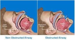
Figure 1: Difference showing Non-Obstructed and Obstructed airway

The two most common varieties of sleep apnea are central sleep apnea, which occurs when the brain fails to deliver vital signals to the breathing muscles, and obstructive sleep apnea, which occurs when air cannot pass through the nose or mouth despite the body's attempts to breathe. Obstructive sleep apnea is significantly more common, and dentists can readily treat it [2].

Obstructive Sleep Apnea and Upper Airway Resistance Syndrome (UARS) are included in Sleep Disordered Breathing (SDB). OSA is a sleep breathing condition characterized by long-term partial upper airway blockage, intermittent total or partial obstruction (obstructive apnea or hypopnea), or both long-term and intermittent obstruction that disrupts normal ventilation, normal sleep patterns, or both [3].

Various studies have shown that sleep-disordered breathing and obstructive sleep apnea (OSA) have a variety of health-related complications, including hypertension, myocardial infarction, stroke, diabetes, depression, excessive daytime fatigue, and an increased risk of car accidents [4,5]. Untreated OSA is linked to poor work performance and a lower quality of life, and it can have a psychological, social, and professional impact on the patient [6]. 

Rapid Eye Movement (REM) sleep, which accounts for 20 to 25% of total sleep time, is connected with physiological changes that affect upper airway function. The pharyngeal dilator muscles' adrenergic and serotonergic regulation is reduced during REM sleep, with cholinergic-mediated inhibition of genioglossus activity. As a result, there is a greater risk of upper airway collapse [7,8].

Furthermore, OSA events during REM sleep are usually longer, more frequent, and associated with higher oxyhemoglobin desaturation than those during non-REM (NREM) sleep. Furthermore, REM sleep is linked to increased sympathetic activity and myocardial demand, as well as significant hemodynamic variability [9]. Apneas and/or hypopneas occur exclusively during REM sleep in 10-37 percent of individuals submitted for OSA examination. OSA during REM sleep has been linked to objective drowsiness in a few clinic-based investigations, and therapy may improve subjective sleepiness and quality of life [9].

REM sleep is linked to OSA and has been linked to both prevalent and incident hypertension as well as glucose metabolism problems. While hypertension and metabolic dysfunction can increase the risk of cardiovascular disease, the impact of REM sleep-specific OSA on endpoints like myocardial infarction, stroke, coronary revascularization, and congestive heart failure is yet unknown [9].

ijmrr_1403_02.JPG
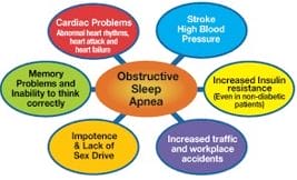
Figure 2: Risks Of Obstructive Sleep Apnea

Prevalence: OSA is characterized by recurrent bouts of upper airway blockage during sleep, which are generally accompanied by a drop in blood oxygen saturation [10]. The apnea-hypopnea index (AHI), which is defined as the number of apneas plus hypopneas per hour of sleep, is commonly used to assess the presence and severity of OSA (or hour of recording for home tests). OSA affects 4% of men and 2% of women


in the middle-aged population (30-60 years) [11]. Prevalence, on the other hand, grows considerably with age, with estimates ranging from 28% to 67% for senior men and 20% to 54% for elderly women [12].

After large-scale epidemiological research, the adult prevalence rates of sleep-disordered breathing are now available in a variety of nations. In the general population, it is estimated to be between 3 and 7 percent for adult men and 2-5 percent for adult women in various nations. As a result, men are 2 to 3 times more likely than women to suffer from OSA [13]. 

Etiology and pathogenesis: OSA is a multi-factorial disorder that is primarily a sleep-related anatomical dysfunction. However, some degree of anatomical damage to the upper airway is required. Anatomic abnormalities that narrow or restrict the airway can cause obstructive sleep apnea. Investigations on the location of a functional occlusion in the upper airway have revealed that there is rarely a single occlusion site. During periods of hypopnea and apnea, obstruction occurs at different levels of the upper airway [14].

Upper airway patency is maintained in all patients by a complex network of anatomic and physiological variables [15]. During inhalation, a negative intra pharyngeal pressure is normal. The pharyngeal abductor and dilator muscles work together to prevent the airway from collapsing. The nasopharynx, oropharynx, and hypopharynx are the three segments of the pharynx. During sleep, the muscles become hypotonic, and the pharyngeal size and pharyngeal tissue compliance in these three segments become more important for airway stability [15,16].

Airway obstruction occurs when the soft tissue compliance in the narrower segments of the passive airway is insufficient to offset the negative intra-luminal pressure caused during inspiration. As a result, the central nervous system changes to a lighter sleep stage by raising muscle tone, allowing the airway to open and the breathing cycle to resume [16].

The potential relevance of elements other than pharyngeal architecture and craniofacial structure in OSA pathophysiology has been highlighted in recent years. OSA can be caused by a variety of factors, the combination of which is likely to

differ significantly between patients. Impaired pharyngeal dilator muscle function, premature waking to modest airway restriction (low respiratory arousal threshold), and unstable control of breathing (high loop gain) are non-anatomical contributions [1].

Impairment in one or more of these non-anatomical factors can sustain OSA severity when paired with a pharyngeal airway that is vulnerable to closure during sleep. Given that OSA only occurs while sleeping, the interaction of an anatomical predisposition with state-dependent changes in non-anatomical components is critical in causing this common disorder [1]. 

Upper airway collapsibility: The Pharyngeal Critical Closing Pressure (PCRIT) is a well-known method for measuring upper airway collapsibility while sleeping. PCRIT is a term that has been used to describe changes in upper airway collapsibility across the spectrum of sleep disordered breathing (from snoring to OSA). It is widely regarded as the gold standard method for determining “functional anatomy” during sleeping. When compared to wakefulness, the PCRIT approach permits the pharyngeal airway to be evaluated under conditions of reduced, but not nonexistent, neuromuscular input.

Once a therapeutic Continuous Positive Airway Pressure (CPAP) level that prevents airway obstruction or narrowing is established, brief reductions (5 breaths) in the holding pressure are applied during stable sleep. This procedure is repeated at different levels of mask pressure until airflow limitation and closure occur [1].

Respiratory Arousal Threshold: Because most respiratory events are coupled with cortical arousal, arousals have long been thought to be essential for reopening the upper airway after a respiratory event in OSA. However, about 20% of respiratory episodes end without cortical arousal, and another 20% happen after the upper airway has already been reopened and the airflow has been restored. Indeed, 75% of adults with OSA have respiratory episodes that end without arousal or arousal that occurs after airway closure at some point throughout the night. As a result, airway reopening can take place without arousal. [1] 

Loop gain: The phrase "loop gain" refers to the stability of a feedback control


system. The ventilatory response to ventilatory disturbance ratio is known as loop gain in respiratory physiology. It is made up of three main components:

1. plant gain (i.e., CO2 storage in tissues, blood, and lungs),

2. circulation delays (i.e., the time it takes for a change in CO2 to mix with existing blood and be sensed by chemoreceptors), and

3. controller gain (i.e., chemosensitivity). Loop gain is affected by any medical condition that changes one or more of these components (e.g., heart failure). Intermittent hypoxia, which is a component of OSA, can also affect respiratory regulation. Exaggerated ventilatory reactions to minor CO2 increases are seen in those with high loop gain. This is a symptom of a faulty control system. CPAP therapy can help to minimize this [1].

Risk Factors: Advanced age, male sex, and obesity are all substantial risk factors for OSA, while the underlying mechanisms are unknown [17].

Age: After 65 years of age, the rising prevalence of SDB breathing appears to level at 10% [18]. Increasing fat deposition in the parapharyngeal area, lengthening of the soft palate, and changes in body tissues around the pharynx have all been hypothesized as mechanisms for increased sleep apnea prevalence in the elderly [19].

Sex: OSA is more frequent in men than women for unknown reasons. It may be due to the upper airway's anatomical and functional properties, as well as the ventilator's response to sleep arousals [20].

Obesity: Obesity/ visceral obesity is the leading cause of OSA, and it is assumed to be linked to anatomical changes that predispose to upper airway blockage during sleeping by increasing adiposity around the pharynx and body. Family history and genetic predisposition: It is considered that genetic factors and familial aggregation play a role in the development of OSA [21].

Family history and genetic predisposition: It is considered that genetic factors and familial aggregation play a role in the development of OSA. When compared to individuals without OSA, first-degree relatives of people with OSA increase their risk by 1.5-2.0, and familial susceptibility

to OSA increases directly with the number of affected relatives [22,23].

Craniofacial and maxillofacial anomalies: Craniofacial and maxillofacial anomalies can both play a role in obstructive sleep apnea instances. Patients with a receding chin and bimaxillary or mandibular retrognathism are more likely to develop indications of obstructive sleep apnea.

Smoking and alcohol consumption: Cigarette smoking and alcohol intake have both been identified as risk factors for OSA. Smoking is linked to an increased risk of snoring and sleep-disordered breathing [24,25].

Clinical Features: some of the common signs and symptoms associated with obstructive sleep apnea are given in Table 1.

Table:1 Signs and Symptoms of Obstructive Sleep Apnea 

Nocturnal Signs And Symptoms Day Time Signs And Symptoms
Drooling Excessive Sleepiness Xerostomia
Xerostomia Morning headaches
Sleep restlessness Non-restorative Sleep
Witnessed apnea Gastroesophageal reflux disease
Choking or Gasping Impaired concentration
Diaphoresis Depression, Impotence

ijmrr_1403_03.JPG
Figure 3:
Symptoms of Obstructive Sleep Apnea

The most sensitive symptom of OSA is snoring. To distinguish between simple snoring and OSA, patients should be asked about their snoring. Patients with OSA report loud, nightly snoring that is disturbing to others. Extreme daytime drowsiness is a generic characteristic that is important in determining therapeutic options and monitoring therapy response [26]. 


Physical examination [2]

The physical examination should include the respiratory, cardiovascular, and neurologic systems.

1. The presence and degree of obesity should be noted.

2. Blood pressure monitoring should be done.

3. Neck circumference measurement (>17” in males and >15.5” in females is significant),

4. Upper airway examination to evaluate:

a. Low hanging bulky soft palate, large tonsils.

b. Tongue scalloping and tongue fat.

c. Low hyoid bone position or maxillomandibular deficiency

d. Dental malocclusion

e. Wear facets (as bruxism is frequently associated with OSA)

f. Signs of nasal obstruction (polyps, septal deviation, turbinate hypertrophy, significant congestion).

Diagnosis

Medical Diagnosis:

1. Polysomnography: A complete overnight sleep study in a laboratory measures the frequency of obstructed breathing events, apneas, and hypopneas during sleep [26]. In general, hypopneas are characterised as reductions in airflow of more than 30% with concurrent reductions in oxyhemoglobin saturation of at least 3% or arousals from sleep [27].

The Apnea Hypopnea Index (AHI) is the average number of apneas and hypopneas per hour of sleep. OSA severity is determined by the AHI score of the patient and is divided into mild, moderate, and severe (AHI score greater than 30) [28]. Not all researchers follow these numerical parameters. De-saturation, quality of life, and daytime sleepiness are all factors that determine OSA severity.

A 5-minute polysomnographic trace of OSA occurrences. The lack of airflow is accompanied by paradoxical breathing (out-of-phase movement of the chest and abdomen) and a drop in oxygen saturation.

Due to the lung-to-finger circulation duration, the nadir oxygen saturation is delayed relative to the apnea. Normal breathing and oxygen saturation return following EEG arousals returning to sleep after each arousal causes upper airway collapse and recurrent obstructive apnea [29].

2. The multiple sleep latency test (MSLT) is used to establish how rapidly the patient falls asleep to distinguish it from narcolepsy.

3. The Epworth sleepiness scale is an 8-item scale that quantifies the propensity for dozing off during everyday activities. [26,30].

4. The Mallampati score (grade 1-4) can be used as a predictor of sleep apnea, particularly in cases where an enlarged tongue seems to be the cause of airway obstruction [31].

5. Lateral cephalometric radiographs reveal the diversion of the airway column, position of hyoid bone and craniofacial skeleton for any maxillo-mandibular deficiencies.

6. Fibro optic naso-pharygoscopy to examine the three-dimensional structure of the airway revealing any anatomic site of obstruction.

Dental diagnostics: A dentist is also the first healthcare professional to recognise an individual with a sleep disorder, as the sleep disorder's distinctive signs and symptoms are frequently observable in the dental office [2].

The following are common clinical findings: 

1. Excess fat accumulation in the palate, tongue, and pharynx (potentially resulting in true macroglossia) — restricting the airway.

2. Those who have small (micrognathia) or retrognathic jaws.

3. Macroglossia with decreased functional space that is driven posteriorly toward the pharyngeal wall and superiorly above the occlusion plane.

4. Because the soft palate is extended and expanded and posteriorly positioned during phonation, the uvula is hidden beneath the base of the posteriorly positioned tongue.

With 3D reconstructed models from computed tomography (CT) data, researchers investigated the influence of the tongue and mandible size ratio (T/M ratio) on the upper airway, concluding


that as tongue volume increases with BMI (body mass index), the posterior airway is affected, and thus is likely to engage in the improvement of OSA [32]. 

Management of the patient with OSA 

Lifestyle modification:

  • This includes positional therapy, which involves placing the patient in a non-supine position to avoid supine slumber. In some circumstances, a non-supine position may be enough to clear the impediment. However, because most patients with sleep disordered breathing (SDB) experience apnea in all positions, this approach is only effective for snoring.
  • Drinking alcohol in the evening should be avoided because it relaxes the airway, making it more susceptible to obstruction in vulnerable areas.
  • All overweight or obese individuals should lose weight in conjunction with other treatments to reduce sleep apnea [33,29].
  • Losing more than 10 kg of weight can improve cardiometabolic health and eliminate obstructive sleep apnea in more than half of people with the mild condition [34].
  • Regular exercise, regardless of weight loss, may help with OSA. The exercise was linked to a 24 percent to 34 percent reduction in OSA severity without considerable weight loss in small randomised clinical studies of patients with moderate to severe OSA. It's unknown what causes this weight-independent advantage. Potential processes include fat redistribution, decreased nightly fluid resorption from the legs, greater pharyngeal muscle strength, and improved sleep quality [29].
  • Medications and medications that relax muscles or reduce respiratory drive (e.g., alcohol, benzodiazepines, and opioids) might worsen obstructive sleep apnea and should be avoided [34]. 

Continuous Positive Airway Pressure (CPAP):

Sullivan and colleagues described the treatment of OSA using nasal continuous positive airway pressure (CPAP). Continuous positive airway pressure (CPAP) is the primary treatment for persons with any severity

of symptomatic OSA. Through a mask worn over the nose or the nose and mouth, PAP devices apply pressure to the airway. This pressure works as a splint during inspiration, preventing the airway from collapsing [29]. Nasal CPAP preserves the patency of the upper airway during sleep using a pneumatic stent. It is the most frequently prescribed treatment for moderate to severe instances of OSA [36]. While wearing the device, CPAP normalises AHI in more than 90% of patients. Benefits are contingent upon therapeutic adherence, with more hours of use per night related to better symptom alleviation and blood pressure reduction. Although subjective, acceptable adherence is typically described as using PAP for at least four hours per night for at least five nights per week, a requirement that the Center for Medicare & Medicaid Services uses to authorise continuing funding for PAP after the initial 90 days of therapy [29]. Factors that contribute to improved PAP adherence include education about the risks of OSA and the expected benefits of CPAP therapy; monitoring of CPAP use with reinforcement and support for technical difficulties; and behavioural interventions, such as cognitive behavioural therapy and motivational enhancement therapy. Each of these factors boosts PAP adherence by more than 30 minutes per night, with behavioural intervention having a mean effect of up to 80 minutes per night [29].

The ability of most contemporary CPAP machines to broadcast adherence data over cellular networks for remote viewing simplifies monitoring PAP adherence. Earlier PAP devices delivered a constant positive pressure and required pressure titration in the laboratory to determine the ideal treatment pressure. Automatic titrating PAP devices, which monitor airflow and alter the pressure in response to variations in flow, have enabled the beginning of PAP therapy without a titration study, lowering costs and enhancing convenience without compromising efficacy or adherence to therapy [29].

Auto-titrating CPAP systems detect flow restriction as a surrogate for upper airway narrowing and adjust pressure automatically to maintain therapeutic levels. In terms of patient adherence and drowsiness reduction, auto-titrating CPAP is comparable to fixed-pressure CPAP. It is not recommended for people with Cheyne–Stokes breathing and has not been well-researched in patients with concomitant pulmonary disease.


A benefit of auto-titrating CPAP is that pressures self-adjust as therapy requirements alter over time, for example, with weight growth [26].

While CPAP is the treatment of choice for patients with symptomatic OSA, it has a low compliance rate of 30-40%. The CPAP machine is bulky and cumbersome, and its use can result in annoying side effects such as nasal congestion, portability issues, pump noise, airway passage dryness, claustrophobia, and nasal leaks with mask discomfort [26,29,30]. 

Oral appliance therapy in the management of OSA:

Simple snoring, upper airway resistance syndrome, and mild to moderate obstructive sleep apnea are the most common indications for oral equipment. Snoring improves in a large proportion of patients, with total relief occurring in a smaller subset. Lowe conducted a comprehensive assessment of the literature and discovered that oral appliances were beneficial in mild to moderate OSA with a compliance rate of 75% [37]. The American Sleep Disorders Association has recognised oral appliance therapy as an approved treatment technique for patients with OSA. Oral appliances can elevate the soft palate (Fig 4), and retain the tongue (Fig 5), or mandible, so facilitating airway opening. In a few instances, a combination of oral appliances and CPAP is employed.

ijmrr_1403_04.JPG

Figure: 4 Soft palate lift appliance

Appliances that raise the soft palate: Due to the gag reflex, discomfort, and success of laser and radio frequency soft palate operations, those that raise the soft palate are rarely used. (Fig 4)

ijmrr_1403_05.JPG
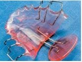
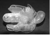
Figure: 5 Tongue retaining devices ( TRD) 

Design consideration of OSA [38, 39]

Over 40 distinct OSAs have been patented to date. Variations in design are determined by the following factors:

  • method of retention
  • material flexibility
  • adjustability
  • vertical opening
  • freedom of jaw movement

These appliances can be manufactured of polyvinyl vacuum moulded thermoplastic or hard acrylic, depending on the material utilised. These may be fixed or adjustable in terms of adjustability. Fixed oral appliances are typically made of one piece and are adjustable in the anteroposterior plane.

One of the recognised designs is a one-piece, non-adjustable soft vinyl vacuum formed mandibular repositioning device made of a thermoplastic material that covers the maxillary and mandibular arches in the appropriate anteroinferior position. A wax bite, silicon bite, or anterior jig with inter-occlusal registration is used to establish and record the occlusal position [40].

Mandibular Repositioning appliance non-adjustable

In the two-part Herbst-style device, the arches are joined by pivoting bars that can be changed in length to taper down the protrusive mandibular posture for the best effectiveness and comfort. Occlusal registration is less necessary with these two-part devices since mandibular repositioning can indeed be gradually increased from the inter-cuspal position [40]. 


Mandibular Repositioning Appliance, Herbst Type TAP (Thornton Adjustable Positioner) The appliance attaches to the mandible through a hook on the maxilla.  

ijmrr_1403_06.JPG
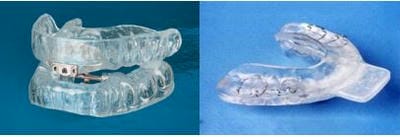
Figure 6: Mandibular repositioning appliance

ijmrr_1403_07.JPG
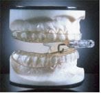
Figure 7: Thornton Adjustable Positioner

For edentulous patients having OSA, a Tongue Stabilizing Device (TSD) that is not attached to the teeth and serves as a pacifier may be employed. It is constructed of soft silicone and gently suctions the tongue forward, preventing it from slipping back. A mandibular repositioning device implanted in the mandible is a feasible therapy option for people with edentulous OSA [41].

ijmrr_1403_08.JPG
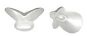
Figure 8: Tongue Stabilizing Device 

Advantages of Oral appliances: Oral appliances provide several advantages over alternative sleep apnea treatment methods, including low cost, high success

rates (equivalent to uvulo-palato-pharyngoplasty but also less effective than CPAP), high compliance, a more benign adverse effect profile, quick effect, and easy cessation without sequelae [42]. In an outpatient situation, OA insertion can be accomplished in a single stage. These can be used effectively for simple snoring and mild to moderate OSA, as recommended by the American Academy of Sleep Medicine [43], but according to a study by Jeffery Pancer, oral appliances appear to be an effective treatment alternative for selected patients with snoring and varying degrees of sleep apnea, including those with severe OSA [44].

In 20% to 75% of patients, OSA improves blood oxygen saturation levels by relieving apnea. They reduce the AHI to ten incidents per hour or achieve a 50% reduction in the AHI. Additionally, they return AHI to normal in 50-60% of patients.

According to a recent study conducted by Kazvya Yoshida, appliance therapy resulted in a significant decrease in mean arterial pressure of approximately 3.7mmHg in a selected cohort [45].

Blood pressure response was substantially linked with AHI reduction, demonstrating oral appliance therapy's efficacy. Reduced blood pressure results in a decreased risk of coronary heart disease and stroke. Oral appliances often have modest side effects, which include excessive salivation, muscle and tooth soreness, and, on rare occasions, temporomandibular joint discomfort. However, symptoms typically improve over time [46]. 

Surgery as a treatment option

Surgery should be considered a last option in the treatment of OSA. Patients who have difficulty with CPAP tolerance owing to anatomical abnormalities may benefit from nasal surgery like septoplasty or turbinate reduction [26]. Although uvulo-palato-pharyngoplasty (UPPP) is a well accepted procedure for managing snoring and sleep apnea, it is only effective in less than half of all patients. Repositioning the hyoid bone is another therapy strategy. Maxillo-Mandibular Advancement (MMA) surgery is another effective method that involves enlarging the upper airway by roughly 10 mm using LeFort I maxillary and bilateral mandibular osteotomies combined with forward stabilisation of the facial bone. Although it is an invasive surgery with a lengthy postoperative recovery period, it cures more than 90% of non-obese


patients with OSA. Additionally, a tracheostomy can be utilised to treat OSA and is occasionally necessary for life-threatening situations. While these two operations may be preferable in certain patients to a lifetime of CPAP therapy, their complexity and associated morbidity preclude their routine usage [26, 29, 30].

While turbinectomy and septoplasty can help reduce AHI, nose surgery is often done in accordance with other surgical procedures. The pillar palatal implant system is a relatively modern therapeutic alternative. This method involves the placement of three mesh polyethylene tetra-phthalate implants within the soft palate muscles under local anaesthesia. By stiffening the palate and minimising the vibratory movements of the palate upon inspiration, these permanent implants assist to decrease snoring [47].

Hypoglossal nerve stimulation is a comparatively recent surgical procedure that improves the tone of the pharyngeal dilator muscle while you sleep. The only device currently approved by the United States Food and Drug Administration involves the unilateral placement of an electrode just on a medial branch of the hypoglossal nerve to stimulate tongue protrusion, a pressure sensor placed between both the internal and external intercostal muscles to detect inspiratory effort, and a small neurostimulator implanted in the chest wall to activate the hypoglossal elect. Patients with a body mass index of less than 32 kg/m2 who exhibit anteroposterior collapse during drug-induced sleep endoscopy should be considered for the procedure [26, 29]. 

Influence of OSA on COVID-19 severity

Sleep deprivation has been proven to significantly increase the production of inflammatory cytokines like interleukin-6, 17 and also tumour necrosis factor-alpha, as well as neutrophilia [48]. Nunes et al. discovered that allergic mice subjected to sleep deprivation while receiving dexamethasone had higher resistance to corticosteroid anti-inflammatory effects, failing to control interleukin-17 and tumour necrosis factor alpha production, as compared to non-allergic animals [49]. Asthma and obstructive sleep apnea might result in sleep loss, exacerbating the inflammatory response of the lung to COVID-19 [48].


Prognosis: OSA treatment often improves sleep apnea and associated behavioural impairment. The degree of improvement is dependent on adherence to therapy. An optimal response is obtained when CPAP therapy is used for more than 6 hours every night. Residual sleepiness is observed in approximately 9% to 20% of people with OSA who use PAP for at least 6 hours per night. This is not much more than the population without OSA and is therefore most likely due to reasons other than OSA, such as sleeping less than the recommended minimum of seven hours per night [29].

CPAP and oral appliances are not curative. Without sufficient weight loss to promote disease remission, lifetime treatment is frequently required. While compliance is not required for surgical procedures, OSA may recur or worsen with additional weight gain [29].

This examination is not without limitations. To begin, it was restricted to English-language articles and sourced mostly from previously published systematic reviews, meta-analyses, and clinical practice guidelines. Second, it is probable that some relevant papers were omitted during the literature search. Third, not all aspects of OSA were addressed. Fourth, certain topics are not adequately covered due to a lack of high-quality data.

Conclusion

Obstructive sleep apnea (OSA) is a prevalent condition that is increasing in prevalence. While excessive daytime sleepiness is a common sign of OSA, many people experience no symptoms. Asymptomatic or mildly symptomatic patients with OSA who do not appear to jeopardise driving safety can be treated with behavioural therapies such as weight loss and exercise. Patients with extreme sleepiness and resistant hypertension may benefit from therapies like CPAP.

Snoring and obstructive sleep apnea are two medical issues that dentistry can significantly help. Due to the relative lack of public and professional attention given to sleep apnea, it is vital to question snoring, daytime fatigue, and other OSA signs and symptoms when they are mentioned. Oral appliance therapy has been recognised by the American Sleep Disorders Association as a viable therapeutic option for certain patients. It is crucial, however, that the dentist cooperate with other members of


the treatment team, including a physician and a sleep specialist, and does not assume entire responsibility for diagnosis and treatment.

Given the enormous increase in risk of heart attack, stroke, and early death associated with obstructive sleep apnea, dentists may be in a unique position to screen, refer, and treat patients, thereby saving lives.

Reference

01. Osman AM, Carter SG, Carberry JC, Eckert DJ. Obstructive sleep apnea: current perspectives. Nat Sci Sleep 2018; 23(10):21-34. . [Crossref][PubMed][Google Scholar]

02. Meenakshi S, Raghunath N. Sleep Apnea: More than Just a Noise. J Orofac Res 2012; 2(2):76-81. . [Crossref][PubMed][Google Scholar]

03. Muhamad A, Azzaldeen A, Nezar W, Firas K. Asian Pac J Health Sci 2014; 1(4): 528-538. . . [Crossref][PubMed][Google Scholar] [Crossref][PubMed][Google Scholar]

04. Waller P C, Bhopal RS. Is snoring a cause of vascular disease: An epidemiological review. Lancet 1989;1:143-146. . [Crossref][PubMed][Google Scholar]

05. Shamsuzzan AS, Gesh BJ Somers VK. Obstructive sleep apnea: Implications for Cardiac and vascular diseases. JAMA- 2003;1906-1914. . [Crossref][PubMed][Google Scholar]

06. Kelly R Magliocca, Joseph I Helman. Obstructive sleep apnea. Diagnosis, medical management and dental implications. J Am Dent. Assoc. 136(8): 1121-1129 [Crossref][PubMed][Google Scholar]

07. Grace KP, Hughes SW, Horner RL. Identification of the Mechanism Mediating Genioglossus Muscle Suppression in REM Sleep. Am J Respir Crit Care Med 2013;187:311-319. . [Crossref][PubMed][Google Scholar]

08. McSharry DG, Saboisky JP, Deyoung P, Jordan AS, Trinder J, Smales E et al. Physiological Mechanisms of Upper Airway Hypotonia During REM Sleep. Sleep 2014;37:561-569. . [Crossref][PubMed][Google Scholar]

09. Aurora RN, Crainiceanu C, Gottlieb DJ, Kim JS, Punjabi NM. Obstructive Sleep Apnea during REM Sleep and Cardiovascular Disease. Am J Respir Crit

Care Med. 2018;197(5):653-660. [Crossref][PubMed][Google Scholar]

10. Diagnostic Classifications Steering Committee: The international classification system of sleep disorders diagnostic and coding manual. Rochester, MN: American Sleep Disorders Association, 1990. . . [Crossref][PubMed][Google Scholar] [Crossref][PubMed][Google Scholar]

11. Young T, Palta M, Dempsey J, Skatrud J, Weber S, Badr S. The occurrence of sleep-disordered breathing in middle-aged adults. N Engl J Med 1993; 328(17):1230-5. . [Crossref][PubMed][Google Scholar]

12. Goodday RH. Nasal respiration, nasal airway resistance, and obstructive sleep apnea syndrome. Oral Maxillofac Surg Clin North Am 1997; 9(2):167-77. . [Crossref][PubMed][Google Scholar]

13. Punjabi NM. The epidemiology of adult obstructive sleep apnea. Proc Am Thorac Soc 2008; 5: 136-43. . [Crossref][PubMed][Google Scholar]

14. Guilleminault C, Hill MW, Simmons FB, Dewent WC. Obstructive sleep apnea: electromyographic and fiberoptic studies. Exp Neurol 1978; 62(1):48-67. . [Crossref][PubMed][Google Scholar]

15. Remmers JE, DeGroot WJ, Sauerland EK, Anch AM. Pathogenesis of upper airway occlusion during sleep. J Appl Physiol 1978; 44(8):931-8. . [Crossref][PubMed][Google Scholar]

16. Lowe AA, Santamaria JD, Fleetham JA, Price C. Facial morphology and obstructive sleep apnea. Am J Orthod Dentofacial Orthop 1986; 90(6):484-91. . [Crossref][PubMed][Google Scholar]

17. Eckert DJ, Malhotra A. Pathophysiology of adult obstructive sleep apnea. Proc Am Thorac Soc 2008; 5:144-53. . [Crossref][PubMed][Google Scholar]

18. Young T, Skatrud J. Peppard PE. Riskfactors for obstructive sleep apnea in adults. JAMA 2004; 291:2013 6. [Crossref][PubMed][Google Scholar]

19. Eikermann M, Jordan AS, Chamberlin NL, gautam S, Wellman A, Lo YL, et al. The influence of aging on pharyngeal collapsibility during sleep. Chest. 2007; 131:1702-9. [Crossref][PubMed][Google Scholar]

20. Jordan AS, McEvoy RD. gender differences in sleep apnea: epidemiology, clinical presentation and pathogenic mechanisms. Sleep Med Rev 2003; 7 : 377-89. . [Crossref][PubMed][Google Scholar]


21. Schwart AR, Patil SP, Laffan AM, Polotsky V, Schneider H, Smith PL. Obesity and obstructive sleep apnea– pathogenic mechanism and therapeutic approaches. Proc Am Thorac Soc2008; 5:185-92. . [Crossref][PubMed][Google Scholar]

22. RedlineS, Tishler PV. The genetics of sleep apnea. Sleep Med Rev 2000; 4 :583-602. . [Crossref][PubMed][Google Scholar]

23. Schwab RJ. Genetic determinants of upper airway structures that predispose to obstructive sleep apnea. Respir Physiol Neurobiol 2005; 147: 289-98. . [Crossref][PubMed][Google Scholar]

24. Khoo SM, Tan WC, Ng TP, Ho CH. Risk factors associated with habitual snoring and sleep-disordered breathing in a multi-ethnic Asian population: a population-based study. Resp Med 2004; 98:557-66. . [Crossref][PubMed][Google Scholar]

25. Ekici M, Ekici A, Keles H, Akin A,Karlidag A, Tunckol M, et al. Risk factors and correlates of snoring and observed apnea. Sleep Med 2008; 9:290-6. . [Crossref][PubMed][Google Scholar]

26. Balachandran JS, Patel SR. In the clinic. Obstructive sleep apnea. Ann Intern Med ;161(9):1-15. [Crossref][PubMed][Google Scholar]

27. Polysomnography Task Force, American sleep Disorders Associaion Standarda of Practice Committee, Practice parameters for the indications for polysomnographu and related procedure. Sleep 1997:20:406-22. . . [Crossref][PubMed][Google Scholar] [Crossref][PubMed][Google Scholar]

28. Flemons WW, Buysse D, Redline S, et al. Sleep related disorders in adults: Recommendations for syndrome definition and measurement techniques in clinical research – the report ofan American Academy of Sleep Medicine Task Force. Sleep 1997:22:667-89. . [Crossref][PubMed][Google Scholar]

29. Gottlieb DJ, Punjabi NM. Diagnosis and Management of Obstructive Sleep Apnea: A Review. JAMA. 2020;323(14):1389-1400. [Crossref][PubMed][Google Scholar]

30. Johns MW. Anew method measuring daytime sleepiness: The Epworth sleepiness scale. Sleep 2001:14:540-45. . [Crossref][PubMed][Google Scholar]

31. Freidman M, Tanyeri H, LaRosa M, Landsberg R, Vaidyanathan K, Pieri S, et al. Clinical predictors of obstructive sleep apnea. Laryngoscope 1999:109:1901-07. . [Crossref][PubMed][Google Scholar]

32. Yuko Shigeta, Takumin Ogawa. Oral surgery, Oral medicine, Oral pathology, Oral Radiology 2011` Feb:111(2):239-43. . . [Crossref][PubMed][Google Scholar] [Crossref][PubMed][Google Scholar]

33. Ramandeep Dugal, M. E. Kothavade, Smita Musani. Role of dentist in management of Obstructive Sleep Apnea-An Overview. IJDA 2010; 2(2) :191-196 [Crossref][PubMed][Google Scholar]

34. Veasey SC, Rosen IM. Obstructive Sleep Apnea in Adults. N Engl J Med. 2019;380(15):1442-1449. [Crossref][PubMed][Google Scholar]

35. Sullivan CE, Issa F G, Berthon Jones M, Eves L. Reversal of obstructive sleep apnea by continuous positive airway pressure applied through the nares. Lancet 1981;1:862-865. . [Crossref][PubMed][Google Scholar]

36. Joo MJ, Hendegen JJ. Sleep apnea in an urban public hospital: Assessment of severity and treatment adherence. J Clin Sleep Med. 2007;3:285-288. [Crossref][PubMed][Google Scholar]

37. Lowe AA; Dental appliances for treatment of snoring and sleep apnea. In: Kruyger M, Roth T, Dement W, eds. Principles and Practice of Sleep Medicine. 2nd ed. Philidelphia: WB Saunders Co., 1994; pp722-735 [Crossref][PubMed][Google Scholar]

38. Isono S, Tanaka A, Sho Y, Konn A, Nishino T. Advancement of mandible improves velopharygeal airway patency. J Appl. Physiol 1995;79:2132-2138. [Crossref][PubMed][Google Scholar]

39. Venkat R, Gopichander N Vasantkumar M. Four novel prosthodontics method for managing upper airway resitance syndrome: An investigative analysis revealing the efficacy of the new nasopharyngeal aperture guard appliance, Indina J Dent Res 2010;21:44-8. . . [Crossref][PubMed][Google Scholar] [Crossref][PubMed][Google Scholar]

40. D. Saee S, S. Sarin P and S Pravin: Obstructive Sleep Apnoea: Dental Implications & Treatment Strategies. Int J Dent Sci. 2009; 7 (1):1-11 [Crossref][PubMed][Google Scholar]


41. Aarnoud Heokema, Frist de Vries, Kees Heydenrijk and Boudewijn Stegenga: Implant-retained oral appliances: a novel treatment for edentulous patients with obstructive sleep apnea-hyponea syndrome. Clin Oral Imp Res 2007; 10-3:383-387. . . 2009; 7 (1):1-11 [Crossref][PubMed][Google Scholar] [Crossref][PubMed][Google Scholar]

42. Padma A, Ramakrishnan N, Narayan V; Management of obstructive sleep apnea: A dental perspective Indian J Dent. Res 2007;18:201-209. . . 2009; 7 (1):1-11 [Crossref][PubMed][Google Scholar] [Crossref][PubMed][Google Scholar] [Crossref][PubMed][Google Scholar]

43. Practice parameters for the treatment of snoring and obstructive sleep apnea with oral appliances. American Sleep Disorders Association 1995;18: 511-513. . . 2009; 7 (1):1-11 [Crossref][PubMed][Google Scholar] [Crossref][PubMed][Google Scholar] [Crossref][PubMed][Google Scholar] [Crossref][PubMed][Google Scholar]

44. Jefferey Pancer, Salem Al Faidi, Mohamed Al Fiadi and Victor Hoffstein: Evaluvation of variable mandibular advancement appliances for treatment of snoring and sleep apnea. Chest 1999; 116:1511-1518. . . 2009; 7 (1):1-11 [Crossref][PubMed][Google Scholar] [Crossref][PubMed][Google Scholar] [Crossref][PubMed][Google Scholar] [Crossref][PubMed][Google Scholar] [Crossref][PubMed][Google Scholar]

45. Kazya Yoshida DDS PhD. Effect on blood pressure of Oral Appliance Therapy for Sleep Apnea Syndrome Int. J Prosthodont 2006;19:61-66. . [Crossref][PubMed][Google Scholar]

46. Kathleen A, Ferguson MD, Rosalind Cartwright, Robert Rogers, Wolfgang Schmidt Nowara. Oral Appliances for snoring and Sleep Apnea: A review Sleep; 2006; 29(2): 244-262. . . [Crossref][PubMed][Google Scholar] [Crossref][PubMed][Google Scholar]

47. Michael Friedman, Paul Schalch. Palatal stiffening after failed uvulopalatopharyngoplasty with the Pillar implant system. Oper Tech Otolaryngol- Head Neck Surg 2007; 18(1):7-10. . [Crossref][PubMed][Google Scholar]

48. Salles C, Mascarenhas Barbosa H. COVID-19 and obstructive sleep apnea. J Clin Sleep Med. 2020;16(9):1647. [Crossref][PubMed][Google Scholar]

49. Nunes JOF, Apostolico JS, Andrade DAG, Ruiz FS, Fernandes ER, Andersen ML et al. Sleep deprivation predisposes allergic mice to neutrophilic lung inflammation. J Allergy Clin Immunol. 2018;141(3):1018-1027. [Crossref][PubMed][Google Scholar]