E-ISSN:2320-8686
P-ISSN:2321-127X

Case Report

Tubercular Osteomyelitis

International Journal of Medical Research and Review

2022 Volume 10 Number 6 November December
Publisherwww.medresearch.in

Tubercular Osteomyelitis of Jaw: A Rare Case Report

Prashant D.1, Prashant V.2*
DOI: https://doi.org/10.17511/ijmrr.2022.i06.01

1 Dolly Prashant, Assistant Professor, Department of Pathology, Govt Dental College, Raipur, Chhattisgarh, India.

2* Viplav Prashant, Associate Professor, Department of Biochemistry, Govt Dental College, Raipur, Chhattisgarh, India.

Tubercular (TB) osteomyelitis of the mandible is a rare and uncommon clinical condition. The clinical appearance of tubercular infection of the temporomandibular joint (TMJ) comprises nonspecific features resembling osteomyelitis, arthritis, or any other kind of chronic joint disease. At times, localized painful swelling of the jaw may be the only manifestation. The presented case is of osteomyelitis of the jaw in a 14-year-old girl patient for whom TB was later suspected.

Keywords: Tubercular Osteomyelitis, Localized Jaw Swelling, Temporomandibular Joint

Corresponding Author How to Cite this Article To Browse
Viplav Prashant, Associate Professor, Department of Biochemistry, Govt Dental College, Raipur, Chhattisgarh, India.
Email:
Dolly Prashant, Viplav Prashant, Tubercular Osteomyelitis of Jaw: A Rare Case Report. Int J Med Res Rev. 2022;10(6):166-170.
Available From
https://ijmrr.medresearch.in/index.php/ijmrr/article/view/1402

Manuscript Received Review Round 1 Review Round 2 Review Round 3 Accepted
2022-11-05 2022-11-07 2022-11-14 2022-11-21 2022-11-28
Conflict of Interest Funding Ethical Approval Plagiarism X-checker Note
Nil Nil Yes 16%

© 2022by Dolly Prashant, Viplav Prashantand Published by Siddharth Health Research and Social Welfare Society. This is an Open Access article licensed under a Creative Commons Attribution 4.0 International License https://creativecommons.org/licenses/by/4.0/ unported [CC BY 4.0].

Introduction

In developed countries, tuberculosis (TB) is uncommon, however, it is not so in developing countries. World Health Organization (WHO) has reported annual mortality of approximately 3 million cases due to TB more commonly in developing countries. Another report shows that there are about 20 million prevalent cases, and 8 million new cases are reported every year [1]. Tuberculous osteomyelitis is a very rare entity occurring more often in young individuals and is usually detected in the late stage of the disease. The sites most involved are dorsal and lumbar vertebrae and epiphysis and diaphysis of long bones. Flat bones of the skull and mandible are rarely affected. The occurrence of tubercular osteomyelitis in the jawbone is very rare [2-6]. We report a case of a 14-year-old female with swelling over the right mandible for 3 months which later proved to be tubercular osteomyelitis in absence of a primary focus. Despite its rare occurrence, tubercular osteomyelitis should be considered a differential diagnosis when routine therapy fails to bring about improvement in the lesion. Early detection of disease results in a complete cure and can even lead to a reversal of all destructive bony changes. If not diagnosed at right time, this can lead to more serious complications like tubercular meningitis. Early diagnosis of tubercular osteomyelitis helps in the reduction of morbidity.

Case report

A 14-year-old girl reported complaining of swelling over the right side of her face in the preauricular region and difficulty in opening her mouth. She had a history of swelling in the mandibular region for 2 months which was slowly increasing in size. She consulted a local dentist who prescribed antibiotics and analgesics, but neither there was a size reduction, nor symptomatic relief. Also, there was no prior medical history of treatment for any chronic infective disease. Her family history was negative for tuberculosis.

The patient was moderately built and well-nourished. She presented with trismus and a diffuse swelling over the right side of her face in the pre-auricular region with no sinus or discharging pus. The overlying skin was normal in appearance.  The ear lobule was not raised.

There was a localized rise in temperature. The swelling was tender and soft on palpation. Tenderness was elicited in the temporalis muscle of the same side with no obvious lymphadenopathy of the head and neck. Examination of the oral cavity was normal [Figure 1]. A provisional diagnosis of chronic osteomyelitis with non-specific inflammation of the condyle was made.

ijmrr_1402_01.JPG
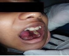
Figure 1: Examination of the oral cavity

In radiographic findings, the panoramic view showed diffuse radiolucency with loss of cortication on the superior and inferior ramus of the condyle, the extent of destruction observed led to the suspicion that a larger lesion may be present. Cone beam computed tomography (CBCT) of the right TMJ showed pronounced rarefaction and destruction of bone in the mandibular condyle with discontinuity of the cortical boundary suggestive of perforation and erosion of inferior ramus [Figure 2]. The radiographic diagnosis came as osteomyelitic changes in relation to the superior and inferior ramus of the mandible.

ijmrr_1402_02.JPG

Figure 2: Radiographic view showing destructive bony changes.


Preoperative chest physician referral and all routine investigations like Hb, CBC, RBS, HbA1c, LFT, RFT, coagulation profile, blood grouping, TSH, viral markers for HIV, HbsAg, and HCV, chest X-ray, and ECG were done. Sequestrectomy and thorough curettage of the necrotic tissue was performed in the right condylar region. The excised tissue was sent for histopathologic examination, which showed epithelioid cell granulomas, histiocytes, and multinucleated Langhans giant cells, with a central acidophilic necrotic focus surrounded by lymphocytes [Figures 3a, 3b, 3c].

The histopathologic report confirmed the diagnosis of TB. Additional GeneXpert TB tests and culture tests were advised. Mycobacterium tuberculosis complex was detected in the GeneXpert TB test and culture test.

ijmrr_1402_03.JPG

Figure 3a: Necrotic osseous fragments (10x view)

ijmrr_1402_04.JPG

Figure 3b: Necrotic osseous fragment with Langhans giant cell (40x view)

ijmrr_1402_05.JPG
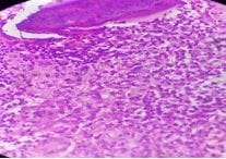
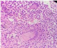
Figure 3c: Area of necrosis with Langhans giant cells (40x view)

Discussion

Oral TB is very rare as low as 1.4%. Oral TB which is commonly seen in patients is a secondary form of TB, where the bacilli present in expectorated sputum from the patient can involve the oral cavity or the upper gastrointestinal/respiratory tract. Secondary TB is comparatively common in adults or geriatric cases. Primary TB of the oral cavity generally occurs in younger patients [7,8]. TB in the young has dissimilar characteristics from those of the secondary form of TB seen in adults. In the present case, we report primary TB of the oral cavity as there was no presence of lung lesion as chest screening and sputum analysis for acid-fast bacilli (AFB) were both negative. 

Primary oral TB is an uncommon disease because of the following reasons: 1) There is the presence of intact stratified squamous epithelium which act as a barrier for invasion of tubercle bacilli into the underlying subepithelial connective tissue, 2) rinsing action of saliva, 3) presence of antimicrobial factors in saliva, 4) presence of salivary enzymes, 5) local pH alteration causing the unfavourable environment for bacterial multiplication, and 6) competition from oral saprophytes for colonization. However, certain systemic factors favour the chances of TB in the oral cavity which includes: 1) lowered host resistance, 2) increased virulence of the organism, 3) local trauma, and 4) pre-existing condition such as cysts, abscesses, leukoplakia, periapical granuloma, periodontitis, extracted sockets and jaw fractures [1,9].


In this case, the child neither had any of the oral problems which can act as a nidus for penetration/localisation of bacilli, nor primaries in the lungs for hematogenous spread; hence, the source of bacilli into bone can only be speculated. 

The common sites of involvement are the tongue, followed by the gingiva, palate, maxillary and mandibular bone, temporomandibular joint, and salivary glands [6,9,10]. Theoretically, the involvement of the mandible is extremely rare as it contains less cancellous bone. However, the mandible is more commonly involved than the maxillary, with the alveolus and angle of the mandible being the commonly involved areas [11,12]. 

Oral TB, as reported by authors, differ in presentations varying from superficial ulcers, patches, indurated soft tissue masses, granulomas, secondary displacement of tooth buds or non-specific involvement of jaws [1]. TB of the jaws can cause gradual necrosis of local osseous tissue and might progress to involve the whole jaw. Presentation of tuberculous involvement of jaws varies from periodontitis with horizontal bone loss, apical osteitis, and widespread destructive lesions which may be misinterpreted for routine dental abscesses. Sequestration and pathological fracture of the bone are also reported [13].

In the present case, there was sequestration and a unilocular destructive bony lesion that elicited a periosteal reaction with cortical expansion. Diagnosis is usually made based on history, clinical presentation, radiography, sputum analysis, histopathology, and serological investigations.

Radiologically, there is no specific appearance of TB osteomyelitis of the mandible. However, findings such as blurring of bone details, diffuse radiolucency of cortical plate and even mixed radiolucent and opaque appearances have been reported by few authors. The normal presentation is indistinguishable from any other pyogenic organism causing non-specific osteomyelitis sometimes associated with periosteal reaction. In the present case, extra-oral radiography showed an ill-defined, smooth, osteolytic, radiolucent, multilocular lesion with onion-peel periosteal reaction over the inferior cortex; intraoral radiography showed a crypt of first permanent molar destroyed in the anteroinferior region with the developing tooth drifted superiorly,

and a CT-scan revealed an expansile multilocular cystic lesion with cortical breaks at places and sequestration at places; all of the signs which are routine to non-specific osteomyelitis; and none specifically indicative of tubercular osteomyelitis [1,9,11]. 

Culture and sensitivity, histopathology, and serological testing are the gold standards for the detection of TB. However, Dimitrakopoulos et al [14] reported primary TB of the oral cavity showing negative smears and cultures for AFB from oral lesion and sputum, but confirmed diagnosis based on history and histopathology, which showed giant cells and epithelioid cells with the absence of classical caseation.

Radiographic findings were consistent with the lytic lesion for which FNA was done; thus, osteosarcoma or Ewing’s sarcoma was to be ruled out. Fine-needle aspiration cytology (FNAC) can distinguish Ewing’s sarcoma which shows round cells containing demarcated nuclear outlines and ill-defined borders [15].

FNAC is a useful tool and the first choice of investigation in India because the cost involved in tests such as PCR is high. Newer diagnostic tests such as PCR are emerging for the diagnosis of TB in children.

According to the current authors, diagnosis of primary TB in oral cavity especially in children is very difficult because of following reasons: 1) Classical history of TB is not present because primary lesion is localised to the bone, 2) there was no pathognomonic bony picture but there was presence of non-specific radiological changes depending on the amount of osseous destruction, 3) sputum analysis was negative due to absence of primary lung lesion, 4) histopathological picture was non-specific because of the absence of classical caseation in the oral lesion, 5) absence of tubercle bacilli in lesions or paucibacilli led to negative reports in Ziehl-Neelsen staining or serum culture sensitivity, 6) there was difficulty in performing specific PCR tests due to costs involved, 7) there was absence of organism in blood cultures as children are subjected to episodic antibiotics that would have decreased the amount of organisms in blood making bacteriological corroboration difficult, 8) due to rarity of lesion, there was low index of suspicion causing misdiagnosis as non-specific osteomyelitis.


Conclusion

Despite its rare occurrence, tubercular osteomyelitis should be considered a differential diagnosis when routine therapy fails to bring about improvement in the lesions. Early detection of the disease results in a complete cure and can lead to a reversal of all destructive bony changes. If not diagnosed at the right time, this can lead to serious complications like tubercular meningitis. Early diagnosis of tubercular osteomyelitis helps in the reduction of morbidity.

Reference

01. Chaudhary S, Kalra N, Gomber S. Tuberculous osteomyelitis of the mandible: a case report in a 4-year-old child. Oral Surg Oral Med Oral Pathol Oral Radiol Endod. 2004 May;97(5):603-6. doi: 10.1016/S1079210404000241 [Crossref][PubMed][Google Scholar]

02. Eng HL, Lu SY, Yang CH, Chen WJ. Oral tuberculosis. Oral Surg Oral Med Oral Pathol Oral Radiol Endod. 1996 Apr;81(4):415-20. doi: 10.1016/s1079-2104(96)80016-3 [Crossref][PubMed][Google Scholar]

03. Gupta MK, Singh M. Primary tuberculosis of mandible. Indian Pediatr. 2007 Jan;44(1):53-4. [Crossref][PubMed][Google Scholar]

04. Dinkar AD, Prabhudessai V. Primary tuberculous osteomyelitis of the mandible: a case report. Dentomaxillofac Radiol. 2008 Oct;37(7):415-20. doi: 10.1259/dmfr/73393014 [Crossref][PubMed][Google Scholar]

05. Chapotel, S. Tuberculose mandibularie. " Rev Odent 51 (1930): 444-5. . [Crossref][PubMed][Google Scholar]

06. Bhatt AP, Jayakrishnan A. Tuberculous osteomyelitis of the mandible: a case report. Int J Paediatr Dent. 2001 Jul;11(4):304-8. doi: 10.1046/j.1365-263x.2001.00276.x [Crossref][PubMed][Google Scholar]

07. Hashimoto Y, Tanioka H. Primary tuberculosis of the tongue: report of a case. J Oral Maxillofac Surg. 1989 Jul;47(7):744-6. doi: 10.1016/s0278-2391(89)80019-9 [Crossref][PubMed][Google Scholar]

08. Ram U, Jha P, Gerland P, Hum RJ, Rodriguez P,

Suraweera W, et al. Age-specific and sex-specific adult mortality risk in India in 2014: analysis of 0·27 million nationally surveyed deaths and demographic estimates from 597 districts. Lancet Glob Health. 2015 Dec;3(12):e767-75. doi: 10.1016/S2214-109X(15)00091-1 [Crossref][PubMed][Google Scholar]

09. Gupta KB, Manchanda M, Yadav SPS et al. Tubercular Osteomyelitis of Mandible. (2005). . [Crossref][PubMed][Google Scholar]

10. Michaud M, Blanchette G, Tomich CE. Chronic ulceration of the hard palate: first clinical sign of undiagnosed pulmonary tuberculosis. Oral Surg Oral Med Oral Pathol. 1984 Jan;57(1):63-7. doi: 10.1016/0030-4220(84)90262-7 [Crossref][PubMed][Google Scholar]

11. Fukuda J, Shingo Y, Miyako H. Primary tuberculous osteomyelitis of the mandible. A case report. Oral Surg Oral Med Oral Pathol. 1992 Mar;73(3):278-80. doi: 10.1016/0030-4220(92)90120-f [Crossref][PubMed][Google Scholar]

12. Ebenezer J, Samuel R, Mathew GC, Koshy S, Chacko RK, Jesudason MV. Primary oral tuberculosis: report of two cases. Indian J Dent Res. 2006 Jan-Mar;17(1):41-4. doi: 10.4103/0970-9290.29893 [Crossref][PubMed][Google Scholar]

13. Erasmus JH, Thompson IO, van der Westhuijzen AJ. Tuberculous osteomyelitis of the mandible: report of a case. J Oral Maxillofac Surg. 1998 Nov;56(11):1355-8. doi: 10.1016/s0278-2391(98)90623-1 [Crossref][PubMed][Google Scholar]

14. Dimitrakopoulos I, Zouloumis L, Lazaridis N, Karakasis D, Trigonidis G, Sichletidis L. Primary tuberculosis of the oral cavity. Oral Surg Oral Med Oral Pathol. 1991 Dec;72(6):712-5. doi: 10.1016/0030-4220(91)90017-7 [Crossref][PubMed][Google Scholar]

15. Lanka, Padmavathy, Lakshmana Rao Lanka, and B. Krishnaswamy. Role of fine needle aspiration cytology of lymphnodes in the diagnosis of cutaneous tuberculosis. (2004). Available at: [Article][Crossref][PubMed][Google Scholar]