E-ISSN:2320-8686
P-ISSN:2321-127X

Research Article

Shear Wave Elastography

International Journal of Medical Research and Review

2022 Volume 10 Number 5 September-October
Publisherwww.medresearch.in

Shear Wave Elastography of Liver: Measurement of normal liver stiffness in healthy population and factors affecting it

Dhar N.1*, Gupta I.2, Gupta K.3
DOI: https://doi.org/10.17511/ijmrr.2022.i05.02

1* Namrita Dhar, Junior Resident, Department of Radiodiagnosis and Imaging, Government Medical College, Jammu, Jammu and Kashmir, India.

2 Ishan Gupta, Lecturer, Department of Radiodiagnosis and Imaging, Government Medical College, Jammu, Jammu and Kashmir, India.

3 Kulbhushan Gupta, Professor, Department of Radiodiagnosis and Imaging, Government Medical College, Jammu, Jammu and Kashmir, India.

Background: Shear Wave Elastography (SWE) is a recent non-invasive method for determining liver stiffness. SWE is a two-dimensional elastography technique in which an amplitude-modulated beam of focused ultrasound is used to generate shear waves which are then transmitted by the transducer to the region of interest (ROI), where the propagation speed of shear waves is measured. The present study is the first attempt to measure the normal range of liver stiffness using SWE in a healthy population from North India and to study the effect of age, gender, and BMI on the liver stiffness values in the healthy population. Methods: This cross-sectional observational study was conducted in the Department of Radiodiagnosis and Imaging, Government Medical College, Jammu on 117 healthy subjects without any known liver pathology or history of any liver disease. B-Mode Ultrasound scan, followed by SWE Examination was performed on all subjects using SAMSUNG RS80EVO using CA1-7A convex array probe with a frequency of 1 to 7 MHz. Results: Successful results were obtained in 98.2%. The mean value of liver stiffness in 115 healthy subjects was 4.74 ± 0.91 kPa, and the 95% confidence interval was 4.58-4.91 kPa. (Range: 2.7-7.8 kPa). There were no statistically significant differences in liver stiffness values regarding age, gender and BMI in the healthy population (all p> 0.05).

Keywords: Shear wave elastography, Liver stiffness measurement, Healthy subjects, Non-invasive diagnosis, Normal

Corresponding Author How to Cite this Article To Browse
Namrita Dhar, Junior Resident, Department of Radiodiagnosis and Imaging, Government Medical College, Jammu, Jammu and Kashmir, India.
Email:
Namrita Dhar, Ishan Gupta, Kulbhushan Gupta, Shear Wave Elastography of Liver: Measurement of normal liver stiffness in healthy population and factors affecting it. Int J Med Res Rev. 2022;10(5):141-146.
Available From
https://ijmrr.medresearch.in/index.php/ijmrr/article/view/1395

Manuscript Received Review Round 1 Review Round 2 Review Round 3 Accepted
2022-09-21 2022-09-23 2022-09-30 2022-10-07 2022-10-14
Conflict of Interest Funding Ethical Approval Plagiarism X-checker Note
Nil Nil Yes 19%

© 2022by Namrita Dhar, Ishan Gupta, Kulbhushan Guptaand Published by Siddharth Health Research and Social Welfare Society. This is an Open Access article licensed under a Creative Commons Attribution 4.0 International License https://creativecommons.org/licenses/by/4.0/ unported [CC BY 4.0].

Introduction

Shear wave elastography (SWE) is a non-invasive quantitative method to measure liver elasticity. Liver fibrosis is characterized by an increase in the deposition of extracellular matrix in the liver and the liver becomes stiffer than normal. An accurate assessment of the degree of fibrosis is essential for the management of liver fibrosis and cirrhosis. Successful detection of fibrosis at an early stage is extremely important to improve the prognosis of these patients as mild to moderate fibrosis is reversible whereas cirrhosis is generally irreversible [1]. Earlier liver biopsy was considered the gold standard method for measurement of fibrosis; however, there are more chances of sampling error due to small tissue samples. In addition, liver biopsy is invasive, costly, has a risk of bleeding and there are more interobserver variations [2,3].

Presently the research focus is on the development of non-invasive methods of assessment of liver fibrosis. Transient Elastography using FibroScan is the most widely used method for the measurement of liver stiffness. However, there are many limitations like difficulty in obese patients, variations in operator experience and it is a one-dimensional technique. Shear Wave Elastography (SWE) is a recent method for determining liver stiffness. The society of Radiologists in Ultrasound has arrived at a consensus for using elastography in the assessment of liver fibrosis [4]. SWE is a two-dimensional elastography technique in which an amplitude-modulated beam of focused ultrasound is used to generate shear waves which are then transmitted by the transducer to the region of interest (ROI), where the propagation speed of shear waves is measured.

Elasticity is calculated by using Young’s Modulus E=3ρc2, where E is the elasticity, ρ is density and c is the shear wave speed [5,6]. Shear waves travel faster in stiffer tissues and loss of elasticity corresponds to the amount of fibrosis. Many studies have evaluated liver stiffness in diffuse liver diseases [7]; however, few studies are available to define the SWE values of liver stiffness in the healthy population. The present study is the first attempt to measure the normal range of liver stiffness in a healthy population from North India and to study the effect of age, gender and Body Mass Index (BMI) on the liver stiffness

values in the healthy population. The present study aimed to determine the normal range of liver stiffness in healthy individuals and to study the effect of age, gender, and BMI on liver stiffness.

Material and Methods

This cross-sectional observational study was conducted in the Department of Radiodiagnosis and Imaging, Government Medical College, Jammu on 117 healthy subjects without any known liver pathology or history of any systemic or liver disease. Subjects with abnormal liver function tests or fatty liver on ultrasound examination were excluded from the study. The study was initiated after approval from the Institutional Ethics Committee. A detailed history was taken and a clinical examination was done. Informed consent was taken from the patients. The various parameters viz., age, sex, weight, height, and BMI were recorded for each subject at the time of the study. B-Mode Ultrasound scan was performed on all subjects using SAMSUNG RS80EVO using CA1-7A convex array probe with a frequency of 1 to 7 MHz. SWE examination was performed on all 117 subjects using the same scanner and transducer. The subjects fasted 4 hours prior to the scan. Imaging was done with subjects in the left lateral decubitus position with their right arm raised overhead. They were required to hold their breath for a few seconds during the examination. The intercostal approach was used for the placement of the transducer. The B- Mode image was optimized to get the best acoustic window. Region of interest (ROI) was placed in the right lobe of the liver perpendicular to the liver capsule, approximately 2 cm beneath the Glisson's capsule, and large blood vessels, bile ducts, and rib shadows were avoided. The SWE image was frozen and the elastic modulus in kPa and shear wave speed in m/s was automatically calculated within the ROI. At least 10 valid measurements were taken with an interquartile range ratio of less than 30% and a Reliability Measurement Index (RMI) of >0.2. The median liver stiffness was recorded and expressed in kPa. (Fig 1).

To study the effect of age, gender, and BMI on liver stiffness, the subjects were divided into six age- groups: < 20 years, 20-29 years, 30-39 years, 40-49 years, 50-59 years, and >59 years; three categories according to WHO Expert


Consultation 2004[8]: underweight <18.5; normal weight 18.5-24.9; overweight >24.9; and two gender categories: males and females. Statistical analysis was done using SPSS 17.0 version and PAST 4.0 version. Statistical significance was set at 0.05 (two-tailed).

ijmrr_1395_01.JPG
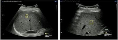
Figure 1:(a) Shearwave Measurements indicating liver stiffness measured in kPa, the depth of region of interest, and the Reliable Measurement Index (RMI).
ijmrr_1395_02.JPG
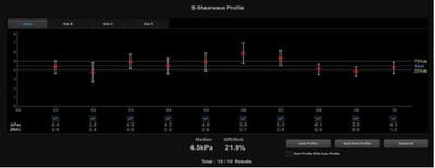
Figure 1:(b) Shear wave profile graph for calculating median liver stiffness.

Results

SWE examination was performed on 117 healthy subjects. Successful results were obtained in 115 cases (62 males and 53 females) with a success rate of 98.2%, as 2 subjects were not able to do successful breath control. The mean value of liver stiffness in 115 healthy subjects was 4.74 ± 0.91 kPa, and the 95% confidence interval was 4.58-4.91 kPa. The range was 2.7-7.8 k Pa.

Influence of age: The value of liver stiffness for healthy adults in each age group is shown in Table 1. The p-value was 0.163, so the difference among various age groups was not statistically significant. (Fig 2)

Table 1: SWE values (kPa) in different age groups

Age (in years) n Mean±SD 95% CI
<20 13 4.34±0.73 3.90-4.78
20-29 37 4.61±0.77 4.36-4.87
30-39 31 5.02±1.06 4.64-5.42
40-49 17 4.81±0.99 4.30-5.32
50-59 9 4.42±0.74 3.86-4.99
>59 8 4.95±1.04 4.09-5.81

ijmrr_1395_03.JPGFigure 2. Liver stiffness according to different age groups

Influence of Gender: The mean shear wave elastography values for males and females are shown in Table 2. The difference was not statistically significant (p-value: 0.097) (Fig 3)

Table 2: SWE value (kPa) in different gender groups

Gender n Mean±SD 95% CI
Male 62 4.84±0.91 4.60-5.07
Female 53 4.64±0.90 4.40-4.89

ijmrr_1395_04.JPG
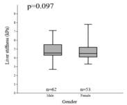
Figure 3. Liver stiffness according to gender

Influence of BMI: The characteristics of liver stiffness among the different BMI groups are shown in Table 3. The p value was 0.084 which was not statistically significant (Fig 4)

Table 3: SWE value (kPa) in different BMI groups

Body mass index (BMI) n Mean±SD 95% CI
<18.5 20 4.63±1.03 4.14-5.11
18.5-24.9 78 4.68±0.86 4.49-4.87
>24.9 17 5.19±0.90 4.73-5.65

ijmrr_1395_05.JPG
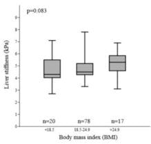
Figure 4: Liver stiffness according to BMI

Discussion

In the present study, liver stiffness was successfully measured in 115 subjects using shear wave elastography, with a success rate of 98.2%. This is comparable to the study by Huang Z et al 2014 [9]. in which the success rate was 98.6%. So, the shear wave elastography can be used successfully to measure liver stiffness as compared to transient elastography in which the rate of uninterpretable results is around 20% [10], and Acoustic Radiation Force Impulse (ARFI) elastography in which valid measurements were found in around 78.83% [11].

The mean value of liver stiffness in the present study was found to be 4.74 ± 0.91 kPa. The results are similar to that studied by Z Haung et al 2014[9]. in which they calculated liver stiffness using SWE and found the mean stiffness to be 5.10±1.02 kPa. Similarly, Mulabecirovic et al 2018[12]. revealed a normal liver stiffness of 4.1±0.8 kPa using SWE. In the studies by Madhok et al 2013[11] and Popescu et al 2011[13], the normal mean liver stiffness using ARFI was 1.197±0.25 m/s and 1.15±0.21m/s respectively. Various studies performed using transient elastography have found mean liver stiffness to be 4.4 kPa [14] ,3.2 to 8.5 kPa [15], 1.08 m/s [16].

In the present investigation, it was found that liver stiffness was not significantly affected by age of the subjects, which is consistent with the previous studies [9,16]. in which there was no influence of age on liver elasticity values. Similarly, Popescu et

al 2011[13], Sirli et al 2013[17], and Fung et al 2013[18]. did not find any statistically significant relationship between age and liver elasticity values.

In the present study, liver stiffness was unaffected by the BMI of the subjects. Similar results were obtained in studies done by Kim et al 2010 [16], Popescu et al 2011[13], Sirli et al 2013[17]. and Z Huang et al 2014 [9]. where no relation was observed between BMI and liver stiffness. However, Das et al 2012[15] found liver stiffness to be higher in both lean and obese individuals.

No statistically significant differences were noted in the liver stiffness between healthy males and females in the present work. This is similar to the studies done by Madhok et al 2013[11]. in which no difference was noted between males and females. Similarly, Popescu et al 2011 [13]. and Sirli et al 2010[17]. also found no difference in liver stiffness between healthy males and females. Higher liver stiffness was reported in males by several researchers [9,14,18].

Conclusion

SWE is a non-invasive and effective method for the measurement of liver stiffness. The mean value of liver stiffness in the north Indian population was 4.74 ± 0.91 kPa, with no statistically significant effect of age, gender and BMI in a healthy population. The present study indicates the importance of the fact that the values of liver stiffness are not universal. This variation can be attributed to different geographic localities, nationalities of the populations, and even the different sample sizes.

Author’s Contribution: Dr Namrita Dhar, Dr Ishan Gupta, and Dr Kulbhushan Gupta contributed equally to the conduct of the study and in the preparation of the manuscript.

Reference

01. Belongia EA, Costa J, Gareen IF, Grem JL, Inadomi JM, Kern ER, et al. NIH consensus development statement on management of hepatitis B. NIH Consens State Sci Statements. 2008 Oct 22-24;25(2):1-29. [Crossref][PubMed][Google Scholar]

02. Froehlich F, Lamy O, Fried M, Gonvers JJ. Practice and complications of liver biopsy. Results of a nationwide survey in Switzerland. Dig Dis Sci.


1993 Aug;38(8):1480-4. doi: 10.1007/BF01308607 [Crossref][PubMed][Google Scholar]

03. Persico M, Palmentieri B, Vecchione R, Torella R, de SI. Diagnosis of chronic liver disease: reproducibility and validation of liver biopsy. Am J Gastroenterol. 2002 Feb;97(2):491-2. doi: 10.1111/j.1572-0241.2002.05507.x [Crossref][PubMed][Google Scholar]

04. Barr RG, Ferraioli G, Palmeri ML, Goodman ZD, Garcia-Tsao G, Rubin J, et al. Elastography Assessment of Liver Fibrosis: Society of Radiologists in Ultrasound Consensus Conference Statement. Ultrasound Q. 2016 Jun;32(2):94-107. doi: 10.1097/RUQ.0000000000000209 [Crossref][PubMed][Google Scholar]

05. Sporea I, Lie I. Shear wave elastography. Ultraschall Med. 2012 Aug;33(4):393-4. [Crossref][PubMed][Google Scholar]

06. Cosgrove D, Piscaglia F, Bamber J, Bojunga J, Correas JM, Gilja OH, et al. EFSUMB guidelines and recommendations on the clinical use of ultrasound elastography. Part 2: Clinical applications. Ultraschall Med. 2013 Jun;34(3):238-53. doi: 10.1055/s-0033-1335375 [Crossref][PubMed][Google Scholar]

07. Bavu E, Gennisson JL, Couade M, Bercoff J, Mallet V, Fink M, et al. Noninvasive in vivo liver fibrosis evaluation using supersonic shear imaging: a clinical study on 113 hepatitis C virus patients. Ultrasound Med Biol. 2011 Sep;37(9):1361-73. doi: 10.1016/j.ultrasmedbio.2011.05.016 [Crossref][PubMed][Google Scholar]

08. WHO Expert Consultation. Appropriate body-mass index for Asian populations and its implications for policy and intervention strategies. Lancet. 2004 Jan 10;363(9403):157-63. doi: 10.1016/S0140-6736(03)15268-3. Erratum in: Lancet. 2004 Mar 13;363(9412):902 [Crossref][PubMed][Google Scholar]

09. Huang Z, Zheng J, Zeng J, Wang X, Wu T, Zheng R. Normal liver stiffness in healthy adults assessed by real-time shear wave elastography and factors that influence this method. Ultrasound Med Biol. 2014 Nov;40(11):2549-55. doi: 10.1016/j.ultrasmedbio.2014.05.008 [Crossref][PubMed][Google Scholar]

10. Castéra L, Foucher J, Bernard PH, Carvalho

F, Allaix D, Merrouche W, et al. Pitfalls of liver stiffness measurement: a 5-year prospective study of 13,369 examinations. Hepatology. 2010 Mar;51(3):828-35. doi: 10.1002/hep.23425 [Crossref][PubMed][Google Scholar]

11. Madhok R, Tapasvi C, Prasad U, Gupta AK, Aggarwal A. Acoustic radiation force impulse imaging of the liver: measurement of the normal mean values of the shearing wave velocity in a healthy liver. J Clin Diagn Res. 2013 Jan;7(1):39-42. doi: 10.7860/JCDR/2012/5070.2665 [Crossref][PubMed][Google Scholar]

12. Mulabecirovic A, Mjelle AB, Gilja OH, Vesterhus M, Havre RF. Liver elasticity in healthy individuals by two novel shear-wave elastography systems-Comparison by age, gender, BMI and number of measurements. PLoS One. 2018 Sep 14;13(9):e0203486. doi: 10.1371/journal.pone.0203486 [Crossref][PubMed][Google Scholar]

13. Popescu A, Sporea I, Sirli R, Bota S, Focşa M, Dănilă M, et al. The mean values of liver stiffness assessed by Acoustic Radiation Force Impulse elastography in normal subjects. Med Ultrason. 2011 Mar;13(1):33-7. [Crossref][PubMed][Google Scholar]

14. Colombo S, Belloli L, Zaccanelli M, Badia E, Jamoletti C, Buonocore M, et al. Normal liver stiffness and its determinants in healthy blood donors. Dig Liver Dis. 2011 Mar;43(3):231-6. doi: 10.1016/j.dld.2010.07.008 [Crossref][PubMed][Google Scholar]

15. Das K, Sarkar R, Ahmed SM, Mridha AR, Mukherjee PS, Das K, et al. Normal liver stiffness measure (LSM) values are higher in both lean and obese individuals: a population-based study from a developing country. Hepatology. 2012 Feb;55(2):584-93. doi: 10.1002/hep.24694 [Crossref][PubMed][Google Scholar]

16. Kim SU, Choi GH, Han WK, Kim BK, Park JY, Kim DY, et al. What are 'true normal' liver stiffness values using FibroScan?: a prospective study in healthy living liver and kidney donors in South Korea. Liver Int. 2010 Feb;30(2):268-74. doi: 10.1111/j.1478-3231.2009.02172.x [Crossref][PubMed][Google Scholar]

17. Sirli R, Bota S, Sporea I, Jurchis A, Popescu A, Gradinaru-Tascău O, et al. Liver stiffness


measurements by means of supersonic shear imaging in patients without known liver pathology. Ultrasound Med Biol. 2013 Aug;39(8):1362-7. doi: 10.1016/j.ultrasmedbio.2013.03.021 [Crossref][PubMed][Google Scholar]

18. Fung J, Lee CK, Chan M, Seto WK, Wong DK, Lai CL, et al. Defining normal liver stiffness range in a normal healthy Chinese population without liver disease. PLoS One. 2013 Dec 26;8(12):e85067. doi: 10.1371/journal.pone.0085067 [Crossref][PubMed][Google Scholar]