E-ISSN:2320-8686
P-ISSN:2321-127X

Case Report

Strongyloidiasis

International Journal of Medical Research and Review

2022 Volume 10 Number 2 March April
Publisherwww.medresearch.in

Parasitic Hypoproteinemia: A Diagnostic Dilemma in an Immunocompetent Patient

Maitra S.1*, Pal D.2, Ray K.3, Adnan Yusuf S.4
DOI: https://doi.org/10.17511/ijmrr.2022.i02.05

1* Somnath Maitra, Associate Professor, Department of General Medicine, JIMSH, Budge, Kolkata, West Bengal, India.

2 Debes Pal, Associate Professor, Department of Pathology, JIMSH, Budge, Kolkata, West Bengal, India.

3 Koushik Ray, Assistant Professor, Department of Anatomy, JIMSH, Budge, Kolkata, West Bengal, India.

4 Sk Adnan Yusuf, Senior Resident, Department of General Medicine, JIMSH, Budge, Kolkata, West Bengal, India.

Strongyloidiasis is caused by a nematode helminth which causes multisystem involvement with signs and symptoms related to the gastrointestinal, pulmonary, dermatological and nervous systems. The index case discussed here presented with edema, hypoalbuminemia, malnutrition and anemia with urease positive duodenal ulcer. Duodenal biopsy suggested malabsorption, and strongyloidiasis was detected in the biopsy, which clinched the diagnosis and treatment was given with ivermectin, after which the patient improved and responded to treatment. The importance lies in the fact that parasitic infections may cause malabsorption even in immunocompetent patients, which is a rare entity but must not be missed as it responds to treatment promptly, which is cheap and effective.

Keywords: Strongyloidiasis, hypoalbuminemia, malabsorption, anemia

Corresponding Author How to Cite this Article To Browse
Somnath Maitra, Associate Professor, Department of General Medicine, JIMSH, Budge, Kolkata, West Bengal, India.
Email:
Somnath Maitra, Debes Pal, Koushik Ray, Sk Adnan Yusuf, Parasitic Hypoproteinemia: A Diagnostic Dilemma in an Immunocompetent Patient. Int J Med Res Rev. 2022;10(2):81-84.
Available From
https://ijmrr.medresearch.in/index.php/ijmrr/article/view/1379

Manuscript Received Review Round 1 Review Round 2 Review Round 3 Accepted
2022-02-28 2022-03-01 2022-03-08 2022-03-15 2022-03-22
Conflict of Interest Funding Ethical Approval Plagiarism X-checker Note
Nil Nil Yes 17%

© 2022by Somnath Maitra, Debes Pal, Koushik Ray, Sk Adnan Yusufand Published by Siddharth Health Research and Social Welfare Society. This is an Open Access article licensed under a Creative Commons Attribution 4.0 International License https://creativecommons.org/licenses/by/4.0/ unported [CC BY 4.0].

Introduction

Strongyloidiasis is a disease caused by Strongyloides stercoralis, which is a nematode. It may present with pain abdomen, diarrhea, vomiting, indigestion along with loss of weight. Rarely it can cause malabsorption syndrome, protein-losing enteropathy and appendicitis.[1]. We present an interesting case of anemia, hypoalbuminemia and pain abdomen associated with strongyloidiasis, which is a rare presentation and causes a diagnostic dilemma.

Case Report

A 45 years Hindu female residing in South 24 Parganas district in West Bengal presented with two months history of generalized weakness with pedal edema for the same duration. She had attained menopause and had no history of fever. There was a history of vomiting with ill-defined abdominal pain for the last one week with a history of intermittent loose motions. There was neither history of facial puffiness, decreased urine volume, hematuria, orthopnoea, PND, jaundice, nor any history suggestive of bleeding from any site. There was no history of photosensitivity, malar rash, abdominal swelling or any heaviness of the chest. She had one son, and there was no history of addiction or drug intake. There was no history of blood transfusion or exposure, and the patient was non-diabetic and non-hypertensive.

Signs and symptoms-On examination, the patient was alert, conscious and cooperative. She was malnourished with severe anemia with koilonychia and leukonychia. There was glossitis with oral ulcers and bilateral pitting pedal edema. There was no icterus or lymphadenopathy. Tachycardia was present with normal blood pressure. Epigastric tenderness was present on superficial palpation without any organomegaly or ascites. There was no scar mark on the abdomen. A provisional diagnosis of malabsorption syndrome with iron deficiency anemia and hypoproteinemia was made with a differential diagnosis of nephrotic syndrome.

Laboratory Investigations-On investigation Hemoglobin was 7.7 gm/dl with microcytic hypochromic anemia and mild eosinophilia on peripheral blood smear with normal WBC and platelet count. Urea and creatinine levels were normal, with mild hyponatremia and hypokalemia.

On LFT, albumin was 1.8 g/dl with a mild rise of transaminases, and alkaline phosphatase was normal. USG WA, chest x-ray, ECG, and Echocardiography were normal. Serology was non-reactive, and fasting plasma glucose, postprandial plasma glucose and HbA1c were normal. Stool examination for OPC revealed mucus and 1-2 pus cells/HPF but did not reveal any organism. Fecal Calprotectin was negative, so was serum ANF by the HEP 2 method. Urine ACR was normal, and there was no proteinuria on urine RE. Serum iron was low with reduced ferritin and increased TIBC. IgA tissue transglutaminase was negative. On upper GI Endoscopy, Urease positive active duodenal ulcer was found in D1 with antral gastritis. Vitamin D3 levels were low with normal vitamin B12 and RBC Folate levels. Duodenal biopsy clinched the diagnosis by detecting larva of Strongyloides stercoralis and flattening of villi altering the crypt villi ratio with mucosal ulceration and moderate chronic inflammation in the lamina propria (Figure 1, 2). Patient refused to do CECT Whole abdomen and colonoscopy.

Diagnosis and treatment

A diagnosis of malabsorption syndrome due to Strongyloides stercoralis was made with duodenal ulcer and iron deficiency anemia with koilonychia and leukonychia due to iron deficiency and hypoalbuminemia respectively. (Figure 3). Initially the patient was treated with an injection of PPI, Ondansetron, drotaverine with judicious iv fluid and two units of PRBC transfusion. Injection metronidazole was also given with Vitamin D3 oral supplementation and injection multivitamin. Triamcinolone acetonide paste was given for oral ulcers, and the patient was started on HP Kit therapy for two weeks after receiving the endoscopy report. Finally, Ivermectin 12 mg OD for two days was given for Strongyloidiasis, after which the patient started improving and serum albumin started to increase. The pain abdomen reduced, and the patient felt better, proving Strongyloides stercoralis as the causative organism. Hookworm was also a differential diagnosis, but a duodenal biopsy revealed the morphology of Strongyloides stercoralis.

Discussion

Strongyloidiasis is an infection of the intestine caused by parasitic nematodes, mostly S stercoralis.


It can stay and multiply within the host for many years without any symptoms in immune-competent individuals but causes life-threatening disseminated infection or hyperinfection syndrome in immunocompromised cases with high mortality. [2,3,4].Risk factors are corticosteroid therapy, HTLV1infection, HIV infection, malignancy, diabetes, malabsorption, chronic alcohol consumption etc.[5,6,7,8,9,10,11]. The gastrointestinal symptoms are anorexia, weight loss, epigastric pain, vomiting, and nausea which were present in our case, but stool examination was unremarkable. Strongyloides hyperinfection presenting as diarrhea was described in a diabetic patient by Shreekant Tiwari et al. [12]. But our case was non-diabetic. Another case was described by Niranjan Tachamo et al. where Strongyloides presented as itching and abdominal pain in an immunocompetent patient [13], but not as malabsorption syndrome. Strongyloides stercoralis presenting as malabsorption is a rare entity.

ijmrr_1379_1.JPG
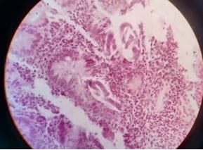
Figure 1: Strongyloides in duodenal biopsy

ijmrr_1379_2.JPG
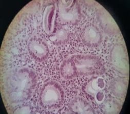
Figure 2: Strongyloides in duodenal biopsy

ijmrr_1379_3.JPG
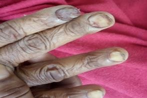
Figure 3: Koilonychia and leukonychia

Conclusion

Strongyloidiasis rarely causes malabsorption syndrome. Our patient presented with anemia, hypoalbuminemia and duodenal ulcer with malabsorption and malnutrition, which responded to ivermectin therapy after a duodenal biopsy revealed strongyloidiasis. The importance of the case lies in the fact that clinicians must remember the treatable causes of malabsorption syndrome before proceeding with complicated investigations and making undue delays in diagnosing and treating parasitic malabsorption which can lead to fatal complications.

Reference

01. Patel J, Gupta D, Rathi C, Parikh P, Ingle M, Sawant P. Uncommon presentation of Strongyloidiasis: chronic malabsorption, multiple small bowel strictures and appendicitis in HTLV-1 positive patient. Trop Gastroenterol. 2015 Jul-Sep;36(3):212-5. doi: 10.7869/tg.293 [Crossref][PubMed][Google Scholar]

02. Arsić-Arsenijević, Valentina, et al. Fatal Strongyloides stercoralis infection in a young woman with lupus glomerulonephritis. Journal of nephrology 18. 6 (2005): 787-790. [Crossref][PubMed][Google Scholar]

03. Weller PF, Leder KL. Strongyloidiasis. Available at http://www. uptodate. com/contents/strongyloidiasis?source=search_result&selectedTitle=1%7E4. Accessed: January 29, 2008 [Crossref][PubMed][Google Scholar]

04. Asdamongkol N, Pornsuriyasak P, Sungkanuparph S. Risk factors for strongyloidiasis


hyperinfection and clinical outcomes. Southeast Asian J Trop Med Public Health. 2006 Sep;37(5):875-84. [Crossref][PubMed][Google Scholar]

05. Fardet L, Généreau T, Poirot JL, Guidet B, Kettaneh A, Cabane J. Severe strongyloidiasis in corticosteroid-treated patients: case series and literature review. J Infect. 2007 Jan;54(1):18-27. doi: 10.1016/j.jinf.2006.01.016 [Crossref][PubMed][Google Scholar]

06. Guyomard JL, Chevrier S, Bertholom JL, Guigen C, Charlin JF. Découverte d'une infestation à Anguillule, 25 ans après retour de zone d'endémie, à l'occasion d'une corticothérapie pour traumatisme oculaire [Finding of Strongyloides stercoralis infection, 25 years after leaving the endemic area, upon corticotherapy for ocular trauma]. J Fr Ophtalmol. 2007 Feb;30(2):e4. French. doi: 10.1016/s0181-5512(07)89571-4 [Crossref][PubMed][Google Scholar]

07. Porto AF, Neva FA, Bittencourt H, Lisboa W, Thompson R, Alcântara L, Carvalho EM. HTLV-1 decreases Th2 type of immune response in patients with strongyloidiasis. Parasite Immunol. 2001 Sep;23(9):503-7. doi: 10.1046/j.1365-3024.2001.00407.x [Crossref][PubMed][Google Scholar]

08. Satoh, M. , et al. Reduced efficacy of treatment of strongyloidiasis in HTLV-I carriers related to enhanced expression of IFN-γ and TGF-β1. " Clinical & Experimental Immunology 127. 2 (2002): 354-359 [Crossref][PubMed][Google Scholar]

09. Porto, Maria Aurelia F. , et al. Atypical clinical presentation of strongyloidiasis in a patient co-infected with human T cell lymphotrophic virus type I. The American journal of tropical medicine and hygiene 72. 2 (2005): 124-125 [Crossref][PubMed][Google Scholar]

10. Carvalho, E. M. , and A. Da Fonseca Porto. Epidemiological and clinical interaction between HTLV‐1 and Strongyloides stercoralis." Parasite immunology 26.11‐12 (2004): 487-497 [Crossref][PubMed][Google Scholar]

11. Thompson BF, Fry LC, Wells CD, Olmos M, Lee DH, Lazenby AJ, Mönkemüller KE. The spectrum of GI strongyloidiasis: an endoscopic-pathologic study. Gastrointest Endosc. 2004 Jun;59(7):906-10. doi: 10.1016/s0016-5107(04)00337-2 [Crossref][PubMed][Google Scholar]

12. Tiwari S, Rautaraya B, Tripathy KP. Hyperinfection of Strongyloides stercoralis in an immunocompetent patient. Trop Parasitol. 2012 Jul;2(2):135-7. doi: 10.4103/2229-5070.105182 [Crossref][PubMed][Google Scholar]

13. Tachamo N, Nazir S, Lohani S, Karmacharya P. Strongyloidiasis in the immunocompetent: an overlooked infection. J Community Hosp Intern Med Perspect. 2016 Sep 7;6(4):32038. doi: 10.3402/jchimp.v6.32038 [Crossref][PubMed][Google Scholar]