E-ISSN:2320-8686
P-ISSN:2321-127X

Research Article

Cervix cancer

International Journal of Medical Research and Review

2022 Volume 10 Number 2 March April
Publisherwww.medresearch.in

Impact on Undefined Normal Tissue by using the different combination of Normal Tissue Objective and Dose Control Ring in IMRT Plans for Cervix cancer

Muthukumar S.1, Navitha S.2*, Nigam J.3, Silambarasan N.4, Kumar P.5
DOI: https://doi.org/10.17511/ijmrr.2022.i02.03

1 S Muthukumar, Intern Medical Physicist, Department of Radiation Oncology, SRMS Institute of Medical Sciences, Bareilly, U.P, India.

2* S Navitha, Medical Physicist, Department of Radiation Oncology, SRMS Institute of Medical Sciences, Bareilly, U.P, India.

3 Jitendra Nigam, Medical Physicist, Department of Radiation Oncology, SRMS Institute of Medical Sciences, Bareilly, U.P, India.

4 NS Silambarasan, Medical Physicist, Department of Radiation Oncology, SRMS Institute of Medical Sciences, Bareilly, U.P, India.

5 Piyush Kumar, Professor and Head, Department of Radiation Oncology, SRMS Institute of Medical Sciences, Bareilly, U.P, India.

Introduction: Normal tissue objective is tool used in inverse planning of Intensity Modulated Radiation Therapy reduce dose spreading surrounding normal tissues. Multitude of potential NTO setting combinations challenges optimal NTO tunning. Aim and Objective: Find impact on Undefined Normal Tissue (UNT) by using different combinations of NTO and DCR in IMRT Plans for Cervical cancer plans. Materials and Methods: Our sample consists of 30 patients with similar treatment prescription doses. Varian Eclipse Treatment Planning System Version13.6 was used in study. 5 different plans were created each patient.Every plan beam energy, several beams, Beam angle, Optimization algorithm - Photon optimizer (PO), Calculation algorithm – Anisotropic analytic algorithm, evaluation methods were maintained constant. 5 plans were different only in optimization process. Before generating plans DCR thickness 1.0 cm and 0.5 cm away from Planning Target Volume was created. Plan with different combinations between NTO, DCR were A. Without NTO, B. Automatic NTO, C. Manual NTO, D. Automatic NTO + DCR, E. Manual NTO + DCR generated. Plan quality was evaluated by comparing PTV: Conformity Index (CI), Homogeneity Index, OAR Doses and mean dose to UNT. Results: HI was better without NTO plans compared to all other plans. CI and OAR doses show significant difference in Manual NTO along with DCR plans. Conclusion: Study shows manual NTO + Dose Control Ring gives better plan quality terms PTV coverage, less dose to Undefined Normal Tissue by maintaining Organ at Risk dose within tolerance limits.

Keywords: Intensity Modulated Radiation Therapy, Normal tissue objective, Undefined Normal Tissue, Radiotherapy, Cervix Cancer

Corresponding Author How to Cite this Article To Browse
S Navitha, Medical Physicist, Department of Radiation Oncology, SRMS Institute of Medical Sciences, Bareilly, U.P, India.
Email:
S Muthukumar, S Navitha, Jitendra Nigam, NS Silambarasan, Piyush Kumar, Impact on Undefined Normal Tissue by using the different combination of Normal Tissue Objective and Dose Control Ring in IMRT Plans for Cervix cancer. Int J Med Res Rev. 2022;10(2):69-74.
Available From
https://ijmrr.medresearch.in/index.php/ijmrr/article/view/1376

Manuscript Received Review Round 1 Review Round 2 Review Round 3 Accepted
2022-02-08 2022-02-10 2022-02-17 2022-02-24 2022-03-03
Conflict of Interest Funding Ethical Approval Plagiarism X-checker Note
Nil Nil Yes 17%

© 2022by S Muthukumar, S Navitha, Jitendra Nigam, NS Silambarasan, Piyush Kumarand Published by Siddharth Health Research and Social Welfare Society. This is an Open Access article licensed under a Creative Commons Attribution 4.0 International License https://creativecommons.org/licenses/by/4.0/ unported [CC BY 4.0].

Introduction

Cervical cancer is the second most common malignant tumour in women and is the third leading cause of cancer-related death among women worldwide [1]. Radiotherapy alongside surgery and chemotherapy is an important part of the therapeutic process in women suffering from these cancers. Radiotherapy is one of the main techniques to control the tumour [2]. The aim of radiotherapy is that more dose to the target volume and less dose to the surrounding normal tissues [3].

In the past few decades, conventional two-dimensional RT (2D-RT) has been widely used in the treatment of cervical cancer, but this treatment option suffers from a high frequency of acute and chronic complications, which affect the treatment efficacy as well as patient quality of life [4]. Three-dimensional conformal RT (3D-CRT) based on computed tomography is becoming a critical part of RT. This approach is relatively favourable in terms of the radiation dose and toxicity to organs in the exposure field [5].

Intensity-modulated RT (IMRT) is a precise RT that has been developed based on 3D-CRT. An advantage of IMRT is that it can deliver a relatively large radiation dose over a target area while minimising the radiation dose to adjacent non-cancerous tissue, thereby offering greater loco-regional control and leading to fewer side effects [6].

In IMRT planning it is classified into two types by optimization method. Those are forward and inverse planning. Forward IMRT is like a 3DCRT, manually can change the fluence by altering the Multi-Leaf Collimator (MLC) according to plan. During Inverse planning optimization, Treatment Planning System (TPS) will generate fluence concerning Planning Target Volume (PTV), Organ at Risk (OAR) Volumes and dose constraints. Usually, medical physicists get the CT structure with delineated PTV and OARs, Other than this structure some soft tissues there within the body. This soft tissue is called Undefined Normal Tissue (UNT) and these soft tissues receive a dose. However, normal tissue that is exposed to radiotherapy can also be affected, leading to toxic effects. In the majority of patients, radiotherapy is delivered to a small, well-defined part of the body. Moreover, radiotherapy is delivered in small doses (fractions), which target and kill comparatively more cancer cells than normal cells [7].

Materials and Methods

Study Setting: Department of Radiation oncology, Shri Ram Murti Smarak Institute of Medical Sciences.
Study Design: Retrospective study
Duration and type of study: A total of thirty patients, diagnosed with cervix cancer, treated between March 2021 and December 2021 were included in this study. For each patient, we have generated five plans with a combination of DCR and NTO for study purposes.
Inclusion criteria:
1. Carcinoma Cervix patients
2. Dose fractionation schedule 50.4Gy/28 Fractions

Radiotherapy: Simulation and Volume Delineation: All the patients underwent simulation in a supine position using the 4-point thermoplastic cast. Contrast-enhanced CT scans of 3mm slice thickness were obtained.

The following volumes were delineated by Oncologist.
Clinical Target Volume (CTV) primary, Clinical Target Volume (CTV) nodal – nodal volumes were delineated as per the guidelines given by RTOG [8]. Planning Target Volume (PTV)- 5mm isotropic margin to the CTV to account for setup errors. The OAR’S were delineated as per RTOG guidelines. This included the Urinary bladder, Rectum, Femoral heads, Bone marrow and Bowel. Undefined normal tissue is a structure within the body excluding target and OARs which is shown in Fig.1. Contouring and dose reporting to UNT are routinely not practised.

Dose prescription: A total of 50.4 Gy in 28 fractions was prescribed to the PTV. All the patients received treatment using the IMRT technique. The constraints are given for the OARs were:-Femoral head Dmax ≤50Gy; Urinary Bladder V50<50; Rectum V50<50; Bowel V45<195cc; Bone Marrow Dmean<30Gy. Dose tolerance for these OARs followed as per RTOG.

Planning
1. The plans were created in the Eclipse Treatment Planning System of Version13.6 (Varian Medical Systems, Palo Alto, CA) for Truebeam which is having the Millenium MLC.
2. Inverse planning with one or multiple optimizations and running was done to


achieve the target dose distribution and OAR sparing. AAA algorithm was used for dose calculation after the optimization process.
3. In IMRT, 7 fields were used such as 0°, 51°, 102°, 153°, 204°, 255°and 306° to achieve the goals.
4. In every plan Beam energy, Number of beams, Beam angle, Optimization algorithm - Photon optimizer (PO), Calculation algorithm – Anisotropic analytic algorithm (AAA) and evaluation methods were maintained constant.
5. To reduce the dose of UNT we have used two methods. In Varian Eclipse Treatment Planning System (TPS) has a feature called Normal Tissue Objective (NTO) as shown in Fig 2. It will also reduce the dose to the surrounding normal tissue during the inverse planning optimization process.

Normal Tissue Objective (NTO): This is a function of the inverse planning optimization that penalizes high dose levels to suppress hotspots and generate a rapid dose fall-off in the surrounding normal tissue. It has a few parameters that are Priority, Distance from target border, Start dose, End dose, and fall-off. By altering this we can change the dose fall-off outside the PTV.

ijmrr_1376_01.JPG
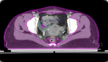
Figure 1: Undefined Normal Tissue (UNT)

ijmrr_1376_02.JPG
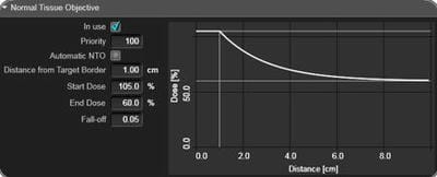
Figure 2: Normal Tissue Objective (NTO)

6. In another method, Dose Control Ring (DCR) is a structure created around the PTV as shown in Fig. 3, to reduce the dose to the surrounding normal tissue dose.

Dose Control Ring (DCR): In this method, we are

creating one virtual structure around the PTV to reduce the dose to surrounding normal tissues. Here we have created 5mm away from the PTV and the Thickness of the DCR is 10mm. During optimization, objectives are given to this structure.

ijmrr_1376_03.JPG
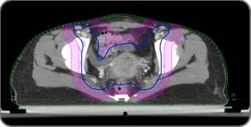
Figure 3: Dose Control Ring (DCR)

7. Combination of DCR and NTO we have created five plans for each patient. That five plans are:
I. Without NTO: In this method, we were given the objective of the target volume (PTV) and OAR structures. During optimization, we didn’t use the NTO for this type of planning.
II. Automatic NTO: In this method, we were given the objective of the target volume (PTV) and OAR structures. During optimization, we used the NTO and NTO values are in-build with the eclipse system.
III. Manual NTO: In this method, we were given the objective of the target volume (PTV) and OAR structures. During optimization we used the NTO and NTO values are changed, distance from the target border to 0.3cm and fall-off to 0.3.
IV. Automatic NTO+DCR: In this method, we were given the objective of the target volume (PTV), OAR structures and DCR. For DCR we have given two objectives one upper and the mean objective. During optimization, we used the NTO and NTO values are in-build with the eclipse system.
V. Manual NTO+DCR: In this method, we were given the objective of the target volume (PTV), OAR structures and DCR. For DCR we have given two objectives one upper and the mean objective. During optimization we used the NTO and NTO values are changed, distance from the target border to 0.3cm and fall-off to 0.3.
8. The OAR dose constraints and target dose parameters were followed as per RTOG guidelines and evaluated as per ICRU 83 recommendations.

Data collection procedure: The following dosimetric parameters were assessed and compared amongst the two planning techniques:


1. PTV- D95%, D50%, D2% (Dnear max), D98% (Dnear min), Conformity Index (CI) (ICRU 62) [9], Homogeneity Index (HI) (ICRU 83) [10].
2. Organs at risk (OARs)
Dmax was calculated for serial organs (Femoral head) and Dmean was calculated for parallel organs (Bone marrow)

Statistical Analysis: Statistical significance was calculated using the ANOVA test. A p-value of <0.05 was considered as statistically significant.

Results

All the plans were evaluated by Homogeneity Index and Conformity Index. HI shows 0.03 for Without NTO plans and 0.06 for Manual NTO along with DCR plans. CI shows 1.08 for Manual NTO along with DCR and 1.5 for Without NTO plans. It shows all the plans were clinically acceptable plans according to the PTV coverage. The observed OAR doses were tabulated below.

Table 1: Mean values of bladder and rectum doses for 30 patients

Target/ OAR Prescription/ Tolerance Without NTO Automatic NTO Manual NTO Automatic NTO+DCR Manual NTO+DCR P-value
Bladder V50<50% 65.05 39.43 35.31 37.76 34.85 <0.001
Rectum V50<50% 56.95 34.75 31.69 32.84 31.68 <0.001

Table 2: Mean values of femur and bone marrow doses for 30 patients

Target/ OAR Prescription/ Tolerance Without NTO Automatic NTO Manual NTO Automatic NTO+DCR Manual NTO+DCR P-value
Bone Marrow DMean<30 30.89 30.47 30.14 30.23 30.07 0.552
Right Femur DMax<50 45.73 45.6 44.95 45.34 44.92 0.255
Left Femur DMax<50 46.19 46.03 45.43 45.92 45.42 0.282

Table 3: The mean dose of Undefined Normal tissue for 30 patients 

Target/ OAR Prescription/ Tolerance Without NTO Automatic NTO Manual NTO Automatic NTO+DCR Manual NTO+DCR P-value
UNT DMean 16.11 14.90 14.45 14.42 14.35 <0.001

ijmrr_1376_04.JPG
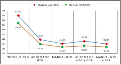
Graph 1: Comparison of Urinary Bladder (V50<50%) and Rectum (V50<50%) for all the plans. Here we indicated the mean values of the 30 patients, for each type of planning.

The Urinary Bladder (V50<50%) and Rectum (V50<50%) were achieved in all the planning method except without NTO plans for cervix cancer patients as shown in table 1 and graph 1.

However, statistically significant differences were observed (p>0.05).


ijmrr_1376_05.JPG
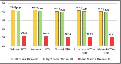
Graph 2: Comparison of Left Femur (DMax<50Gy) for all the plans. Here we indicated the mean values of the 30 patients, for each type of planning.

The Left femoral head (Dmax<50Gy), Right Femoral head (Dmax<50Gy) and Bone marrow (Dmean<30Gy) were achieved in all the planning methods and in that comparatively less dose in Manual NTO along with DCR plans for cervix cancer patients as shown in table 2 and graph 2.

However, no statistically significant differences were observed (p>0.05).

Table 3 and graph 3showed the mean dose of UNT (Dmean) and it was less in the Manual NTO along with DCR planning method comparatively to all the other planning methods. However, significant results were observed (p<0.005).


ijmrr_1376_06.JPG
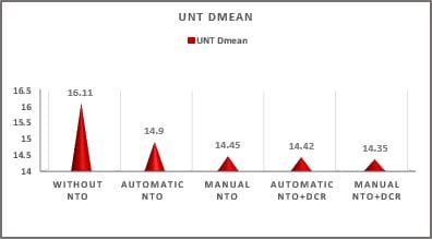
Graph 3: Comparison of Undefined Normal Tissue (DMean) for all the plans. Here we indicated the mean values of the 30 patients, for each type of planning.

Discussion

In the present study conformity index, CI was shown better in manual NTO along with DCR plan. More constraints were added during optimization, to reduce the dose distribution outside the target volume.  A similar trend was observed in the inhomogeneity index HI was shown better without an NTO plan. Fewer constraints are added during optimization, so homogeneous dose distribution over the target volume.

Similar results were shown in A Caldeira et al [2] study found that CI varied between 1.05 to 1.30 with NTO plans and without NTO plans 1.1 to 1.41. In our study, we find the CI index for without NTO and Automatic NTO was 1.56 and 1.13 respectively. Compare to these plans Manual NTO along with DCR gives a better CI was 1.08.

Bell JP et al [11] the study conclude that an automatic NTO is not recommended for lung SBRT planning because of poor performance at reducing low-dose spillage. Plans with well-tuned NTO settings (priority of 500, fall-off of 0.15mm−1) compared favourably with prior plans created with ring structures, achieving significantly lower R50% and lung V20 values. Compare to our study we got a better plan with Manual NTO (distance from target border of 0.3cm and fall-off of 0.3) gives a better plan compared to other plans. In term terms of OAR dose, Urinary bladder (V50<50%) was observed significant (p<0.05) difference in manual NTO along with DCR plan. Without NTO plan shows 65.05% and it was reduced in manual NTO along with DCR plan as 34.85%. A similar difference was observed in Rectum (V50<50%) also, 56.95% and 31.68% respectively for without NTO and Manual NTO along with DCR plan.

Non-Significant (p>0.05) was observed in terms of Bone marrow, Right femoral head and Left femoral head. Bone marrow (Dmean<30) was 30.87Gy without NTO plans and it was reduced to 30.07Gy in Manual NTO along with DCR. The right femoral head (Dmax<50) was 45.73Gy without NTO plans and it was reduced to 44.92Gy in Manual NTO along with DCR. A similar, trend was observed in the left femoral head (Dmax<50) was 46.19Gy without NTO plans and it was reduced to 45.42Gy in Manual NTO along with DCR. In GerdánM et al [12] the study concludes that analysis of the fall-off of 0.15 and the priority of 500 have satisfied our institutional criteria best and plans were calculated with AAA and AXB. In our study, we calculated all the plans with the AAA algorithm.

Undefined Normal Tissue (Dmean) shows 16.11Gy in without NTO plan and it was reduced in manual NTO along with DCR to 14.35Gy. This study shows dosimetric differences but needs to assess the clinical outcomes. Normal tissue that is exposed to radiotherapy can also be affected, leading to toxic effects. Reducing the dose to normal tissue may prevent secondary malignancy.

Conclusion

This study shows that a manual NTO + Dose Control Ring gives better plan quality in terms of PTV coverage and less dose to Undefined Normal Tissue by maintaining Organ at Risk dose within the tolerance limits. 

What does the study add to existing knowledge?

The present study demonstrates the dosimetric characteristic of the different planning methods to reduce the doses of OAR and undefined normal tissues. This may prevent secondary malignancy in the future. Also, it shows that it is important to delineate and report dose to UNT. Because the difference in the planning methods significantly affects dose deposition in the UNT.

Author’s contribution: Mr Muthukumar S: Statistical analysis, drafting and editing of the manuscript, Mrs Navitha S: Study designing, drafting and editing the manuscript, Mr Jitendra Nigam: verification of data, Mr.Silambarasan NS: Treatment Planning, Dr Piyush Kumar: Study designing, manuscript editing, finalising and intellectual content.


Reference

01. Lin Y, Chen K, Lu Z, Zhao L, Tao Y, Ouyang Y, et al. Intensity-modulated radiation therapy for definitive treatment of cervical cancer: a meta-analysis. Radiat Oncol. 2018 Sep 14;13(1):177. doi: 10.1186/s13014-018-1126-7 [Crossref][PubMed][Google Scholar]

02. Caldeira A, Trinca WC Mr, Flores TP Ms, Obst FM, Brito CS, Grüssner MM, et al. The Influence of Normal Tissue Objective in the Treatment of Prostate Cancer. J Med Imaging Radiat Sci. 2020 Jun;51(2):312-316. doi: 10.1016/j.jmir.2020.02.006 [Crossref][PubMed][Google Scholar]

03. Khan, Faiz M. , and John P. Gibbons. Khan's the physics of radiation therapy. Lippincott Williams & Wilkins, 2014 [Crossref][PubMed][Google Scholar]

04. Gallagher MJ, Brereton HD, Rostock RA, Zero JM, Zekoski DA, Poyss LF, et al. A prospective study of treatment techniques to minimize the volume of pelvic small bowel with reduction of acute and late effects associated with pelvic irradiation. Int J Radiat Oncol Biol Phys. 1986 Sep;12(9):1565-73. doi: 10.1016/0360-3016(86)90279-8 [Crossref][PubMed][Google Scholar]

05. Bucci MK, Bevan A, Roach M 3rd. Advances in radiation therapy: conventional to 3D, to IMRT, to 4D, and beyond. CA Cancer J Clin. 2005 Mar-Apr;55(2):117-34. doi: 10.3322/canjclin.55.2.117 [Crossref][PubMed][Google Scholar]

06. Bryant AK, Huynh-Le MP, Simpson DR, Mell LK, Gupta S, Murphy JD. Intensity Modulated Radiation Therapy Versus Conventional Radiation for Anal Cancer in the Veterans Affairs System. Int J Radiat Oncol Biol Phys. 2018 Sep 1;102(1):109-115. doi: 10.1016/j.ijrobp.2018.05.044 [Crossref][PubMed][Google Scholar]

07. De Ruysscher, Dirk, et al. Radiotherapy toxicity. " Nature Reviews Disease Primers 5. 1 (2019): 1-20. [Crossref][PubMed][Google Scholar]

08. RTOG Dose templates for cervical cancer, Available via the National Cancer Institute (NCI) Cancer Trials Support Unit (CTSU) website. . . 1 (2019): 1-20. [Crossref][PubMed][Google Scholar] [Crossref][PubMed][Google Scholar]

09. Stroom, Joep C. , and

Ben JM Heijmen. Geometrical uncertainties, radiotherapy planning margins, and the ICRU-62 report. " Radiotherapy and oncology 64. 1 (2002): 75-83 [Crossref][PubMed][Google Scholar]

10. Hodapp, N. The ICRU Report 83: prescribing, recording and reporting photon-beam intensity-modulated radiation therapy (IMRT). " Strahlentherapie und Onkologie: Organ der Deutschen Rontgengesellschaft. .[et al] 188.1 (2012): 97-99 [Crossref][PubMed][Google Scholar]

11. Bell JP, Patel P, Higgins K, McDonald MW, Roper J. Fine-tuning the normal tissue objective in eclipse for lung stereotactic body radiation therapy. Med Dosim. 2018 Winter;43(4):344-350. doi: 10.1016/j.meddos.2017.11.004 [Crossref][PubMed][Google Scholar]

12. Gerdán M, Pócza T, Polgár C, Major T. Normálszöveti optimalizálási paraméterek hatása tüdősztereotaxiás besugárzási tervek dóziseloszlására [The effects of normal tissue objective parameters on lung stereotactic body radiotherapy dose distributions]. Magy Onkol. 2021 Mar 17;65(1):14-22. Hungarian [Crossref][PubMed][Google Scholar]

13. Fogliata A, Nicolini G, Clivio A, Vanetti E, Cozzi L. Critical appraisal of Acuros XB and Anisotropic Analytic Algorithm dose calculation in advanced non-small-cell lung cancer treatments. Int J Radiat Oncol Biol Phys. 2012 Aug 1;83(5):1587-95. doi: 10.1016/j.ijrobp.2011.10.078 [Crossref][PubMed][Google Scholar]