E-ISSN:2320-8686
P-ISSN:2321-127X

Research Article

COVID-19

International Journal of Medical Research and Review

2022 Volume 10 Number 2 March April
Publisherwww.medresearch.in

Laboratory profile of COVID-19 Patients at the time of admission

Basak I.1, Krishnamurthy V.2*
DOI: https://doi.org/10.17511/ijmrr.2022.i02.04

1 Indrakshi Basak, Postgraduate, pathology, JSS Medical College, JSS Academy of Higher Education and Research, Mysuru, Karnataka, India.

2* Vani Krishnamurthy, Associate professor, Pathology, JSS Medical College, JSS Academy of Higher Education and Research, Mysuru, Karnataka, India.

Background: COVID-19 is a recent pandemic, the complete picture of which is yet to be described. Recent advances in the treatment and improved outcomes have been contributed mainly by knowing the pathological and biochemical status of the patients. Further improvement in understanding this disease at all levels will help in formulating appropriate management plans. Objective: To describe the various biochemical and haematological parameters of COVID – 19 positive patients at the time of admission to the hospital for treatment. Method: We analysed the biochemical and haematological parameters of 100 COVID patients admitted to our hospital from June 2020 to September 2020. Results: 60.6% of our subjects were between 40 and 70 Years. 74.7% of the cases were males. Fever and cough were the most frequent complaints and 49. 5% of them had pneumonia at the time of admission. 19% of the patients needed ICU management. The most common haematological abnormality was lymphopenia seen in 79.9% followed by Eosinopenia seen in 46.5%. Reactive lymphocytes were seen in the majority of the patients (84 %). Toxic granules in the neutrophils, fragmented RBCs and significant left shift of neutrophils are found in small proportions. CRP was elevated in 92% of our patients, followed by elevated Ferritin in 78.2%. D-Dimer was elevated in 44.4% of the patients. Conclusion: The majority of Covid patients at presentation have deranged coagulation and increased D-Dimer. Lymphopenia is the most common haematological abnormality.

Keywords: COVID-19, Haematological parameters, Lymphopenia, Eosinopenia, D-dimer

Corresponding Author How to Cite this Article To Browse
Vani Krishnamurthy, Associate professor, Pathology, JSS Medical College, JSS Academy of Higher Education and Research, Mysuru, Karnataka, India.
Email:
Indrakshi Basak, Vani Krishnamurthy, Laboratory profile of COVID-19 Patients at the time of admission. Int J Med Res Rev. 2022;10(2):75-80.
Available From
https://ijmrr.medresearch.in/index.php/ijmrr/article/view/1371

Manuscript Received Review Round 1 Review Round 2 Review Round 3 Accepted
2022-02-28 2022-03-01 2022-03-08 2022-03-15 2022-03-22
Conflict of Interest Funding Ethical Approval Plagiarism X-checker Note
Nil Nil Yes 17%

© 2022by Indrakshi Basak, Vani Krishnamurthyand Published by Siddharth Health Research and Social Welfare Society. This is an Open Access article licensed under a Creative Commons Attribution 4.0 International License https://creativecommons.org/licenses/by/4.0/ unported [CC BY 4.0].

Introduction

The outbreak of COVID-19 was first identified in Wuhan, China after a cluster of unexplained pneumonia cases was reported by the People’s Republic of China to the World Health Organization (WHO) on 31st December 2019. The aetiology of this outbreak was a novel coronavirus named severe acute respiratory distress syndrome coronavirus 2 (SARS-CoV-2).

The WHO declared COVID-19 as a global pandemic on the 11th of March 2019 and addressed it as a public health emergency of international concern [1]. Based on the current information, the most likely mode of transmission is person-to-person transmission, by droplets and to a lesser extent by fomites.

A wide spectrum of clinical manifestations is known in COVID – 19 and common symptoms include fever, shortness of breath, cough with or without sputum, sore throat, nasal congestion, dizziness, chills, muscle ache, arthralgia, weakness, fatigue or myalgia, chest tightness, excessive mucus production with expectoration, hemoptysis, and dyspnea [2-8]. Other less characterized symptoms include headache, diarrhoea, abdominal pain, vomiting, chest pain, rhinorrhoea and loss of taste and smell [9-12].

Most the patients with COVID-19 remain mild in the early days but some progress rapidly to acute respiratory failure, metabolic acidosis, septic shock, ARDS or death [13,14].

Therefore, early diagnosis is critical to prevent rapid progression to death.

COVID 19 is predominantly a multisystem inflammatory disease. Haematological and biochemical tests have a significant role in the early quantification of the severity of the disease. These parameters significantly aid the physicians to plan appropriate management.

These include leukocyte count and characteristics such as neutrophil- or lymphocyte-dominance; inflammatory markers like erythrocyte sedimentation rate (ESR), C-reactive protein; collateral organ damage (acute renal failure, acute liver failure) markers like Lactate dehydrogenase (LDH) and D-dimer for the severity of the disease [15].

Furthermore, severe COVID-19 infection might also lead to a cytokine storm similar to the cytokine profile present in secondary haemophagocytic lymphohistiocytosis, including increased ferritin levels, IL-1, IL-2, IL-6, IL-7, granulocyte-colony stimulating factor, interferon-γ inducible protein 10, monocyte chemoattractant protein 1, macrophage inflammatory protein 1-α, and tumour necrosis factor-α [13,16].

Recent advances in the treatment and improved outcomes have been contributed mainly by understanding the pathological and biochemical status of the patients. Further improvement in the understanding of this disease at all levels will help in formulating appropriate management plans, thereby reducing mortality and improving disease outcomes.

Methods

Study design: Prospective descriptive study conducted over 3 months.

Source of the data: Patients admitted to COVID isolation ward/ICU, JSS Hospital, Mysuru from June 2020 to August 2020.

Subjects: RT-PCR proven cases of SARS-CoV-2, hospitalized after fulfilling the criteria set, as in need of inpatient care, were included in the study. RT-PCR proven cases, who just needed home quarantine were excluded from the study.

Data collection: The presenting complaints of the patients at the time of admission were obtained from the laboratory information system (LIS).

Hematological parameters obtained were Hb, HCT, RBC count, TC, DC, ANC, ALC, AEC, IMG, IPF, IRF, and Platelet count. PS findings analyzed were for the presence of nucleated RBCs (nRBCs), fragmented RBCs, reactive lymphocytes, and toxic changes in the neutrophils. Biochemical parameters recorded were CRP, Ferritin, LDH and D-Dimer.

Statistical Analysis: All the COVID-19 positive patients admitted in the isolation ward of JSSH from 1st of June 2020 to 31st August 2020 were the cases included in the study. Quantitative data were summarized as mean with standard deviation or median with interquartile range and categorical data expressed as proportions.

Ethics: This study was approved by the institutional ethical committee.


Results

We studied 99 patients admitted during the study period. The youngest patient was 13 years old and the oldest one was 92 years. 60.6% of our patients were between 40 and 70 yrs. Three-fourths (74.7%) of the admitted cases were males. Clinical presentation at the time of admission is depicted in table 1 in descending order. Nearly half of our patients (49.5%) were diagnosed with pneumonia and 19.1% of the patients required ICU admission for advanced treatment.

Summarized values of haematological parameters are given in Table 2. Twelve-point one percent of our patients had a hemoglobin of less than 11 gm/dl at admission. Three percent had hemoglobin above 16 gm/dl. NRBC was elevated in 2.4% of our subjects. RBC count of less than 4 million was seen in 17%. Immature reticulocyte fraction of >12% was seen in 18% of our patients. Of these 18 patients, 2 of them (11%) had fragmented RBCs as well in the peripheral smear. Figure 1 depicts other haematological abnormalities seen in COVID 19 patients in decreasing order of frequency. Leukocytosis is defined as total count >11000/cumm, Leucopenia as total count <4000/cumm, Neutrophilia as absolute neutrophil count >7000/cumm, Neutropenia as absolute neutrophil count <1500/cumm, Lymphocytosis as absolute lymphocyte count >3500/cumm, Lymphopenia as absolute lymphocyte count <1500/cumm, Eosinophilia as absolute eosinophil count >400/cumm and Eosinopenia as absolute eosinophil count <10/cumm.

Eighty-two patients had their peripheral blood smear reported. 84.1% of these patients had Reactive lymphocytes, 46.3% had toxic granules in neutrophils, 13.4% had RBC fragments and in 5.5% of the patients, there was a significant left shift. Among the biochemical parameters, C-Reactive Protein (CRP) was elevated in 92% of the patients at admission, Ferritin was elevated to a variable extent in 78.2% of the patients, and LDH was raised in 66.3% and D-Dimer was elevated in 44.4%.

Of the 99 patients admitted, 29 (29.2%) did not have any comorbidities. The rest of them had either one or more than one comorbidity. The comorbidities we studied included Diabetes, Hypertension, Chronic lung disease, and chronic kidney disease. 45.5% had Diabetes mellitus,

52.5% had hypertension, 11.1% had chronic kidney disease, 11.1% had chronic lung disease and 3% had other illnesses needing long term treatment such as hypothyroidism, psychiatric illness etc. Out of 9 deaths, five (56%) had two or more two morbidities. The Median (IQR) duration of stay in the hospital was 9 (6 – 15) days. The maximum duration of stay was 43 days. Among 99 cases, 9 patients (9.1%) succumbed to the disease course.

Table 1: Predominant clinical symptoms at presentation

Clinical presentation  % of pts
Fever 67.7%
Cough 50.5%
Dyspnoea 34.7%
Myalgia 13.1%
Headache 4.0%
Diarrhoea 2.0%

Table 2: Summarized values of haematological parameters

Haematological parameter Median (Interquartile range)
Hemoglobin (gm/dl) 13.4 (11.9 – 14.5)
Total Leucocyte count (cells/cumm) 6710 (4900 – 8803)
Absolute neutrophil count (cells/cumm) 4920 (3246 – 6983)
Absolute Lymphocyte count (cells/cumm) 1140 (715 – 1578)
Absolute Eosinophil count (cells/cumm) 12 (0 – 47)
Platelet count (lakh/cumm) 2.2 (1.7 – 2.8)

ijmrr_1371-01.JPG
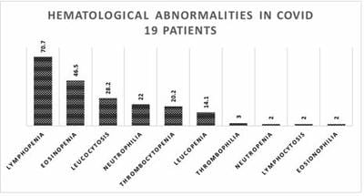
Figure 1: Haematological abnormalities (excluding RBC) seen in COVID -19 patients in the order of frequency

Discussion

During the study period, more than 153 million cases had been reported in 192 countries and territories, resulting in more than 3.2 million deaths [17].

Our study conducted over 3 months, with a sample size of 99 cases, has demonstrated a 9.1% death


rate with the recovery of the rest. In the present study group, more than half of the patients were adults between 40 to 70 years. Similar age-related incidence is reported by Jian-Min Jin et al [18]. Fever, cough and pneumonia were the common presenting features in our study, followed by shortness of breath. Very few patients have also presented with myalgia, headache, and diarrhea. As per the WHO‑China Joint Commission Report, based on 55924 laboratory-confirmed cases, fever and dry cough were the most common presenting features, followed by fatigue, sputum production and shortness of breath [19].

Complete blood counts (CBC) being quick, easily performed, and economical blood tests will be done on all the patients as a baseline investigation at the time of admission. The various parameters included in the study are total white blood cells, absolute neutrophil count, absolute lymphocyte count, absolute eosinophil count and platelet count (PLT), These can be used as inflammatory markers. CRP is the most common parameter elevated. D-Dimer, the marker of microthrombi, was elevated in slightly more than half of all the patients and majority of the patients, it was well within 2 µg/ml. Klok et al have mentioned in their research paper that thrombotic complications can affect up to 31% of adults in the ICU with SARS-CoV-2 pneumonia, thus supporting our finding that D-Dimer may not show a significant elevation in the early stage of the illness [20].

On peripheral blood smear, reactive lymphocytes were seen in most of our patients. Vanessa et al have reported reactive lymphocytes in 23 out of 32 (72%) cases of RT-PCR proven COVID-19 cases [21]. Toxic changes in neutrophils which are suggestive of possible secondary bacterial infection were seen in nearly half of our patients. A similar observation is reported by Aminder Singh et al [22]. Lymphopenia is the next common hematological abnormality seen which is typical of many viral infections. Interestingly, Neutropenia is a less common finding. Li tan et al have concluded that lymphopenia is an effective and reliable indicator of the severity and hospitalization in COVID-19 patients [23]. Thrombocytopenia was observed in 20% of our subjects which is less compared to other viral illnesses such as Dengue infection which is common in our region. Fragmented RBCs, suggestive of intravascular hemolysis, were observed in a small proportion of the cases which is

an important hematological parameter to follow during the illness.

Conclusion

Individuals of all age groups were affected by SARS-CoV-2 and hospitalization was seen more commonly in the males. The most common complaints at the time of admission were found to be fever followed by cough and the most common presenting feature being pneumonia. All the biochemical markers were elevated during the illness, but CRP was the most common acute phase reactant to be raised. The most common haematological finding was lymphopenia while thrombocytopenia was less common when compared to other viral illnesses. On peripheral blood smear, reactive lymphocytes were seen in the majority of the patients, and it is common to find toxic changes in the neutrophils.

Author’s contribution: IB collected the data and did the literature search. Helped with manuscript preparation, VK designed the study, did the analysis, and prepared the manuscript.

What does this study add to existing knowledge?

The lab parameters of the covid disease have a pattern that is different from many other common viral illnesses. Neutrophilic leucocytosis predominates over lymphocytosis which is common in viral infections. Thrombocytopenia is less common and lymphopenia and eosinopenia is the predominant feature in covid infection.

Acknowledgement: We thank Dr Vijaya B, HOD of Pathology for her active encouragement in conducting this study. We also acknowledge Dr Srinivasa Murthy D, professor in Pediatrics for helping with data analysis.

Reference

01. World Health Organization. WHO Director-General’s opening remarks at the media briefing on COVID-19-11 March 2020. (2020). [Crossref][PubMed][Google Scholar]

02. Guan WJ, Ni ZY, Hu Y, Liang WH, Ou CQ, He JX, et al. China Medical Treatment Expert Group for Covid-19. Clinical Characteristics of Coronavirus Disease 2019 in China. N Engl J Med.


2020 Apr 30;382(18):1708-1720. doi: 10.1056/NEJMoa2002032 [Crossref][PubMed][Google Scholar]

03. Lei S, Jiang F, Su W, Chen C, Chen J, Mei W, et al. Clinical characteristics and outcomes of patients undergoing surgeries during the incubation period of COVID-19 infection. EClinicalMedicine. 2020 Apr 5;21:100331. doi: 10.1016/j.eclinm.2020.100331 [Crossref][PubMed][Google Scholar]

04. Xie J, Tong Z, Guan X, Du B, Qiu H. Clinical Characteristics of Patients Who Died of Coronavirus Disease 2019 in China. JAMA Netw Open. 2020 Apr 1;3(4):e205619. doi: 10.1001/jamanetworkopen.2020.5619 [Crossref][PubMed][Google Scholar]

05. Wang Z, Chen X, Lu Y, Chen F, Zhang W. Clinical characteristics and therapeutic procedure for four cases with 2019 novel coronavirus pneumonia receiving combined Chinese and Western medicine treatment. Biosci Trends. 2020 Mar 16;14(1):64-68. doi: 10.5582/bst.2020.01030 [Crossref][PubMed][Google Scholar]

06. Kim ES, Chin BS, Kang CK, Kim NJ, Kang YM, Choi JP, et al. Korea National Committee for Clinical Management of COVID-19. Clinical Course and Outcomes of Patients with Severe Acute Respiratory Syndrome Coronavirus 2 Infection: a Preliminary Report of the First 28 Patients from the Korean Cohort Study on COVID-19. J Korean Med Sci. 2020 Apr 6;35(13):e142. doi: 10.3346/jkms.2020.35.e142 [Crossref][PubMed][Google Scholar]

07. Tu H, Tu S, Gao S, Shao A, Sheng J. Current epidemiological and clinical features of COVID-19; a global perspective from China. J Infect. 2020 Jul;81(1):1-9. doi: 10.1016/j.jinf.2020.04.011 [Crossref][PubMed][Google Scholar]

08. Shi F, Yu Q, Huang W, Tan C. 2019 Novel Coronavirus (COVID-19) Pneumonia with Hemoptysis as the Initial Symptom: CT and Clinical Features. Korean J Radiol. 2020 May;21(5):537-540. doi: 10.3348/kjr.2020.0181 [Crossref][PubMed][Google Scholar]

09. Qian GQ, Yang NB, Ding F, Ma AHY, Wang ZY, Shen YF, et al. Epidemiologic and clinical characteristics of 91 hospitalized patients with COVID-19 in Zhejiang, China: a retrospective, multi-centre case series.

QJM. 2020 Jul 1;113(7):474-481. doi: 10.1093/qjmed/hcaa089 [Crossref][PubMed][Google Scholar]

10. Xu XW, Wu XX, Jiang XG, Xu KJ, Ying LJ, Ma CL, et al. Clinical findings in a group of patients infected with the 2019 novel coronavirus (SARS-Cov-2) outside of Wuhan, China: retrospective case series. BMJ. 2020 Feb 19;368:m606. doi: 10.1136/bmj.m606 [Crossref][PubMed][Google Scholar]

11. Mi B, Chen L, Xiong Y, Xue H, Zhou W, Liu G. Characteristics and Early Prognosis of COVID-19 Infection in Fracture Patients. J Bone Joint Surg Am. 2020 May 6;102(9):750-758. doi: 10.2106/JBJS.20.00390 [Crossref][PubMed][Google Scholar]

12. Wu J, Liu J, Zhao X, Liu C, Wang W, Wang D, et al. Clinical Characteristics of Imported Cases of Coronavirus Disease 2019 (COVID-19) in Jiangsu Province: A Multicenter Descriptive Study. Clin Infect Dis. 2020 Jul 28;71(15):706-712. doi: 10.1093/cid/ciaa199 [Crossref][PubMed][Google Scholar]

13. Huang C, Wang Y, Li X, Ren L, Zhao J, Hu Y, et al. Clinical features of patients infected with 2019 novel coronavirus in Wuhan, China. Lancet. 2020 Feb 15;395(10223):497-506. doi: 10.1016/S0140-6736(20)30183-5 [Crossref][PubMed][Google Scholar]

14. Wang D, Hu B, Hu C, Zhu F, Liu X, Zhang J, et al. Clinical Characteristics of 138 Hospitalized Patients With 2019 Novel Coronavirus-Infected Pneumonia in Wuhan, China. JAMA. 2020 Mar 17;323(11):1061-1069. doi: 10.1001/jama.2020.1585 [Crossref][PubMed][Google Scholar]

15. Baj J, Karakuła-Juchnowicz H, Teresiński G, Buszewicz G, Ciesielka M, Sitarz R, et al. COVID-19: Specific and Non-Specific Clinical Manifestations and Symptoms: The Current State of Knowledge. J Clin Med. 2020 Jun 5;9(6):1753. doi: 10.3390/jcm9061753 [Crossref][PubMed][Google Scholar]

16. Mehta P, McAuley DF, Brown M, Sanchez E, Tattersall RS, Manson JJ; HLH Across Speciality Collaboration, UK. COVID-19: consider cytokine storm syndromes and immunosuppression. Lancet. 2020 Mar 28;395(10229):1033-1034.


doi: 10.1016/S0140-6736(20)30628-0 [Crossref][PubMed][Google Scholar]

17. COVID-19 Dashboard by the Center for Systems Science and Engineering (CSSE) at Johns Hopkins University (JHU)". ArcGIS. Johns Hopkins University. Retrieved 22 September 2020. Available at: [Article][Crossref][PubMed][Google Scholar]

18. Jin JM, Bai P, He W, Wu F, Liu XF, Han DM, et al. Gender Differences in Patients With COVID-19: Focus on Severity and Mortality. Front Public Health. 2020 Apr 29;8:152. doi: 10.3389/fpubh.2020.00152 [Crossref][PubMed][Google Scholar]

19. Report of the WHO-China Joint Mission on Coronavirus Disease 2019 (COVID-19). Available at:https://www. who. int/publications/i/item/report-of-the-who-china-joint-mission-on-coronavirus-disease-2019-(covid-19). [Crossref][PubMed][Google Scholar]

20. Klok FA, Kruip MJHA, van der Meer NJM, Arbous MS, Gommers DAMPJ, Kant KM, et al. Incidence of thrombotic complications in critically ill ICU patients with COVID-19. Thromb Res. 2020 Jul;191:145-147. doi: 10.1016/j.thromres.2020.04.013 [Crossref][PubMed][Google Scholar]

21. Chong VCL, Lim KGE, Fan BE, Chan SSW, Ong KH, Kuperan P. Reactive lymphocytes in patients with COVID-19. Br J Haematol. 2020 Jun;189(5):844. doi: 10.1111/bjh.16690 [Crossref][PubMed][Google Scholar]

22. Singh A, Sood N, Narang V, Goyal A. Morphology of COVID-19-affected cells in peripheral blood film. BMJ Case Rep. 2020 May 27;13(5):e236117. doi: 10.1136/bcr-2020-236117 [Crossref][PubMed][Google Scholar]

23. Tan L, Wang Q, Zhang D, Ding J, Huang Q, Tang YQ, et al. Lymphopenia predicts disease severity of COVID-19: a descriptive and predictive study. Signal Transduct Target Ther. 2020 Mar 27;5(1):33. doi: 10.1038/s41392-020-0148-4. Erratum in: Signal Transduct Target Ther. 2020 Apr 29;5(1):61 [Crossref][PubMed][Google Scholar]