E-ISSN:2320-8686
P-ISSN:2321-127X

Research Article

Radiotherapy

International Journal of Medical Research and Review

2022 Volume 10 Number 2 March April
Publisherwww.medresearch.in

Volumetric and dosimetric effects of different slice thickness in radiotherapy planning Computed Tomography for Head and Neck cancer

Fazilathunnisa J.1, Navitha S.2*, Nigam J.3, Silambarasan N.4, Kumar P.5
DOI: https://doi.org/10.17511/ijmrr.2022.i02.01

1 J Fazilathunnisa, Intern, Medical Physicist, Department of Radiation Oncology, SRMS Institute of Medical Sciences, Bareilly, UP, India.

2* S Navitha, Medical Physicist, Department of Radiation Oncology, SRMS Institute of Medical Sciences, Bareilly, UP, India.

3 Jitendra Nigam, Medical Physicist, Department of Radiation Oncology, SRMS Institute of Medical Sciences, Bareilly, UP, India.

4 NS Silambarasan, Medical Physicist, Department of Radiation Oncology, SRMS Institute of Medical Sciences, Bareilly, UP, India.

5 Piyush Kumar, Professor & Head, Department of Radiation Oncology, SRMS Institute of Medical Sciences, Bareilly, UP, India.

Introduction: Accurate estimation of target and Organ at Risk Volume is required to ensure treatment efficacy and minimal normal tissue toxicity in radiotherapy planning. Computed Tomography slice thickness plays a vital role in volume estimation. It highly impacts smaller volume organs such as 1-3cm3.Materials and Methods: CT datasets of 20 head and neck cancer patients were recruited for this study in each CT data three CT series with a slice thickness of 1.5mm, 3mm and 5mm were imported to the TPS. Eclipse TPS of version 13.6 was used for delineation and treatment planning. Results: The variability of volumes with CT slice thickness was significant, especially for small volume structures. The maximum volume error of 63% was found in >3cc volume structures with 5mm slice thickness. Whereas in larger volume structures the differences were observed 2%in terms of volume and mean dose. And in terms of homogeneity and conformity, there is no significant difference was found. Conclusion: This study concludes that for head and neck cancer which has many smaller volume structures 1.5mm slice thickness will accurately estimate the volume which is clinically useful for OAR near the PTV.

Keywords: Radiotherapy, IMRT, Head and Neck cancer, Slice thickness, Small volume organs

Corresponding Author How to Cite this Article To Browse
S Navitha, Medical Physicist, Department of Radiation Oncology, SRMS Institute of Medical Sciences, Bareilly, UP, India.
Email:
J Fazilathunnisa, S Navitha, Jitendra Nigam, NS Silambarasan, Piyush Kumar, Volumetric and dosimetric effects of different slice thickness in radiotherapy planning Computed Tomography for Head and Neck cancer. Int J Med Res Rev. 2022;10(2):57-62.
Available From
https://ijmrr.medresearch.in/index.php/ijmrr/article/view/1362

Manuscript Received Review Round 1 Review Round 2 Review Round 3 Accepted
2021-12-23 2021-12-25 2022-01-01 2022-01-08 2022-01-15
Conflict of Interest Funding Ethical Approval Plagiarism X-checker Note
Nil Nil Yes 16%

© 2022by J Fazilathunnisa, S Navitha, Jitendra Nigam, NS Silambarasan, Piyush Kumarand Published by Siddharth Health Research and Social Welfare Society. This is an Open Access article licensed under a Creative Commons Attribution 4.0 International License https://creativecommons.org/licenses/by/4.0/ unported [CC BY 4.0].

Introduction

Accurate estimation of target volume and Organ at Risk (OAR) is required to ensure treatment efficacy and minimal normal tissue toxicity in radiotherapy planning. It is due to the rapid development of higher modalities like Intensity Modulated Radiotherapy (IMRT), stereotactic radiosurgery (SRS), and stereotactic radiotherapy (SRT) Treatments. Defining target volumes is the most critical steps in higher modalities [1]. Volume definition is a fundamental prerequisite for successful Advanced Radiotherapy (RT) treatment. Even though Magnetic resonance imaging and positron emission tomography imaging helps to define volumes, Computed tomography (CT) imaging remains an essential modality for RT planning [2]. CT acquisition modalities slice thickness need to be more clearly defined and optimized. Small organ such as optic nerves, cochlea and lenses, the volume estimation could have significant clinical implications [3]. CT slice thickness of <4mm is suggested for smaller target volume (<20cm3) in IMRT of thoracic cancer patients [4].CT slice thickness plays a vital role in volume estimation. It highly impacts smaller volume organs such as between 1-3cm3.

Materials and Methods

Study Setting: Department of Radiation Oncology, Shri Ram Murti Smarak Institute of Medical Sciences.

Study Design: Retrospective study.

Study population: Head and neck cancer patients treated between 2020-2021.

Duration and type of study: A total of twenty head and neck cancer patients who had been treated with 3mm slice thickness were recruited. For each patient, three CT series with the reconstructed thickness of 1.5mm, 3mm, and 5mm were imported into the Treatment Planning System (TPS) as DICOM RT.

Volume Delineation: Varian Eclipse TPS (version 13.6) was used. For each patient with the help of Somatom scope 32 slice CT machine which takes the slice thickness of 0.6mm a three CT series with a reconstructed thickness of 1.5mm, 3mm, and 5mm were imported into the Treatment Planning System (TPS) as DICOM RT. In

each CT series Target, Volume and OARs were delineated by radiation oncologists.

Target volumes; Gross Tumor Volume (GTV), Clinical Target Volume (CTV), Planning Target Volume (PTV), Organ at Risk volume; Right eye lens, Left eye lens, Left eye, Right eye, Right optic nerve, Left optic nerve, Right cochlea, Left cochlea, Optic chiasma, Planning Risk Volume (PRV) brainstem and combined parotids were delineated. International Commission on Radiation Units and Measurements (ICRU) 83 recommendation is used for volume delineation [5].

Dose prescription: Dose to PTV has varied from 60Gy in 30# to 70Gy in 35#. All patients were treated with a 3mm slice thickness CT image. The remaining CT series was used to collect data for this study. Dose constraints to OARs were: PRV Spine ≤50Gy; Mandible Dmax≤ 70Gy PRV Brain Stem≤54Gy; Left and Right Cochlea Dmean ≤ 45Gy; Left and Right Parotid≤ 26Gy; Left and Right Eye≤ 50Gy; Left and Right Eye Len ≤ 7 Gy; Left and Right Optic Nerve ≤ 55Gy. All these tolerance values followed from RTOG Guidelines [6-7].

In every CT series clinically acceptable Intensity Modulated Radiotherapy Treatment plans were generated. The difference in the delineated volume and dosimetric values were compared with the help of the evaluation tool Dose Volume Histogram (DVH).

Statistical analysis: An independent sample t test was used to examine the changes calculated between CT slice thickness variation for each patient volume delineation and dosimetry data. A test result with less than or equal to 0.05 was regared as statistically significant.

Results

Delineation difference in smaller objects:

Delineation difference in the smaller organs such as optic nerve, cochlea, lens with 1.5mm, 3mm and 5mm slice thickness in (Fig 1,2,3).

In 1.5mm slice thickness the optic nerve was contoured in four slices and 3mm slice thickness it is contoured in 2 slices whereas in a 5mm slice it is contoured only in one slice which means when the slice thickness is reduced more the delineation will be accurate. Same results were observed in Cochlea and lens.


ijmrr_1362_01.JPG
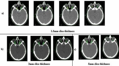
Figure 1: Contour of optic nerve in different CT slice thickness.

ijmrr_1362_02.JPG
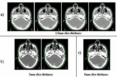
Figure 2: Contour of cochlea.

ijmrr_1362_03..JPG
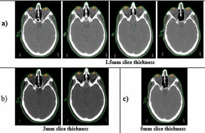
Figure 3: Contour of the lens.

ijmrr_1362_04.JPG
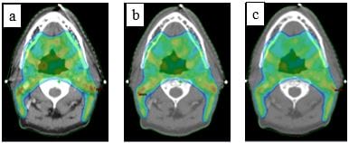
Figure 4: Dose color wash of 95% dose coverage to the Planning target volume: a) 1.5mm b) 3mm and c) 5mm slice thickness.

The dose color wash of 95% dose coverage to the PTV in 1.5mm, 3mm and 5mm slice thickness which

is almost similar in all slice thickness which means there is no change in the PTV coverage (Fig 4).

ijmrr_1362_05.JPG
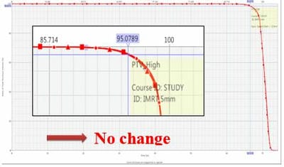
Figure 5: Dose-volume Histogram curve of planning target volume with 1.5mm, 3mm and 5mm slice thicknesses.

DVH evaluation for planning target volume for three slice thickness all three curves are overlapping which means in all slice thicknesses there is no significant difference was found in (Fig 5).

Volume and dose analysis:

Table 1: comparison of volume and mean dose for larger volume structures GTV, CTV and PTV.

Structures Slice thickness1.5mm Slice thickness 3mm Slice thickness 5mm p value 
Volume (cc) Mean dose (Gy) Volume (cc) Mean dose (Gy) Volume (cc) Mean dose (Gy)
GTVa 28.17 69.83 27.44 69.51 26.24 70.05 0.9
CTVa 300.62 64.02 300.22 63.86 293.67 64.12 0.9
PTVa 609.81 63.84 609.63 63.64 602.76 63.91 0.9

a - Mean dose: It is observed that there is no significant (p-0.9) difference in terms of dose and volume in a larger volume structure. (Table 1). 

Table 2: Comparison of volume and mean/max dose to the larger volume OAR’s.

Structures Slice thickness1.5mm Slice thickness 3mm Slice thickness 5mm p value 
Volume (cc) Mean/max dose (Gy) Volume (cc) Mean/max dose (Gy) Volume (cc) Mean/max dose (Gy)
LT eyeb 8.56 3.34 8.23 3.33 8.02 3.21 0.2
RT eyeb 8.6421 3.9384 8.305 3.82 10.93 4.18 0.2
Opticchiasmb 0.5263 2.4536 0.645 2.463 0.3 2.386 0.2
Brainstema 26.25 21.844 25.99 22.271 25.57 21.668 0.15
Parotidsa 51.03 35.668 50.525 35.290 49.46 35.545 0.72

a – Mean dose,b – Max dose:


In >3cm3 volume organs which are not showing any significant difference in 1.5mm, 3mm and 5mm slice thickness so, depending on the slice thickness there will not be much change in terms of volume as well as dose in the larger volumes in (Table 2). 

Table 3: Comparison of volume and mean/max dose for smaller volume structures.

Structures Slice thickness1.5mm Slice thickness 3mm Slice thickness 5mm p-value 
Volume (cc) Mean/max dose (Gy) Volume (cc) Mean/max dose (Gy) Volume (cc) Mean/max dose (Gy)
LT cochleaa 0.12   16.54   0.1   16.13   0.045   15.31   0.00001
RT cochleaa 0.125   18.712   0.105   18.162   0.045   17.17   0.00001
LT optic nerveb 0.473   2.593   0.355   2.621   0.22   2.528   0.00007
RT optic nerveb 0.726   2.725   0.335   2.730   0.17   2.619   0.0001
LT lensb 0.173   1.815   0.15   1.839   0.095   1.864   0.001
RT lensb 0.173   10.933   0.145   1.876   0.07   1.845   0.0002

a- Mean dose, b- Max dose: The significant variability of volumes with different slice thickness mainly in the smaller volume structures which have a volume of <3cm3 and the volume difference is less significant in the larger volumes. In this study, the variation of volume in the cochlea (p-0.00001), optic nerve (p – 0.00007), and lens (p – 0.001) are showing highly significant results. This study shows that 1.5mm slice thickness is optimum for IMRT of nasopharynx cancer in (Table 3).

Discussion

Volume difference for targets structures like GTV, CTV and PTV has insignificant difference. So, same results observed in mean dose of GTV, CTV and PTV. Haunli Luo et al study on thoracic cancer also observed no significant difference in larger volume target IMRT plans. So author suggested that CT slice thickness of less than 4mm for small targets plans in thoracic cancer [4]. Our study suggests that less than 3mm slice thickness is minimum requirement as for as smaller targets concerned. And also shows that the selection of slice thickness depends on the distance of the OAR from the PTV.

OAR volumes like LT eye, Rt eye, Optic chiasm, brainstem and parotids

shows insignificant difference in terms of volume and dose respectively. Eventhough there is no significant difference observed our study suggest less than 3mm slice thickness for head and neck cancer. Because a phantom study done by S. P. Srivastava et al shows volume underestimation in 3mm slice thickness compared with 1mm slice thickness. It is due to reconstruction algorithm of CT machine and contouring algarithm in TPS [3].and author suggested that the smallest possible slice thickness should be used for IMRT planning , since smaller slice thickness provides superior dosimetry with improved Tumor Control probability (TCP). Our study used real patients, and we had observed the same volume difference in the smaller structures and also in dose.

Prabhakar R et al done a study on 3D conformal planning of brain tumors. Author observed that for volume less than 25cc, most of the cases were underdosed by 18% with 5mm slice thickness. Greater than 25cc volume target underdosage was less than 6.7% for same slice thickness. So author concluded that 2.5mm slice thickness is optimum for tumor volumes less than 25cc [8]. our study suggest that less than 3mm slice thickness is required in head and neck cancer since it contains many small volume organs which are less than 25cc. and 1.5mm slice thickness is optimum for the cases where the OARs are near to the PTV and it will estimate the volume accurately. This study also shows that depending on slice thickness there is no change in the target coverage.

Alirezaei et al done a study on phantom and real patient comparison of different slice thicknesses in low-grade glioma compared 1.5, 3, 5, and 10mm slice thickness. Phantom study finds insignificant result between 1.5mm and 3mm slice thickness. But in patient study found significant increase in the volume of retina, chiasma, genu and splenium and left optic nerve [9]. Our study showes similar results for smaller volume organs and especially for serial organ present near the PTV.

Another study on slice thickness effects on brain metastasis done by S L thrower shows that the volume difference between 1 mm and 2mm thickness CT was 0.5cc maximum and the mean difference was .055cc for brain lesions. Our study results also show that significant difference in the smaller organs volume estimation. Importance of accuracy in volume delineation and radiotherapy delivery studied by several authors [11-18].


Eventhough smaller slice thickness provides accurate estimation of volume and dose, it also increases contouring time of an oncologist. It is important to optimize the slice thickness between volume accuracy and clinical necessity for decimal accuracy in volume estimation. So 1.5 mm slice thickness is optimum for head and cancer patient when the serial organ at risk is very close to the target volume.

Conclusion

This study suggests that for the head and neck cancer which has many smaller volume structures, 1.5mm slice thickness will accurately estimate the volume which is clinically useful for OAR near the PTV.

What does the study add to the existing knowledge?

The present study demonstrates that a smaller slice thickness of planning CT will be highly effective to precisely estimate the volume and accurate dose reporting of serial organs which are present near to the target volume.

Author’s contribution: FazilathunnisaJavid: Statistical analysis, drafting and editing the manuscript, S Navitha:, Study designing, drafting and editing the manuscript, Jitendra Nigam: verification of data, Silambarasan NS: Treatment Planning, Piyush Kumar: Study designing, manuscript editing, finalising and intellectual content.

Reference

01. Giraud, P. ; Kantor, G. ; Loiseau, H. ; et al. Target definition in the thorax and central nervous system. Semin.adiat. Oncol.15:146–56; 2005 [Crossref][PubMed][Google Scholar]

02. Caivano R, Fiorentino A, Pedicini P, Califano G, Fusco V(2014) The impact of computed tomography slice thicknesson the assessment of stereotactic, 3D conformal and intensity-modulated radiotherapy of brain tumors. Clinicaland Translational Oncology, 16(5): 503-508. . Target definition in the thorax and central nervous system. Semin.adiat. Oncol.15:146–56; 2005 [Crossref][PubMed][Google Scholar] [Crossref][PubMed][Google Scholar]

03. Srivastava SP, Cheng CW, Das IJ. The effect

of slice thickness on target and organs at risk volumes, dosimetric coverage and radiobiological impact in IMRT planning. Clin Transl Oncol. 2016 May;18(5):469-79. doi: 10.1007/s12094-015-1390-z [Crossref][PubMed][Google Scholar]

04. Luo H, He Y, Jin F, Yang D, Liu X, Ran X, et al. Impact of CT slice thickness on volume and dose evaluation during thoracic cancer radiotherapy. Cancer Manag Res. 2018 Sep 20;10:3679-3686. doi: 10.2147/CMAR.S174240 [Crossref][PubMed][Google Scholar]

05. ICRU Report 83, Prescribing, Recording, and Reporting Photon-Beam Intensity-Modulated Radiation Therapy (IMRT), Journal of the ICRU Vol 10 No 1 (2010) Report 83. doi:10. 1093/jicru/ndq003. [Crossref][PubMed][Google Scholar]

06. RTOG 0920, A phase iii study of postoperative radiation therapy (imrt) +/- cetuximab for locally-advanced resected head and neckCancer. Available via the National Cancer Institute (NCI) Cancer Trials Support Unit (CTSU) website. . [Crossref][PubMed][Google Scholar] [Crossref][PubMed][Google Scholar]

07. RTOG 0619,A randomized phase ii trial of chemo radiotherapy versus chemo radiotherapy and vandetanib for high-risk postoperative advanced squamous cell carcinoma of the head and neck. Available via the National Cancer Institute (NCI) Cancer Trials Support Unit (CTSU) website. . [Crossref][PubMed][Google Scholar] [Crossref][PubMed][Google Scholar] [Crossref][PubMed][Google Scholar]

08. Prabhakar R, Ganesh T, Rath GK, Julka PK, Sridhar PS, Joshi RC, et al. Impact of different CT slice thickness on clinical target volume for 3D conformal radiation therapy. Med Dosim. 2009 Spring;34(1):36-41. doi: 10.1016/j.meddos.2007.09.002 [Crossref][PubMed][Google Scholar]

09. Alirezaei, Z. , et al. Optimization of CT slice thickness in 3D-CRT and IMRT planning of low grade glioma. " International Journal of Radiation Research 19. 2 (2021): 291-298 [Crossref][PubMed][Google Scholar]

10. Thrower SL, Al Feghali KA, Luo D, Paddick I, Hou P, Briere T, et al. The Effect of Slice Thickness on Contours of Brain Metastases


for Stereotactic Radiosurgery. Adv Radiat Oncol. 2021 Apr 11;6(4):100708. doi: 10.1016/j.adro.2021.100708 [Crossref][PubMed][Google Scholar]

11. Dutreix A. When and how we can improve precision in radiotherapy? Radiother Oncol. 1984;2:275–92. [Crossref][PubMed][Google Scholar]

12. Mah K, Van Dyk J, Keane T, Poon PY. Acute radiation-induced pulmonary damage: a clinical study on the response to fractionated radiotherapy. Int J Radiat Oncol Biol Phys. 1987;13:179–88. [Crossref][PubMed][Google Scholar]

13. International Commission on Radiation Units and Measurements. Determination of absorbed dose in a patient irradiated by beams of X or Gamma rays in radiotherapy procedures. Bethesda, MD: International Commission on Radiation Units and Measurements. 1976, Report No. 24 [Crossref][PubMed][Google Scholar]

14. Mijnheer BJ, Batterman JJ, Wambersie A. What degree of accuracy is required and can be achieved in photon and neutron therapy? Radiother Oncol. 1987;8:237–52. [Crossref][PubMed][Google Scholar]

15. International Commission on Radiation Units and Measurements. Prescribing, Recording and Reporting Photon Beam Therapy. Bethesda, MD: International Commission on Radiation Units and Measurements. 1993, Report No. 50 [Crossref][PubMed][Google Scholar]

16. International Commission on Radiation Units and Measurements. Prescribing, recording and reporting photon beam therapy (supplement to ICRU report 50). Bethesda, MD: International Commission on Radiation Units and Measurements. 1999, Report No. 62 [Crossref][PubMed][Google Scholar]

17. Schlegel W, Bortfeld T, editors. A new approach for improved tumor volumetry. Proceedings of the XIIIth International conference on the use of computers in radiation therapy: 2000 May 22–25; Heidelberg, Germany. Berlin: Springer; 2000. [Crossref][PubMed][Google Scholar]

18. Jansen EP, Dewit LG, van Herk M, Bartelink H. Target volumes in radiotherapy for high-grade malignant glioma of the brain. Radiother

Oncol. 2000;56:151–6. [Crossref][PubMed][Google Scholar]