E-ISSN:2320-8686
P-ISSN:2321-127X

Research Article

Chronic suppurative otitis media

International Journal of Medical Research and Review

2022 Volume 10 Number 1 January February
Publisherwww.medresearch.in

To Study the Computed Tomography Temporal Bone Findings in Patients with Chronic Suppurative Otitis Media

Mandal P.1, Vasanthan R.2*, Kumar K.3, Prabu V.4, Govardhanan .5
DOI: https://doi.org/10.17511/ijmrr.2022.i01.02

1 Priyanka Mandal, Senior Resident, Department of Radio-Diagnosis, Sri Venkateshwaraa MCH and Research Center, Puducherry, , India.

2* Ratna Vasanthan, Associate Professor, Department of Radio-Diagnosis, Sri Venkateshwaraa MCH and Research Center, Puducherry, , India.

3 Krishna Kumar, Professor, Department of Radio-Diagnosis, Sri Venkateshwaraa MCH and Research Center, Puducherry, , India.

4 Velayutham Prabu, Professor, Department of Otorhinolaryngology, Sri Venkateshwaraa MCH and Research Center, Puducherry, , India.

5 Govardhanan, Professor, Department of Radio-Diagnosis, Sri Venkateshwaraa MCH and Research Center, Puducherry, , India.

Background: Chronic suppurative otitis media is a chronic inflammation of middle ear cleft. It can present with discharging ear and decreased hearing. Diagnosis is mostly on clinical examination with an otoscope or otoendoscope. Our study was undertaken to evaluate role of computed tomography in defining extent and severity of various pathological changes occurring in temporal bone in case of chronic suppurative otitis media. Materials and Methods: Current study was a prospective cross-sectional study was conducted in Sri Venkateshwaraa Medical College and Research Center, Ariyur, Pondicherry from January 2019 to May 2020. Study included 60 patients clinically diagnosed with a chronic middle ear infection and who were referred to Department of Radiodiagnosis and Imaging for an HRCT scan of temporal bone. All obtained data were entered in Microsoft Excel and analyzed with SPSS 23.0 software. Results: In our study, a total number of 60 patients were involved. Youngest patient was aged 7 years and oldest patient’s age was 63 years. Majority (28.33%) of patients were aged between 21-30 years followed by 26.67% in 11-20 years age group. Our study found site and extent of involvement of middle ear and mastoid air cell system as follows: protympanum (20.00%); meso-tympanum (26.67%); posterior tympanum (30.00%); epitympanum (60.00%); hypo-tympanum (25.00%); peri-labyrinthine cells (1.67%); aditus (53.33%); antrum (40.00%); mastoid air cells (58.33%). Conclusion: HRCT scan is a unique method to detect early cholesteatoma and also to detect cholesteatoma in hidden areas.

Keywords: Chronic suppurative otitis media; High Resolution Computed tomography; Cholesteatoma

Corresponding Author How to Cite this Article To Browse
Ratna Vasanthan, Associate Professor, Department of Radio-Diagnosis, Sri Venkateshwaraa MCH and Research Center, Puducherry, , India.
Email:
Priyanka Mandal, Ratna Vasanthan, Krishna Kumar, Velayutham Prabu, Govardhanan, To Study the Computed Tomography Temporal Bone Findings in Patients with Chronic Suppurative Otitis Media. Int J Med Res Rev. 2022;10(1):10-16.
Available From
https://ijmrr.medresearch.in/index.php/ijmrr/article/view/1360

Manuscript Received Review Round 1 Review Round 2 Review Round 3 Accepted
2021-12-15 2021-12-17 2021-12-24 2021-12-31 2022-01-07
Conflict of Interest Funding Ethical Approval Plagiarism X-checker Note
Nil Nil Yes 17%

© 2022by Priyanka Mandal, Ratna Vasanthan, Krishna Kumar, Velayutham Prabu, Govardhananand Published by Siddharth Health Research and Social Welfare Society. This is an Open Access article licensed under a Creative Commons Attribution 4.0 International License https://creativecommons.org/licenses/by/4.0/ unported [CC BY 4.0].

Introduction

Chronic Suppurative Otitis Media is a chronic inflammation of middle ear cleft. It can present with discharging ear and decreased hearing. The diagnosis is mostly on clinical examination with an otoscope or otoendoscope. Short and long-term sequelae of otitis media may be devastating due to strategic location of tympanomastoid compartment, separated from middle and posterior cranial fossa by a thin bony partition, otitis media has got easy access and maximum potential for intracranial extension. It can be avoided if recognized early and properly treated. [1]. Therefore before surgical treatment, it is essential to know exact size and extent of disease. As such, radiological examination of temporal bone plays an important role in providing crucial information to surgeon in this regard. [2,3]. The incidence rate of chronic suppurative otitis media (CSOM) is 4.76% (31 million cases) with 22.6% of under-five cases occurring every year. [4]. The World Health Organisation estimated that globally 65-300 million people are affected by CSOM. Among them, 50% of people have a hearing impairment and 28000 deaths per year are attributed to complications of otitis media. [5]. Chronic otitis media can be due to chronic mucosal disease or cholesteatoma. The differentiation is generally made by clinical examination. Although Otorhinolaryngologist can diagnose most of cholesteatoma with thorough clinical examination, it may not be possible to determine extent and size of lesion. Conventional techniques of temporal bone imaging like X-Ray Mastoid bones have been replaced by High-Resolution Computerised Tomography (HRCT) at present. HRCT of mastoids is done to evaluate extent of disease and its complications. [6]. With advent of thin section HRCT, it can allow imaging of ossicular chain structures up to a spatial resolution of 0.45 to 0.65mm. [7]. A block of ultra-thin sections of CT imaging (thickness of 0.35mm) can be taken in less than a minute to cover entire temporal bone. [8]. HRCT scan of an acquired cholesteatoma of temporal bone imaging could reveal homogeneous soft tissue mass with erosion of local bone, scutum and ossicles, middle ear opacification, and labyrinthine fistula extending to tympanum and widening audits and antrum. HRCT can detect exposed dura, lateral canal fistulae, and facial canal dehiscences, and demonstrate ossicular chain. [9]. Lateral semicircular

canal dehiscence can increase risk of labyrinthitis and dehiscence in dural can raise risk of extending pathology to brain. Having preoperative knowledge of anatomy and anomalies of dehiscence in facial canal is important to prevent postoperative morbidity among patients requiring surgery for middle ear disorders. [10].  HRCT imaging of temporal bone has been playing an increasingly important role in diagnosis, surgical decision, and further follow-up. The decision for choice of surgical technique is of particular importance to preserve a higher hearing rate and prevent complications. [11] Hence this study was undertaken to evaluate role of computed tomography in defining extent and severity of various pathological changes occurring in temporal bone in case of chronic suppurative otitis media.

Materials and Methods

This was a prospective cross-sectional study to evaluate the role of CT of the temporal bone in 60 patients clinically diagnosed with a chronic middle ear infection and who were referred to the Department of Radiodiagnosis and Imaging, Sri Venkateshwaraa Medical College and Research Center, Ariyur, Pondicherry for HRCT scan of temporal bone between Jan 2019- May 2020.

Sample size: Using statistics and sample size calculation software and with the absolute precision of 10% and an alpha error of 5%, the sample size was estimated to be 60.

Inclusion criteria: All the patients with a chronic middle ear infection who were referred to the Department of Radiology for HRCT Temporal bone were taken into the study.

Exclusion criteria: Pregnant women, patients with electric devices at the skull base, such as cochlear implants, those who have undergone previous temporal bone surgeries, and those with a history of trauma to the temporal bone were excluded from the study. Patients who were not willing to undergo CT were also excluded.

All the CT scans were performed at our institute on the Siemens Somatom Emotion 6 Slice Ct. No extra financial burden was incurred on the patients. After written informed consent, patients were scanned in the axial plane. Topograms were taken routinely in all patients before starting the scan. Scanning commenced from the lower margin


of the external auditory meatus including the inferior mastoid and extended upward to the arcuate eminence of the superior semicircular canal as seen on the lateral topogram. The patients were positioned with a slightly extended head to avoid gantry tilt and thereby protect the lens from radiation. Reformatted coronal images were obtained perpendicular to the axial plane from the cochlea to the posterior semicircular canal. Scanning parameters: Each section thickness with 0.63 mm applying 130 kV, 240 mA s, and 0.6:1 pitch.  The images were reconstructed with a bone algorithm. All images were interpreted using source images, multiplanar reformations, and required window settings.

Data analysis: All obtained data were entered in Microsoft Excel and analyzed with SPSS 23.0 software. Continuous variables (like age) were expressed as mean and standard deviation (or) median with the inter-quartile range depending on normality distribution. Categorical variables (like sex, temporal bone findings) were expressed as proportion and percentage.

Results

Table 1: Distribution of age among the study participants. 

Age group (Years) Number (n=60) Percentage
<10 4 6.67
11-20 16 26.67
21-30 17 28.33
31-40 8 13.33
41-50 9 15
51-60 4 6.67
>60 2 3.33
Total 60 100.00

Table 2: Distribution of gender among the study participants. 

Gender Number (n=60) Percentage
Male 34 56.67
Female 26 43.33
Total 60 100.00

Table 3: Distribution of the CSOM in relation to the side of the ear affected. 

Side Number (n=60) Percentage
Left 30 50
Right 27 45
Bilateral 3 5

Table 4: Distribution of presenting symptoms among the study participants. 

Chief Complaints Number of Patients Percentage
Ear discharge 53 88.33
Decreased Hearing 38 63.33
Earache 18 30.00
Headache 10 16.67
Tinnitus 4 6.67
Giddiness 6 10.00
Facial Paralysis 1 1.67

ijmrr_1360_01.JPG
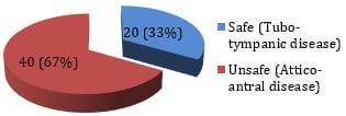
Figure 1: Distribution of the impression of HRCT findings regarding CSOM type.

ijmrr_1360_02.JPG
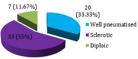
Figure 2: Distribution of the HRCT findings of temporal bone regarding mastoid pneumatization (n=60).

ijmrr_1360_03.JPG
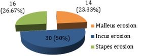
Figure 3: Distribution of the HRCT findings regarding ossicular involvement.

Table 5: Distribution of findings from the HRCT. 

Findings from the HRCT (n=60) Yes (%) No (%)
Non-dependent soft tissue mass 36(60) 24(40)
Scutum erosion 27(45) 33(55)
Ossicular involvement 32(53.33) 28(46.67)
Labyrinthine fistula 2(3.33) 58(96.67)
Sigmoid sinus plate 7(11.67) 53(88.33)
Mastoid cortex erosion 5(8.33) 55(91.67)
Tegmen erosion 4(6.67) 56(93.33)
Mastoiditis with sub-periosteal abscess 2(3.33) 58(96.67)

Table 6: Distribution of site and extent of involvement of the middle ear and mastoid air cell system from the HRCT images. 

  Number Percentage
Protymanum 12 20.00
Mesotympanum 16 26.67
Posterior tympanum 18 30.00
Epitympanum 36 60.00
Hypotympanum 15 25.00
Peri-labyrinthine cells 1 1.67
Aditus 32 53.33
Antrum 24 40.00
Mastoid air cells 35 58.33

Discussion

Imaging techniques in the medical field have undergone significant changes in the technical aspect as well as the clinical ground since the discovery of X-ray on 8th November 1895 by Wilhelm Conrad Roentgen, a German Physicist. Conventional tomograms used an image of a section of the patient orienting parallel to the film. Since it lacked enough clarity, HRCT has taken over by image reconstruction to provide sharp and clear images. Conventional radiography has a limited value to assess mastoid pneumatization. The HRCT of the temporal bone can provide excellent details of the temporal bone and display the soft tissue mass within the air spaces of the mastoid, middle ear, and external auditory canal. Bony structures like scutum, ossicles and labyrinthine capsules cannot be viewed by magnetic resonance imaging. Hence, HRCT is the modality of choice to assess middle ear pathology. [12]. In our study, a total number of 60 patients were involved. The youngest patient was aged 7 years and the oldest patient’s age was 63 years. The majority (28.33%) of patients were aged between 21-30 years followed by 26.67% in the 11-20 years age group. This finding was similar to a study by Sharma et al where the majority of the patients belonged to the 21-30 years age group (40%) followed by 11-20 years (30%)(36). Dhulipalla et al also observed the majority of the patients (49%) the patients aged between 11 to 30 years. [11].

In this study, the mean age of the patients was 27.2 ±13.1 years. Thukral et al [19]. found that the mean of the patients was 27.95 years which was similar to our study finding [7]. Jadia et al, Chatterjee et al and Kanotra et al observed

the mean age of the patients to be 23.2 years, 23.17 years, and 36.38 years respectively. [7,15,16]. Thus most of the studies which targeted to study the temporal bone pathologies had mentioned the mean age of the patients to be between 20 to 30 years.

In our study, the males (53.67%) were commonly affected than females (43.33%). Sharma et al identified more male patients (60%) than female patients (40%). [14]. Dhulipalla et al also observed more males (58%) than females (42%) in their study. [11]. Chatterjee et al also observed more male patients (68.86%) than female patients. [15]. Kanotra et al observed male patients (57.44%) be more than female patients (42.55%). [16]. Our study had the chief complaints in the following order: ear discharge (88.33%); decreased hearing (63.33%); earache (30%); headache (16.67%); giddiness (10%); tinnitus (6.67%); facial paralysis (1.67%).  Jadia et al observed that otorrhoea (100%) was the common presenting finding followed by hearing loss (98.1%), tinnitus (67.3%), dull headache (21.2%), and facial weakness (1.92%). [7] Tamilarasan et al [13]. reported that the symptoms in their study population were otorrhoea (91.95%), hearing loss (59.77%), otalgia (29.89%), nausea and vomiting (11.49%), headache (10.34%), tinnitus (9.20%), fever with chills and rigors (9.20%), swelling behind the ear (8.05%) and facial weakness (5.75%). Dhulipalla et al [11]. observed that the common presenting symptoms of these patients in decreasing order were ear discharge (83%), decreased hearing (70%), otalgia (25%), headache (13%), tinnitus (8%), giddiness (8%) and facial palsy (2%).

The present study identified sclerosed mastoid in 55.0%, well pneumatised in 33.33%, and diploic mastoid in 11.67%. The finding of sclerosed mastoid pneumatization (100%) in the HRCT of the temporal bone was observed by Jadia et al. [7]. The characteristics of mastoid bone included well-pneumatised bone in 40.23%, sclerotic bone in 43.68%, and diploic bone in 9.20% as reported by Tamilarasan et al. [13]. Regarding the pneumatization of the mastoid, the HRCT revealed sclerotic mastoid in all the patients (100%) and pneumatic in none of the patients as identified by Kanotra et al. [16]. The current study found the presence of non-dependent soft tissue mass in 60.00% of the patients since the current had considered both safe and unsafe ear


pathologies in the selection criteria. The cholesteatoma is highly suggestive with the presence of non-dependent soft tissue mass in the middle ear cavity. Payal et al observed the presence of soft tissue mass in 86.67% of the temporal bone study. [18]. Chatterjee et al found the presence of soft tissue mass in all 100% of the patients. [15].  Our study found eroded scutum in 45% of the CSOM patients which was much similar to what Thukral et al [42]. study found. scutum erosion in 42% of the CT findings. Tamilarasan et al identified eroded scutum in 34.48%. [13]. Sharma et al identified 84% and Chatterjee et al found it in 67.07% of the cases. [14, 15]. Ossicular erosion in our study was 53.33% which was similar studies finding ossicular involvement was observed in 90%, 62.28%, 73%, and 54.57% by Sharma et al, Chatterjee et al, Bathla et al and Gul et al respectively. [14, 15, 17, 20].

In our study, malleus erosion (23.33%), incus erosion (50.00%), and stapes erosion (28.33%) were observed in HRCT images which correlate with the findings of Tamilarasan et al and Payal et al. [13, 18]. Destruction of the ossicles is commonly seen in cholesteatoma which enlarges and erodes on touching the contiguous structures in the middle ear cleft. Prior knowledge of the ossicular involvement can help the surgeon in the decision-making to preserve the hearing.

The present study observed labyrinthine fistula in 3.33% of the patients. Tamilarasan et al observed it in 3.8% of the images. [13]. Mandal et al observed this finding in 4% of the images [21]. Sharma et al found this in 6% of cases. [14]. The current study had this finding mastoid erosion in 8.33% of the cases. Tamilarasan et al had observed mastoid cortex erosion in 8.05% of the cases. [13]. Sharma et al found this in 10% of the cases. [14]. Prakash et al mentioned this erosion as 16.67% in their study. [22]. Our study found tegmen erosion in 6.67% of the cases in HRCT images. Jadia et al observed the same tegmen erosion in 23.1% of the cases. [7]. Kataria et al found tegmen erosion in 7.14% of the cases. [23]. Keskin et al mentioned it as 19.6% in their study. [24].

Mastoiditis was found in our study in 60% of the cases in HRCT images. This finding corroborated with the study finding of Chatterjee et al and Thukral et al., [15,19]. observed mastoiditis in 50.3% and 76% of the cases

respectively. Our study found the site and extent of involvement of middle ear and mastoid air cell system as follows: protympanum (20.00%); meso-tympanum (26.67%); posterior tympanum (30.00%); epitympanum (60.00%); hypo-tympanum (25.00%); peri-labyrinthine cells (1.67%); aditus (53.33%); antrum (40.00%); mastoid air cells (58.33%). Jadia et al found the extension in epi-tympanum (100%), meso-tympanum (78.85%), hypo-tympanum (55.77%), pro-tympanum (9.62%), sinus tympani (9.62%), aditus (78.85%), antrum (84.62%) and mastoid (19.23%). [7]. Srigiri et al. observed epitympanum, antrum, aditus, mastoid air cells, posterior tympanum, mesotympanum, hypotympanum, protympanum and perilabyrinthine cells as 88%, 88%, 84%, 76%, 52%, 44%, 44%, 36% and 24% respectively. [25]. The decreasing order of the involvement was much similar to our study.

Conclusion

The High Resolution Computed Tomography scan has become the standard imaging technique for the temporal bone. The HRCT findings of the temporal bone in patients with chronic suppurative otitis media were the presence of non-dependent soft tissue mass more commonly followed by ossicle erosion, scutum erosion, sigmoid sinus plate erosion, mastoid cortex erosion, tegmen erosion, labyrinthine fistula, and mastoiditis with sub-periosteal abscess. HRCT scan is a unique method to detect early cholesteatoma and also to detect cholesteatoma in hidden areas.

What does the study add to existing knowledge?

This study shows that HRCT imaging plays a gold standard role in the diagnosis of CSOM and its complications. Furthermore, it plays a major role in deciding the surgical technique, which is of great importance to preserve hearing and prevent complications.

Author contributions: Mandal P collected the data, conducted this study, Vasanthan R did data analysis. Mandal P, Kumar K, Prabu V and Govardhanan did manuscript drafting. All authors were involved in revising and approved the final version of the manuscript.

Reference


01. Nadol JB Jr. Revision mastoidectomy. Otolaryngol Clin North Am. 2006 Aug;39(4):723-40, vi-vii. doi: 10.1016/j.otc.2006.05.003 [Crossref][PubMed][Google Scholar]

02. Kangsanarak J, Fooanant S, Ruckphaopunt K, Navacharoen N, Teotrakul S. Extracranial and intracranial complications of suppurative otitis media. Report of 102 cases. J Laryngol Otol. 1993 Nov;107(11):999-1004. doi: 10.1017/s0022215100125095 [Crossref][PubMed][Google Scholar]

03. Karki S, Pokharel M , Suwal S , Poudel R. Correlation between Preoperative High Resolution Computed Tomography (CT) Findings with Surgical Findings in Chronic Otitis Media (COM) Squamosal Type. Kathmandu Univ Med J (KUMJ). 2017 Jan. -Mar;15(57):84-87 [Crossref][PubMed][Google Scholar]

04. Monasta L, Ronfani L, Marchetti F, Montico M, Vecchi Brumatti L, Bavcar A, et al. Burden of disease caused by otitis media: systematic review and global estimates. PLoS One. 2012;7(4):e36226. doi: 10.1371/journal.pone.0036226 [Crossref][PubMed][Google Scholar]

05. Browning GG, Weir J, Kelly G, Swan lain R. Chronic otitis media. In: Watkinson JC, Clarke RW, editors. Scott-Brown’s Otorhinolaryngology-Head and Neck Surgery. 8th ed. Suite: CRC Press; 2018. p. 977–1021 [Crossref][PubMed][Google Scholar]

06. Majeed J, Sudarshan Reddy L. Role of CT Mastoids in the Diagnosis and Surgical Management of Chronic Inflammatory Ear Diseases. Indian J Otolaryngol Head Neck Surg. 2017 Mar;69(1):113-120. doi: 10.1007/s12070-016-1023-z [Crossref][PubMed][Google Scholar]

07. Jadia S, Qureshi S, Sharma S, Mishra K. Correlation of Preoperative 'HRCT Temporal Bone' Findings with 'Surgical Findings' in Unsafe CSOM. Indian J Otolaryngol Head Neck Surg. 2021 Mar;73(1):33-40. doi: 10.1007/s12070-020-01932-8 [Crossref][PubMed][Google Scholar]

08. Rai T. Radiological study of the temporal bone in chronic otitis media: Prospective study of 50 cases. Indian J. Otol. 2014;20(2):48-55 [Crossref][PubMed][Google Scholar]

09. Rogha M, Hashemi SM, Mokhtarinejad F, Eshaghian A, Dadgostar A. Comparison

of Preoperative Temporal Bone CT with Intraoperative Findings in Patients with Cholesteatoma. Iran J Otorhinolaryngol. 2014 Jan;26(74):7-12. [Crossref][PubMed][Google Scholar]

10. Vallabhaneni, Rohit, C Srinivasa Babu. HRCT temporal bone findings in CSOM: our experience in rural population of South India. IOSR J Dent Med Sci. 2016;15(3):49-53. [Crossref][PubMed][Google Scholar]

11. Dhulipalla S, Swetha C, Madala N. A Study To Evaluate The Role Of High Resolution Ct Temporal Bone In Preoperative Assessment Of Ossicular Chain Status In Chronic Suppurative Otitis Media Patients. IOSR-JDMS. 2018 Jan;17(1):33-37. DOI: 10.9790/0853-1701143337 [Crossref][PubMed][Google Scholar]

12. Valvassori GE. Imaging of the temporal bone. In: Glasscock ME, Gulya AJ, editors. Glasscock-Shambaugh Surgery of the Ear. 5th ed. Hamilton, Ontario: BC Decker Inc; 2003. p. 227–63 [Crossref][PubMed][Google Scholar]

13. V. Tamilarasan, G. Yuvabalakumaran, Md. Ameen, Vinay Jadhav. Role of CT in documenting the extent of temporal bone and middle ear involvement among patients with chronic otitis media. IJCMSR. 2019 Dec;4(4):D1-D5. DOI: [Article][Crossref][PubMed][Google Scholar]

14. Sharma VK, Prajapati N, Sharma R, Iqbal Z, Dadoo S. Radiological changes in anatomy of temporal bone in cases of unsafe chronic suppurative otitis media: A retrospective study. Indian J. Otol. 2017;23(3):176 [Crossref][PubMed][Google Scholar]

15. Chatterjee P, Khanna S, Talukdar R. Role of High Resolution Computed Tomography of Mastoids in Planning Surgery for Chronic Suppurative Otitis Media. Indian J Otolaryngol Head Neck Surg. 2015 Sep;67(3):275-80. doi: 10.1007/s12070-015-0873-0 [Crossref][PubMed][Google Scholar]

16. Kanotra S, Gupta R, Gupta N, Sharma R, Gupta S, Kotwal S. Correlation of high-resolution computed tomography temporal bone findings with intra-operative findings in patients with cholesteatoma. Indian J. Otol. 2015;21(4):280 [Crossref][PubMed][Google Scholar]

17. Gül A, Akdağ M, Kiniş V, Yilmaz B, Şengül E,


Teke M, et al. Radiologic and surgical findings in chronic suppurative otitis media. J Craniofac Surg. 2014 Nov;25(6):2027-9. doi: 10.1097/SCS.0000000000001017 [Crossref][PubMed][Google Scholar]

18. Payal G, Pranjal K, Gul M, Mittal MK, Rai AK. Computed tomography in chronic suppurative otitis media: value in surgical planning. Indian J Otolaryngol Head Neck Surg. 2012 Sep;64(3):225-9. doi: 10.1007/s12070-011-0325-4 [Crossref][PubMed][Google Scholar]

19. Thukral CL, Singh A, Singh S, Sood AS, Singh K. Role of High Resolution Computed Tomography in Evaluation of Pathologies of Temporal Bone. J Clin Diagn Res. 2015 Sep;9(9):TC07-10. doi: 10.7860/JCDR/2015/12268.6508 [Crossref][PubMed][Google Scholar]

20. Bathla M, Doshi H, Kansara A. Is Routine Use of High Resolution Computerized Tomography of Temporal Bone in Patients of Atticoantral Chronic Suppurative Otitis Media without Intracranial Complications Justified? Indian J Otolaryngol Head Neck Surg. 2018 Mar;70(1):79-86. doi: 10. 1007/s12070-017-1103-8 [Crossref][PubMed][Google Scholar]

21. Mandal S, Muneer K, Roy M. High Resolution Computed Tomography of Temporal Bone: The Predictive Value in Atticoantral Disease. Indian J Otolaryngol Head Neck Surg. 2019 Nov;71(Suppl 2):1391-1395. doi: 10.1007/s12070-018-1465-6 [Crossref][PubMed][Google Scholar]

22. Lyngwa, G Y, Mohanty J, Singh, M, Swain, S. Role of high-resolution computed tomography temporal bone in the evaluation of unsafe chronic suppurative otitis media. EJMS. 2020 Jun;5(2):30-34. DOI: [Article][Crossref][PubMed][Google Scholar]

23. . . EJMS. 2020 Jun;5(2):30-34. DOI: [Article][Crossref][PubMed][Google Scholar] [Crossref][PubMed][Google Scholar]

24. Kataria T, Sehra R, Grover M, Sharma S, Verma N, Sharma MP. Correlation of Preoperative High-resolution Computed Tomography Temporal Bone Findings with Intra-operative Findings in Various Ear Pathologies. Indian J Otolaryngol Head Neck Surg. 2020 Jul. DOI: [Article][Crossref][PubMed][Google Scholar]

25. Keskin S, Çetin H, Töre HG. The Correlation of Temporal Bone CT With Surgery Findings in Evaluation of Chronic Inflammatory Diseases of The Middle Ear. EUR J GEN MED. 2011 Jan;8(1):24-30. DOI: [Article][Crossref][PubMed][Google Scholar]

26. Sirigiri RR, Dwaraknath K. Correlative Study of HRCT in Attico-Antral Disease. Indian J Otolaryngol Head Neck Surg. 2011 Apr;63(2):155-8. doi: 10.1007/s12070-011-0162-5 [Crossref][PubMed][Google Scholar]