E-ISSN:2320-8686
P-ISSN:2321-127X

Research Article

Ultrasonographic Markers

International Journal of Medical Research and Review

2021 Volume 9 Number 6 November December
Publisherwww.medresearch.in

First Trimester Screening by Ultrasonographic Markers for Prediction of Pre-eclampsia

Pandey S.1, Gupta V.2, Shukla R.3, Gupta D.4*
DOI: https://doi.org/10.17511/ijmrr.2021.i06.04

1 Seema Pandey, Associate Professor, Department of Radiodiagnosis, MLN Medical College, Prayagraj, U.P, India.

2 Veena Gupta, Professor, Department of Obstetrics and Gynaecology, MLN Medical College, Prayagraj, U.P, India.

3 Rakesh Kumar Shukla, Assistant Professor, Anatomy, MVASMC, Mirzapur, U.P, India.

4* Divya Gupta, Resident, Department of Obstetrics and Gynaecology, MLN Medical College, Prayagraj, U.P, India.

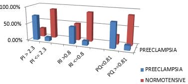
Aim: To correlate the association of ultrasonographic markers in antenatal patients with subsequent development of pre-eclampsia. Material Method: 131 pregnant women in the first trimester of pregnancy were investigated for their uterine artery Doppler and placental volume. These patients were followed up till the term for the development of pre-eclampsia. Results: Out of 131 antenatal cases, 20.61% developed pre-eclampsia. Uterine artery Pulsatility index >95th percentile (>2.3) with mean 2.08±0.43 (p-value 0.0001) predicted pre-eclampsia with a sensitivity of 51.85% with a specificity of 95.19%. Similarly, the Resistance index of the Uterine artery >95th percentile (>0.8) with a mean 0.771±0.084 (p-value 0.018) predicted pre-eclampsia with a sensitivity of 48.15% and specificity of 87.50%. Placental Quotient <10th percentile (<0.81) with mean 0.89 (p-value 0.0001) predicted pre-eclampsia with a sensitivity of 40.74% and specificity of 96%. On combining all three ultrasonographic parameters, the sensitivity for prediction of pre-eclampsia was increased to 70.37% with a specificity of 86.54% with PPV of 36.74% and NPV of 96.34%. Conclusion: Maternal ultrasonographic markers like Uterine artery PI, RI, Placental Quotient levels vary between normal pregnancies and those that subsequently developed pre-eclampsia. Thus our study concludes that first-trimester screening with ultrasonographic markers has high sensitivity, specificity and negative predictive value in detecting pre-eclampsia.

Keywords: Preeclampsia, Pulsatility index, Ultrasonography

Corresponding Author How to Cite this Article To Browse
Divya Gupta, Resident, Department of Obstetrics and Gynaecology, MLN Medical College, Prayagraj, U.P, India.
Email:
Seema Pandey, Veena Gupta, Rakesh Kumar Shukla, Divya Gupta, First Trimester Screening by Ultrasonographic Markers for Prediction of Pre-eclampsia. Int J Med Res Rev. 2021;9(6):374-378.
Available From
https://ijmrr.medresearch.in/index.php/ijmrr/article/view/1357

Manuscript Received Review Round 1 Review Round 2 Review Round 3 Accepted
2021-12-04 2021-12-06 2021-12-13 2021-12-20 2021-12-26
Conflict of Interest Funding Ethical Approval Plagiarism X-checker Note
Nil Nil Yes 19%

© 2021by Seema Pandey, Veena Gupta, Rakesh Kumar Shukla, Divya Guptaand Published by Siddharth Health Research and Social Welfare Society. This is an Open Access article licensed under a Creative Commons Attribution 4.0 International License https://creativecommons.org/licenses/by/4.0/ unported [CC BY 4.0].

Introduction

Pre-eclampsia could be a syndrome that manifests clinically within the last half of pregnancy (usually after 20 weeks) as a replacement onset hypertension blood pressure exceeding 140/90 mmHg on two occasions, at least 4 to six hours apart with associated proteinuria ≥ 300mg per 24 hours of urine collection or with any one of the following features of organ damage, i.e., platelet count<1Lac/mm3, serum creatinine>1.1mg/dl, raised liver enzymes twice the conventional, evidence of pulmonary oedema and cerebrovascular symptoms. Pre-eclampsia is the major contributor to maternal and perinatal mortality and morbidity worldwide [1].

According to the World Health Organization, 16 percent of maternal deaths are attributed to hypertensive disorders [2] and fivefold increase in perinatal mortality, with iatrogenic prematurity being the main culprit. The first identification of patients with increased risk for pre-eclampsia is, therefore, one every of the foremost essential goals in obstetrics [3].

Pre-eclampsia may be a multi-organ disease, which presents with varying degrees of severity. The prevention of pre-eclampsia remains a substantial challenge in obstetrics. Although the symptoms of pre-eclampsia generally manifest in the second to 3rd trimester of pregnancy, their underlying pathology takes place in the trimester between 9 - 13 weeks [4].

Numerous pathophysiological mechanisms, alone or together, are suggested to blame for diverse subsets of pre-eclampsia. They include impaired vascular remodelling of the maternal-fetal interface, excessive reaction to paternal antigens, maternal inflammatory response, very density lipoprotein toxicity, increased trophoblastic apoptosis and imbalance of angiogenic factors of these processes being modulated by genetic and environmental parameters.

Increased resistance to vascular flow may be measured by Doppler flow studies of the myometrial segments of the arteries supplying the spiral arterioles. The indices include systolic to diastolic velocity ratio, pulsatility index and resistance index, which might be investigated to predict pre-eclampsia.

Materials and Methods

The present study was done in Swaroop Rani Nehru Hospital and Kamla Nehru Hospital, in the Department of Obstetrics and Gynaecology, Moti Lal Nehru Medical College Prayagraj on 131 pregnant women, attending outpatient department and indoor cases in the first trimester of pregnancy (9 to 13 weeks six days of gestation) over twelve months (September 2019-August 2020).

This study is a case-control study, and the sample size was calculated using the standard formula:-

ijmrr_1357_02.JPG

Where -Z is that the z score at Confidence Interval 75% is 1.15 -ε is that the margin of error, assumed 5% -p̂ is that the population proportion assumed 50%.

All women included in this study were investigated for their uterine artery Doppler and placental volume at 9 to 13 weeks of gestation. 

Inclusion Criterion: All pregnant women of 9 to 13 weeks six days giving consent.

Exclusion Criterion:

  • Not giving consent
  • Twin pregnancy
  • Missed abortion
  • Chronic hypertension

Ethical clearance was given by an institutional ethical committee of MLN Medical College Prayagraj, and the consent of every individual was recorded as per the standard protocol.

The values of PI >2.3 and RI>0.8, i.e., >95th percentile, were considered high risk. Placental Quotient of th percentile i.e, 0.81 was taken abnormal. Follow up of all pregnant women enrolled in the study was done after 20 weeks of gestation till term for the development of pre-eclampsia. An Independent T-test was applied to find the association of Ultrasonographic markers with the development of pre-eclampsia. Sensitivity, specificity, PPV, NPV for Ultrasonography markers and placental quotient were also calculated and compared for prediction of pre-eclampsia.


Result

Out of a total number of antenatal cases, 14% had a Pulsatility index >95thpercentile (>2.3) of the 73.68% developed pre-eclampsia while 12% of patients with normal levels developed pre-eclampsia. 19% of antenatal cases had Resistivity index>95th percentile (>0.8) of the 50% developed pre-eclampsia while 13% with normal levels developed pre-eclampsia and 11% of antenatal cases had Placental Quotient<10th percentile (<0.81) of the 73.3% developed pre-eclampsia. In comparison, 14% with normal levels developed pre-eclampsia.

In the present study, the mean levels of PI, RI were found to be increased. Placental Quotient was found to be decreased in women who developed pre-eclampsia as compared to those who remained normotensive  [ 0.783+0.285MoM vs 1.489+-1.033 MoM] with (p-value 0.001),[0.969+-0.59MoM  vs 1.29+0.63MoM] with (p-value 0.0184) and [0.54+0.158MoM vs 1.55+-0.619MoM] with (p value<0.0001)respectively.

Table 1: Comparision of Sensitivity, Specificity, Ppv and Npv Of First Trimester Ultrasonographic Markers In Prediction Of Preeclampsia.

Parameters Sensitivity Specificity Positive Predictive Value Negative Predictive Value
Uterine Artery Doppler PI 51.85% 95.19% 54.51% 94.68%
RI 48.15% 87.50% 29.97% 93.82%
Placental Quotient 40.74% 96% 54.06% 93.59%
Uterine Artery Doppler+Placental Quotient 70.37% 86.54% 36.74% 96.34%

The above table shows that at the stage of 1st-trimester prediction of pre-eclampsia Of the total cases in the study, 27 cases that developed pre-eclampsia, 14 (51.85%) had mean Pulsatility index levels >95th percentile and rest 13 (48.14%) had their Pulsatility index levels 95th percentile or below. 13 cases (48.15%) had mean Resistance index levels >95th percentile and rest 14 (51.85%) had their Resistance index levels 95th percentile or below. Eleven cases (40.74%) had Placental Quotient levels<10th percentile

(<0.81 MoM), and the rest, 16 (59.25%), had their Placental Quotient levels 90th percentile or above. Out of 104 cases who remained normotensive, 5 (4.8%) had Pulsatility index levels >95th percentile, and the rest 99 (95.2%) had their Pulsatility index levels 95th percentile and below, 13 (12.54%) had Resistance index levels >95th percentile and rest 91 (87.5%) had their Resistance index levels 95th percentile and down, 4 (3.84%) had Placental Quotient levels <10th percentile and rest 100 (96.15%) had their Placental Quotient levels 90th percentile and above.

ijmrr_1357_01.JPG
Figure 1: Association between Ultrasonographic Markers and Development of Preeclampsia.

Discussion

In studies similar to our research, there was a significant association between increased Pulsatility index in antenatal patients and development of pre-eclampsia in different studies where p-value <0.001 and p-value 0.001 [5, 6], which is similar to our study (p-value 0.0001). Association between increased Resistance index in antenatal patients and development of pre-eclampsia in different studies are p-value <0.001 and p-value <0.01[5,7]  which is similar to our study were p-value 0.018 In our study Sensitivity of Uterine artery PI was 51.08% specificity was 95.19% in prediction of pre-eclampsia. In a study, the pulsatility index had a sensitivity of 44% [13], while in a study had sensitivity-75.9%, specificity-79.6% for prediction of pre-eclampsia [8]. In a study done by Leslie Myatt, were sensitivity and specificity of PGF was 32% and 80% in the first trimester for the prediction of pre-eclampsia [9].

The combination of placental volume measurement and uterine artery Doppler in the first trimester may identify women at risk for subsequent development of pregnancy complications [10]. The combination of abnormal uterine artery Doppler and low placental volume at 11–14 weeks


achieves better results than does either test alone in the prediction of pre-eclampsia [11]. Association between decreased Placental Quotient in antenatal patients and development of pre-eclampsia [12-14]. In a study were p-value <0.003 [15] similar to present study p-value 0.0001. In Our study, the Sensitivity of the Placental Quotient was 40.74%, and specificity 96%, whereas a study has done had a sensitivity of 38.5% [16]. Our study shows that Ultrasonography markers in the first trimester increase the sensitivity for the prediction of pre-eclampsia.  Uterine artery PI, RI and Placental quotient can be considered promising as a potential screening tool. Early identification of high-risk pregnancies may help in increased monitoring and early intervention to decrease fatal complications and to improve maternal and perinatal outcomes.

Conclusion

Our study thus concludes that integrated first-trimester screening shows a high prediction in the detection of pre-eclampsia. Ultrasound examination is a useful tool in pregnancy; with the advent of the high-resolution transvaginal probe, the possibility of using ultrasound uterine artery Doppler and placental volume studies can be used to identify pregnancies at increased risk of subsequent development of complications such as pregnancy-induced hypertension. In the present study, uterine artery Doppler parameters PI and RI were significantly increased. The Placental quotient was reduced in the pre-eclampsia group and had a predictive potential in pre-eclampsia.

The findings of my study are similar to the previous studies, which revealed that placental volume is reduced while artery Doppler parameters PI and RI are increased in antenatal cases who later developed pre-eclampsia. Moreover, combining all three ultrasonography parameters improved the early prediction of pre-eclampsia by increasing the sensitivity to 70.37% with a specificity of 86.54%, PPV of 36.74% and increased NPV to 84.92%. A large population-based study evaluating algorithms combining multiple markers are needed if screening approaches are to be eventually implemented.  The high cost of these tests can be a limiting factor in implementing these tests as a screening modality in the general population.

Outcome of This Study:  This is the first study with such parameters and

methodology in the eastern part of Uttar Pradesh, proving that ultrasonography parameters improved the early prediction of pre-eclampsia.

Disclosure: The authors declare that there is no conflict of interest.

Authors Contribution: Dr Seema Pandey- Performed all ultrasonography and radiological findings in this study. Dr Divya Gupta- Sample collections, data interpretations and manuscript writing. Dr Veena Gupta- Major Guidance and review of literature. Dr Rakesh Kumar Shukla- Anatomical correlations.

Reference

01. Alexander, James M. , et al. Severe pre-eclampsia and the very low birth weight infant: is induction of labor harmful?. " Obstetrics & Gynecology 93. 4 (1999): 485-488 [Crossref][PubMed][Google Scholar]

02. Khan KS, Wojdyla D, Say L, Gülmezoglu AM, Van Look PF. WHO analysis of causes of maternal death: a systematic review. Lancet. 2006 Apr 1;367(9516):1066-1074. doi: 10.1016/S0140-6736(06)68397-9 [Crossref][PubMed][Google Scholar]

03. Pilalis A, Souka AP, Antsaklis P, Daskalakis G, Papantoniou N, Mesogitis S, et al. Screening for pre-eclampsia and fetal growth restriction by uterine artery Doppler and PAPP-A at 11-14 weeks' gestation. Ultrasound Obstet Gynecol. 2007 Feb;29(2):135-40. doi: 10.1002/uog.3881 [Crossref][PubMed][Google Scholar]

04. Kadyrov M, Schmitz C, Black S, Kaufmann P, Huppertz B. Pre-eclampsia and maternal anaemia display reduced apoptosis and opposite invasive phenotypes of extravillous trophoblast. Placenta. 2003 May;24(5):540-8. doi: 10.1053/plac.2002.0946 [Crossref][PubMed][Google Scholar]

05. Harrington K, Cooper D, Lees C, Hecher K, Campbell S. Doppler ultrasound of the uterine arteries: the importance of bilateral notching in the prediction of pre-eclampsia, placental abruption or delivery of a small-for-gestational-age baby. Ultrasound Obstet Gynecol. 1996 Mar;7(3):182-8. doi: 10.1046/j.1469-0705.1996.07030182.x [Crossref][PubMed][Google Scholar]


06. Scazzochio E, Munmany M, Garcia L, Meler E, Crispi F, Gratacos E, et al. Prognostic role of maternal neutrophil gelatinase-associated lipocalin in women with severe early-onset pre-eclampsia. Fetal Diagn Ther. 2014;35(2):127-32. doi: 10.1159/000356499 [Crossref][PubMed][Google Scholar]

07. de Lima TH, Sass N, Mattar R, Moron AF, Torloni MR, Franchim CS, Daher S. Cytokine gene polymorphisms in pre-eclampsia and eclampsia. Hypertens Res. 2009 Jul;32(7):565-9. doi: 10.1038/hr.2009.58 [Crossref][PubMed][Google Scholar]

08. Shiva, Kumar HC, et al. "Study of length of umbilical cord and fetal outcome: a study of 1000 deliveries. " International Journal of Reproduction, Contraception, Obstetrics and Gynecology 6. 9 (2017): 3770-3776. [Crossref][PubMed][Google Scholar]

09. Myatt L, Clifton RG, Roberts JM, Spong CY, Hauth JC, Varner MW, et al. First-trimester prediction of pre-eclampsia in nulliparous women at low risk. Obstet Gynecol. 2012 Jun;119(6):1234-42. doi: 10.1097/AOG.0b013e3182571669 [Crossref][PubMed][Google Scholar]

10. Martin AM, Bindra R, Curcio P, Cicero S, Nicolaides KH. Screening for pre-eclampsia and fetal growth restriction by uterine artery Doppler at 11-14 weeks of gestation. Ultrasound Obstet Gynecol. 2001 Dec;18(6):583-6. doi: 10.1046/j.0960-7692.2001.00594.x [Crossref][PubMed][Google Scholar]

11. O'Gorman N, Wright D, Poon LC, Rolnik DL, Syngelaki A, de Alvarado M, et al. Multicenter screening for pre-eclampsia by maternal factors and biomarkers at 11-13 weeks' gestation: comparison with NICE guidelines and ACOG recommendations. Ultrasound Obstet Gynecol. 2017 Jun;49(6):756-760. doi: 10.1002/uog.17455. Erratum in: Ultrasound Obstet Gynecol. 2017 Dec;50(6):807 [Crossref][PubMed][Google Scholar]

12. Tan MY, Wright D, Syngelaki A, Akolekar R, Cicero S, Janga D, et al. Comparison of diagnostic accuracy of early screening for pre-eclampsia by NICE guidelines and a method combining maternal factors and biomarkers: results of SPREE. Ultrasound Obstet Gynecol. 2018 Jun;51(6):743-750. doi: 10.1002/uog.19039 [Crossref][PubMed][Google Scholar]

13. Poon LC, Rolnik DL, Tan MY, Delgado JL, Tsokaki T, Akolekar R, et al. ASPRE trial: incidence of preterm pre-eclampsia in patients fulfilling ACOG and NICE criteria according to risk by FMF algorithm. Ultrasound Obstet Gynecol. 2018 Jun;51(6):738-742. doi: 10.1002/uog.19019 [Crossref][PubMed][Google Scholar]

14. Francisco C, Wright D, Benkő Z, Syngelaki A, Nicolaides KH. Competing-risks model in screening for pre-eclampsia in twin pregnancy according to maternal factors and biomarkers at 11-13 weeks' gestation. Ultrasound Obstet Gynecol. 2017 Nov;50(5):589-595. doi: 10.1002/uog.17531 [Crossref][PubMed][Google Scholar]

15. Rizzo G, Capponi A, Cavicchioni O, Vendola M, Arduini D. First trimester uterine Doppler and three-dimensional ultrasound placental volume calculation in predicting pre-eclampsia. Eur J Obstet Gynecol Reprod Biol. 2008 Jun;138(2):147-51. doi: 10.1016/j.ejogrb.2007.08.015 [Crossref][PubMed][Google Scholar]

16. Schuchter K, Metzenbauer M, Hafner E, Philipp K. Uterine artery Doppler and placental volume in the first trimester in the prediction of pregnancy complications. Ultrasound Obstet Gynecol. 2001 Dec;18(6):590-2. doi: 10.1046/j.0960-7692.2001.00596.x [Crossref][PubMed][Google Scholar]