E-ISSN:2320-8686
P-ISSN:2321-127X

Case Report

Rhupus

International Journal of Medical Research and Review

2021 Volume 9 Number 6 November December
Publisherwww.medresearch.in

Overlap syndrome of systemic lupus erythematosus and rheumatoid arthritis (“rhupus”) with systemic involvement – A rare case report

Kulkarni P.1*, Rathod J.2, Kamble A.3, Chintale K.4, Bhattacharya M.5
DOI: https://doi.org/10.17511/ijmrr.2021.i06.07

1* Prabhanjan V Kulkarni, JR-II, Department of Medicine, Government Medical College and Hospital, Aurangabad, Maharashtra, India.

2 Jevant Rao Rathod, JR-I, Department of Medicine, Government Medical College and Hospital, Aurangabad, Maharashtra, India.

3 Akash Arjun Kamble, JR-I, Department of Medicine, Government Medical College and Hospital, Aurangabad, Maharashtra, India.

4 Kailas N Chintale, Associate Professor, Department of Medicine, Government Medical College and Hospital, Aurangabad, Maharashtra, India.

5 Meenakshi A Bhattacharya, Professor & Head, Department of Medicine, Government Medical College and Hospital, Aurangabad, Aurangabad, India.

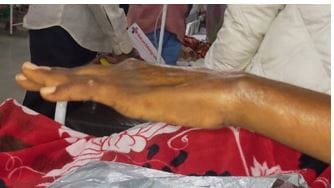
In this study the case of a 43 year old female, known for hypothyroidism, is presented, showcasing chest pain, breathlessness, without fever or rash, since 15 days. She had multiple joint pains since 11 months, however, the distal interphalangeal joints were spared. She had hand deformities and tenderness in the metacarpophalangeal and proximal interphalangeal joints of all the 10 fingers and both hips and knees. 2D-echo showed pulmonary hypertension. High resolution computed tomography of the thorax showed non-specific interstitial pneumonia. She had anemia, raised C - reactive protein and positive indirect Coomb’s test. Her anti-nuclear antibodies, rheumatoid factors, anti-Sm antibodies, anti-Sm/RNPs and anti-Ro antibodies were positive. As she satisfied the diagnostic criteria for both lupus and rheumatoid arthritis, she was diagnosed as a case of rhupus syndrome. She was treated with pulse injectable methylprednisolone for 3 days and then shifted to oral prednisone, along with hydroxychloroquine, oral methotrexate, sildenafil, pirfenidone and torsemide-spironolactone.

Keywords: Systemic lupus erythematosus, Rheumatoid arthritis, Overlap syndrome, Rhupus, pulmonary hypertension, Interstitial lung disease, Hemolytic anemia

Corresponding Author How to Cite this Article To Browse
Prabhanjan V Kulkarni, JR-II, Department of Medicine, Government Medical College and Hospital, Aurangabad, Maharashtra, India.
Email:
Prabhanjan V Kulkarni, Jevant Rao Rathod, Akash Arjun Kamble, Kailas N Chintale, Meenakshi A Bhattacharya, Overlap syndrome of systemic lupus erythematosus and rheumatoid arthritis (“rhupus”) with systemic involvement – A rare case report. Int J Med Res Rev. 2021;9(6):395-398.
Available From
https://ijmrr.medresearch.in/index.php/ijmrr/article/view/1350

Manuscript Received Review Round 1 Review Round 2 Review Round 3 Accepted
2021-12-07 2021-12-09 2021-12-16 2021-12-23 2021-12-30
Conflict of Interest Funding Ethical Approval Plagiarism X-checker Note
Nil Nil Yes 19%

© 2021by Prabhanjan V Kulkarni, Jevant Rao Rathod, Akash Arjun Kamble, Kailas N Chintale, Meenakshi A Bhattacharyaand Published by Siddharth Health Research and Social Welfare Society. This is an Open Access article licensed under a Creative Commons Attribution 4.0 International License https://creativecommons.org/licenses/by/4.0/ unported [CC BY 4.0].

Introduction

Overlap syndromes have been defined as conditions satisfying the classification criteria of at least two connective tissue diseases occurring in the same patient. Rhupus, a term coined by Peter Schur in 1971, is the co-existence of systemic lupus erythematosus (SLE) with rheumatoid arthritis (RA). [1] The syndrome is manifested by patients coincidentally sharing features of both RA and SLE but cannot be combined as a unique clinical, pathologic, or immunologic syndrome. The exact etiology and triggers of rhupus remain unknown till date. The incidence of rhupus in patients with arthritis is 0.01%–0.2% and <2% in patients with connective tissue diseases. [2] It is an extremely rare disease in the Indian population, with only a handful of isolated case reports published so far. Here we report such a case of a 43 year old female with polyarthritis over a duration of 11 months.

Case History

A 43-year-old female came with complaints of right-sided chest pain, sweating and breathlessness for 15 days. There was no history of fever, cough with expectoration, palpitations, vomiting or lower limb swelling.11 months before admission she was treated for infective villonodular synovitis of the right shoulder joint with intravenous antibiotics and discharged on an oral antibiotic course. An RA factor test was negative during that admission.She developed multiple joint pains and morning stiffness after the above episode. The pain was more in the proximal interphalangeal, metacarpophalangeal, wrist, elbow, hip, knee and metatarsophalangeal joints. The distal interphalangeal joint was spared.Her complete blood countsdone repeatedly over 8 months revealed microcytic hypochromic anemia, and she was started on iron and folic acid supplementation, without improvement. She received a diagnosis for hypothyroidism 5 months before admission and was started on levothyroxine supplementation.

15 days before admission she developed right-sided chest pain, sweating and gradually progressive breathlessness which was initially on exertion but later progressed to breathlessness even at rest. At presentation, her pulse rate was 135 beats per minute, her blood pressure was 110/80 mmHg, her respiratory rate was 28 per minute

and spo2 was 98% on room air. There was no rash. Hand deformities were present (Figure 1). There was tenderness in the metacarpophalangeal and proximal interphalangeal joints of all 10 fingers and both hips and knees. There was no pedal edema, lymphadenopathy or hepatomegaly.

ECG showed Q waves in leads II, III and avF. 2D echo showed an ejection fraction of 70%, concentric left ventricular hypertrophy and moderate pulmonary arterial hypertension (PAH). A high resolutioncomputed tomography (HRCT) of the thorax showed peripheral ground-glass opacities in bilateral upper lobes, reticulations and few bronchiectatic changes in bilateral lower lobes, suggestive of non-specific interstitial pneumonia (NSIP). Ultrasonography of the abdomen showed no significant abnormalities. Blood investigations were done which are shown in Table 1.

Table 1: Blood investigations.

Parameter Previous admission This admission
Hemoglobin (g/dL), Total leukocyte count (cells/cumm), Platelet count (cells/cumm), ESR (mm at 1 hr) 8.6, 15,400, 7.85, - 9.7, 16,300, 2.71, 18
CRP (qualitative) - Raised
Urea (mg/dL), Creatinine (mg/dL) 31, 1.3 64, 1.3
Total bilirubin (mg/dL), AST (IU/L), ALT (IU/L), Albumin (g/dL) 1.0, 30, 36, 3.2 0.5, 65, 16, 2.1
Reticulocyte count - 0.5%
Direct Coomb’s test - Negative
Indirect Coomb’s test - POSITIVE
Urine protein, Red cell, casts Trace, nil, nil Trace, nil, nil
Rheumatoid factor (IU/mL)[normal <20] Negative 91.2
ACPA [ECLIA] - Negative
ANA [ELISA], Sm/RNP, Sm, Ro 52kD antibody - POSITIVE
Anti-dsDNA antibody - Negative
24 hour urine protein - 43mg/24hrs

ACPA: Anti-citrullinated peptide antibody, ALT: Alanine transaminase, ANA: Antinuclear antibody, AST: Aspartate transaminase, CRP: C-reactive protein, ECLIA: Electrochemiluminescence immunoassay, ELISA: Enzyme linked immunosorbent assay, ESR: erythrocyte sedimentation rate, RNP: Ribonucleoprotein, Sm: Smith

As her EULAR score for rheumatoid arthritis was 10/10, and she satisfied 5 of the 17 SLICC criteria for SLE (Table 2), she was diagnosed as a case of SLE-RA overlap syndrome, classically referred to


as rhupus. She had predominant features of rheumatoid arthritis with a few features of SLE.

She was given pulse injectable methylprednisolone for 3 days and then shifted to oral prednisone. Hydroxychloroquine and oral methotrexate was started. She was also started on sildenafil, perfenidone and torsemide-spironolactone for her ILD and PAH. Her levothyroxine supplementation was continued.

Table 2: SLICC and ACR/EULAR scoring of the patient.

1.SLICC Criteria for the diagnosis of SLE 2. ACR/EULAR criteria for the diagnosis of RA
Clinical criteria Result (patient-specific) Parameter Point Score (patient-specific)
Acute cutaneous lupus No Joint distribution 1 large joint: 0 2-10 large joints: 1 1-3 small joints: 2 4-10 small joints: 3 >10 small joints: 5 5
Chronic cutaneous lupus No
Oral or nasal ulcers No
Non-scarring alopecia Yes
Arthritis Yes
Serositis No
Renal No Serology Negative RF and ACPA: 0 Low positive RF or ACPA: 2 High positive RF or ACPA: 3 3
Neurologic No
Hemolytic anemia Yes
Leukopenia No
Thrombocytopenia No
Immunologic criteria
ANA Yes
Anti-dsDNA antibody No Symptom duration <6 weeks: 0 >6 weeks: 1 1
Anti-Smith antibody Yes
Antiphospholipid antibody No Acute phase reactants Normal ESR, CRP: 0 Raised ESR, CRP: 1 1
Low complement No
Direct Coomb’s test No
Total 17 [required 4] 5 out of 17
Total score: 10, required for the diagnosis of definite RA: 6 10

ACPA: Anti-citrullinated peptide antibody, ACR/EULAR: American College of Rheumatology/European Alliance of Associations for Rheumatology, ANA: Antinuclear antibody, CRP: C-reactive protein, ESR: erythrocyte sedimentation rate, RA: Rheumatoid arthritis, RF: Rheumatoid factor, SLE: Systemic lupus erythematosus, SLICC: Systemic Lupus Erythematosus International Collaborating Clinics

The final diagnosis was SLE-RA overlap syndrome, non-specific interstitial pneumonia, pulmonary arterial hypertension, indirect coomb’s positive hemolytic anemia and hypothyroidism.

ijmrr_1350_01.JPG
Figure 1: Hand deformities.

Discussion

There are no standard guidelines or criteria to diagnose rhupus. RA-like arthritis is the main presenting feature, with some of the most common antibodies found in rhupus being ANA (89%), RF (84%), dsDNA (74%), and ACPA (58%). [3] A positive ACPA, the presence of the Shared Epitope (SE), and a raised CRP are often seen in a majority of patients. [4]

This patient presented with a longstanding history of small joint arthritis, alopecia, and persistent anemia. Significant findings on investigation were the presence of an indirect Coomb’s positive autoimmune hemolytic anemia (AIHA), raised CRP, positive ANA, RF, Sm/RNP, Sm, and anti-Ro 52kD antibodies. HRCT scan of the chest showed NSIP and 2D echo scan revealed PAH. She did not have a positive ACPA titre. Despite of this, the presence of erosive arthritis, a raised CRP and duration of symptoms led to an ACR/EULAR score of 10 points out of 10, pointing towards the diagnosis of rhupus. Erosive and symmetrical polyarthritis, accompanied by clinical signs and symptoms of SLE and the presence of autoantibodies with high specificity (ie native anti-DNA antibody or anti-Sm antibody) proved sufficient to diagnose her with rhupus. [5]

The study by Antonini et al [3] involving 287 rhupus patients showed that the most common manifestations of rhupus were RA-like polyarthritis, hematological abnormalities, renal involvement, skin and mucosal involvement and serositis. Only 6% of rhupus patients showcase pulmonary involvement. Another study by Li J et al [6]


involving 56 patients showed that 17% of rhupus patients had ILD and 7% had PAH. Both studies showed that rhupus patients had a lower incidence of malar rash, hemolytic anemia, and renal and neurological involvement, as compared to patients suffering from SLE without RA. This patient had interstitial lung disease leading to pulmonary hypertension, and the presence of indirect Coomb’s positive hemolytic anemia, which are also some rare manifestations of the disease. [3, 6]

Conclusion

Rhupus is a special overlap syndrome of RA and SLE that is manifested characteristically by more RA and less SLE-associated damage. It is a very rare disorder. The exact pathogenetic mechanisms of the disease are not yet clear. There are no standardized diagnostic guidelines or treatments available. Treatment is currently guided by the presenting clinical features. Further studies need to be undertaken to find out the exact pathophysiology and formulate a standardized treatment regimen for this rare disease.

Reference

01. Schur PH. Systemic lupus erythematosus. In: Beeson PB, McDermott W, editors. Cecil-loeb Textbook of Medicine. 13th ed. Philadelphia, PA: W.B. Saunders; 1971. p. 821 [Crossref][PubMed][Google Scholar]

02. Sinha, P. , Singhal, A. , Bhattacharjee, S. , & Oberoi, B. (2019). A Case of Rhupus-reporting of a Rare Entity. Indian dermatology online journal, 10(1), 58–60. [Article][Crossref][PubMed][Google Scholar]

03. Luca Antonini, Brigitte Le Mauff, Christian Marcelli, Achille Aouba, Hubert de Boysson, Rhupus: a systematic literature review, Autoimmunity Reviews, Volume 19, Issue 9, 2020, 102612, ISSN 1568-9972, https://doi. org/10. 1016/j. autrev. 2020.102612. ( [Article][Crossref][PubMed][Google Scholar]

04. Overlap Between Systemic Lupus Erythematosus and Rheumatoid Arthritis: Is It Real or Just an Illusion?. (2009). J Rheumatol 2009;36:4–6; doi:10. 3899/jrheum. 081067 (downloaded from: [Article][Crossref][PubMed][Google Scholar]

05. Simon, J Abraham & Granados, Julio & Cabiedes, J & Morales, J & Varela, J. (2002). Clinical and immunogenetic characterization of Mexican patients with 'Rhupus'. Lupus. 11. 287-92. 10.1191/0961203302lu189oa [Crossref][PubMed][Google Scholar]

06. Li J, Wu H, Huang X, et al. Clinical analysis of 56 patients with rhupus syndrome: manifestations and comparisons with systemic lupus erythematosus: a retrospective case-control study. Medicine. 2014 Aug;93(10):e49. DOI: 10.1097/md.0000000000000049. PMID: 25170930; PMCID: PMC4616332 [Crossref][PubMed][Google Scholar]