E-ISSN:2320-8686
P-ISSN:2321-127X

Research Article

Benign bone tumor

International Journal of Medical Research and Review

2022 Volume 10 Number 1 January February
Publisherwww.medresearch.in

Treatment of benign bone tumors: judicious use of adjuvants results in better functional outcomes.

More H.1*, Kundu Z.2, Singh R.3, Khanna M.4, Wadhwani J.5, Kamboj P.6
DOI: https://doi.org/10.17511/ijmrr.2022.i01.01

1* Hemant More, Assistant Professor, Department of Orthopaedics, Pt BD Sharma PGIMS, Rohtak, Haryana, India.

2 Zile S Kundu, Director, Department of Orthopaedics, Positron Multispeciality & Cancer Hospital, Rohtak, Haryana, India.

3 Roop Singh, , Department of Orthopaedics, Pt BD Sharma PGIMS, Rohtak, Haryana, India.

4 Mohit Khanna, , Department of Orthopaedics, Pt BD Sharma PGIMS, Rohtak, Haryana, India.

5 Jitendra Wadhwani, , Department of Orthopaedics, Pt BD Sharma PGIMS, Rohtak, Haryana, Haryana.

6 Pradeep Kamboj, , Department of Orthopaedics, Pt BD Sharma PGIMS, Rohtak, Haryana, India.

Background: While curettage has been a common treatment option for low grade benign lytic bone lesions, a careful extension of curettage enhances its efficacy without compromising integrity of surrounding normal bone. Many adjuvants are used for extension of curettage, but all are not universally available, and each has its drawbacks. We report outcome of extended curettage of benign lytic bone lesions using high-speed burr, electrocautery, hydrogen-peroxide, and pulsatile lavage. Methods: The study was conducted on 25 patients,10 to 40 years in age, with lytic bone lesions proven benign. Tumours belonged to Campanacci Grade 1 and 2 of varying histological types. High-speed burr, electrocautery, hydrogen-peroxide, and pulsatile lavage were extensively used after curetting lesions with sharp curettes. Defects were filled with bone graft or substitute where needed if cavity size was more than 1/3rd of width of bone on radiographic assessment. Patients were reviewed for a minimum of 18 months. Results: All defects reverted to near-normal radiological appearance with excellent functional outcome recorded in most cases. Eighty percent of patients recovered uneventfully. Mild limited complications were noted as a prolonged period of pain in 12% and superficial infection in 8%. No local recurrence was observed. Conclusions: This study demonstrates efficacy of extended curettage in treatment of benign tumors and tumour like lesions of bone using simple cheap modalities which are universally available. In properly selected cases of benign bone tumors, a meticulously performed extended curettage provides an effective treatment option that safeguards functionality without compromising on safety.

Keywords: Benign bone tumor, Lytic lesion, Extended curettage

Corresponding Author How to Cite this Article To Browse
Hemant More, Assistant Professor, Department of Orthopaedics, Pt BD Sharma PGIMS, Rohtak, Haryana, India.
Email:
Hemant More, Zile S Kundu, Roop Singh, Mohit Khanna, Jitendra Wadhwani, Pradeep Kamboj, Treatment of benign bone tumors: judicious use of adjuvants results in better functional outcomes.. Int J Med Res Rev. 2022;10(1):1-9.
Available From
https://ijmrr.medresearch.in/index.php/ijmrr/article/view/1335

Manuscript Received Review Round 1 Review Round 2 Review Round 3 Accepted
2021-12-13 2021-12-14 2021-12-21 2021-12-28 2022-01-04
Conflict of Interest Funding Ethical Approval Plagiarism X-checker Note
Nil Nil Yes 16%

© 2022by Hemant More, Zile S Kundu, Roop Singh, Mohit Khanna, Jitendra Wadhwani, Pradeep Kambojand Published by Siddharth Health Research and Social Welfare Society. This is an Open Access article licensed under a Creative Commons Attribution 4.0 International License https://creativecommons.org/licenses/by/4.0/ unported [CC BY 4.0].

Introduction

While most of the benign lytic bone tumors are amenable to treatment by curettage, there is a higher rate of local recurrence when compared with resection. However, curettage allows for a better functional result. To decrease the recurrence rate, curettage is extended by meticulously removing tissue from the healthy margins of the curetted cavity to reduce the chances of microscopic residual tumor tissue or cells escaping the procedure and acting as a nidus for future recurrence. The curettage can be extended by the use of physical, thermal or chemical adjuvants like liquid nitrogen, phenol, polymethylmethacrylate (PMMA), or thermal cautery that further extend the destruction of tumor cells along the margins. Each of these procedures has its share of advantages and complications [1-6].

To decrease the risk of complication in the form of fractures following extended curettage, and to expedite the bone healing process, in select cases the residual bone cavities can be reinforced with bone grafts or bone graft substitutes. These fillers augment final bone strength, reduce the risk of local recurrence (esp. giant cell tumors) and fill the empty cavities. We evaluated the efficacy, complications and functional outcome in patients with benign bone tumors and tumor like lesions treated with extended curettage using high-speed burr, electric cautery, hydrogen peroxide, and pulsatile lavage.

Methods

The study was conducted in a tertiary care hospital over 3 years on 25 patients ranging from 10 to 40 years in age with benign lytic lesions of bone proven non-malignant by clinical, radiological, and/or histopathological study/biopsy. Successive patients with lytic bone lesions reporting to the Out-Patient Department were screened and patients with solitary benign lesions were included irrespective of age and sex. Patients with osteochondroma, patients who had multiple lesions, malignant lesions, or recurrence, and patients with tumors of Campanacci Grade 3 were excluded from the study [7,8]. Preoperative biopsy was performed in all locally aggressive lesions except those in which clinic-radiological assessment pointed unequivocally to a benign diagnosis. Under spinal, regional

or general anaesthesia, patients and limb/s were placed in an appropriate position as per the location of the lesion. Tourniquet was used wherever possible. A simple curettage was done by first making a large cortical window over the lesion. This window was at least as large as the lesion itself. The bulk of the tumor was scooped out with a large sharp curette. The curetted tissue was sent for histopathological confirmation. The cavity was then enlarged in all directions with a high-speed burr till normal bone was exposed. The cavity was washed with hydrogen peroxide to extend the tumor kill. The curettage was further extended by a few millimetres by the use of power burr and electric cautery to extend the destruction of tumor cells. After each step, the cavity and the wound were washed with hydrogen peroxide and copiously irrigated with pulsatile lavage to remove any debris and tumor cells. In smaller lesions, where the cavity was up to one-third of the width of the bone, the cavity was left as such, while the larger cavities were filled with bone grafts/bone cement. Where required, the metal implant was used for fixation of the preoperative fracture. Wound closure, an antiseptic dressing and external splinting with a brace, slab or cast was done. Sutures were removed after two weeks and patients were discharged. The limb was kept immobilised for four to six weeks. After the removal of the external splint, joint mobilisation exercises were started immediately. In the lower limb, partial weight-bearing was allowed after six weeks and full weight bearing after three months after radiological confirmation of bone consolidation. In the upper limb, the active movement was started after six weeks and full use of the extremity after three months. Patients were followed up at three months intervals in the first year and six months intervals thereafter. Plain radiographs were taken postoperatively and then every 3 months in the first year and every 6 months thereafter. The outcomes were based on serial radiographs as well as subjective clinical assessment and function based on MSTS scoring [9]. Statistical significance was analyzed using the Chi-square test. A value of P < 0.05 was considered significant.

Results

The mean age of the patients was 20.3 years (range 10-40 years); there were 10 females and 15 males. The average period of follow up was 20 months


(range 18-27 months). The most common histological diagnosis was that of giant cell tumor (32%, n=8) followed by aneurysmal bone cyst (28%, n=7), chondroblastoma (12%, n=3), enchondroma (8%, n=2), and fibrous dysplasia, simple bone cyst, chondromyxoid fibroma, and eosinophilic granuloma (4% each, n=1). [Table 1].

Table 1: Tumor profile - Histological diagnosis (n=25). 

Tumor No. of Cases Percentage (%)
Giant Cell Tumor 8 32
Aneurysmal Bone Cyst 7 28
Chondroblastoma 3 12
Enchondroma 2 8
Fibrous Dysplasia 1 4
Simple Bone Cyst 1 4
Non-ossifying Fibroma 1 4
Chondromyxoid Fibroma 1 4
Eosinophilic Granuloma 1 4

Out of 25 tumors, 5 (20%) were radiographic grade-1 lesions, 20 (80%) were grade-2 as per Campanacci grading. The tibia was the most commonly involved bone at (n=8, 32%) followed by the femur (n=5, 20%) and humerus (n=4, 16%). The pelvis, metacarpals and calcaneum were involved in two cases (8%) each while in one case (4%) the lesion was present in the talus. Based on the location of the tumor is within the bone, a majority (n=11, 44%) of the tumors occurred in the metaphyseal region of the respective bones followed by epiphyseal (n=3, 12%), diaphyseal (n=3, 12%), cancellous bones (n=3, 12%), flat bones (n=3, 12%) and short tubular bones (n=2, 8%).

Although several patients reported a vague sense of discomfort on being asked a leading question, only 3 patients (12%) recorded pain as their complaint. There were superficial infections in 2 patients (8%) patients that were easily controlled with the change of oral antibiotics. There was no neurovascular deficit in any of the 25 patients who participated in this study. There was no reported fracture during surgery or the follow-up period. No recurrence was recorded clinically or radiologically during the follow-up period. (P<0.05) (Table 4).

In 20 patients (80%) the MSTS scores (max=30points) were between 26-30 points, while in 4 patients (16%) the scores were between 21-25 points. Only one patient (4%) had a score below 20 points. Twenty-three patients (92%) had excellent

functional outcomes while two patients (8%) had good functional outcomes and all patients could carry out their activities of daily living unrestricted. (P<0.05) (Table 5).

Table 2: Tumor profile - Bone involved (n=25).

Bone involved No. of Cases Percentage (%)
Scapula 1 4
Humerus 4 16
Metacarpals 2 8
Pelvis 2 8
Femur 5 20
Tibia 8 32
Talus 1 4
Calcaneum 2 8

Table 3: Tumor profile - Physeal involvement (n=25).

Location in Bone No. of Cases Percentage (%)
Epiphysis 3 12
Metaphysis 11 44
Diaphysis 3 12
Short Tubular Bones 2 8
Cancellous Bones 3 12
Flat Bones 3 12

Table 4: Complications (n=25).

Complications Persistant Pain Fracture  Infection Neuro-vascular Deficit Recurrence
No. of Cases (out of 25) 3 0 2 0 0
Percentage of Total (%) 12 0 8 0 0

Table 5: Outcome rating (as per MSTS score, n=25).

MSTS Score No. of Cases Percentage (%)
Excellent (>75%) 23 92
Good (50-75%) 2 8
Fair (25-50%) 0 0
Poor (<25%) 0 0

Discussion

Studies have been conducted by various authors who have compared different surgical interventions for treating benign tumorous lesions of bone. These include intralesional excision (curettage – simple and extended), marginal excision, wide resection and amputation. Different combinations of adjuvants also have been used in studies where extended curettage was done. While curettage


is the most commonly applied mode of the treatment in benign lytic lesions of bone, being a simple modality with minimal morbidity for the patient, it does have a fairly high rate of recurrence as compared to marginal or wide excision, especially in lesions like giant cell tumor that have a propensity for recurrence. The majority of the benign bone lesions (44%) in our study are present in the epiphysio-metaphysial area of the bone, and the area around the knee joint is the most commonly involved. These findings are consistent with international literature. [7,10,11]. This tendency of benign bone tumors for the involvement of periarticular regions and more involvement in lower limbs makes it more likely to cause increased morbidity in case radical surgical modalities are used for the treatment. In this context, curettage offers an option of limiting post-surgical morbidity.

Extended curettage provides a bridge that increases the cure rate when used judiciously in the selected profile of cases, while at the same time avoiding the complications or morbidity associated with radical excisional surgery or amputation. Multiple authors have reported greatly reduced recurrence rates of aggressive tumours with the use of adjuvants [12,13]. Although each adjuvant treatment has its proponents, no study has proved that anyone is superior, with each having advantages and disadvantages.

A high-speed burr helps in the removal of hard bony ridges and spurs that prevent the effective removal of tumor tissue lodged between them. This clears the way for curette and other adjuvants to act unhindered on the tumor margins and hence is commonly used alone or in combination with other adjuvants while performing extended curettage. Cryosurgery with liquid nitrogen is effective at extending the tumor kill. It is superior to phenol and methacrylate at creating a rim of necrotic bone. It is usually applied by direct pour technique and associated with greater complications, such as pathological fracture and nerve injury. High rates of such complications have been reported at 30% by Schreuder et al and 79% by Marcove et al including 20% fracture incidence in one study[ 14,15].  Khalil et al have reported a 17% recurrence rate in lesions managed by extended curettage with cryosurgery and bone cement [16]. With the use of cryoprobe instead of direct pour technique, the complication rate has been reported to be

low at 2.2% by Chen et al with a recurrence rate of 5.2% when used in conjunction with PMMA [17]. (Table 6)

Table 6: Recurrence rate in different procedures (in percentage). 

Study No. of patients Curettage Extended curettage Marginal resection Wide resection Amputation Adjuvant used for extended curettage Follow up (Years)
Campanacci et al [7] 327 27 - 8 0 0 - >2
McDonald et al [11] 221 - 34 - 7 - Burr + Phenol/Alcohol >2
Schreuder et al [14] 27 - 4 - - - Burr + Cryotherapy >2
Khalil et al [16] 52 50 17 - - - Cryosurgery + PMMA >2
Gitelis et al [18] 40 - 5 - 0 - Burr + Phenol >2
Stewart & Richardson [19] 34 50 36 27 - 0 Phenol/Alcohol >2
Dreinhofer et al [24] 10 - 20 - - - Burr + H2O2 +PMMA >2
Lim & Tan [25] 16 - 28 - 50 - Burr + H2O2/Phenol/ Cryotherapy >2
Marsh et al [27] 172 20 - 0 - - - >1.5
Blackley et al [28] 59 - 12 - - - Burr >2
Durr et al [29] 29 43 9 - 9 - Burr + Phenol >2
Zhen et al [30] 92 - 13 - - - Burr + Zinc chloride >2
Suneja et al [31] 53 - 13 - - - Burr >2
Wolfgang et al [32] 384 49 15 - - - Burr + Phenol >2
O’Donnell et al [33] 60 42 17 - - - Burr + Phenol >2
Present Study 25 - 0 - - - Burr+ Electric cautery+ Hydrogen peroxide >2

Phenol, conversely, has relatively poor bone penetration, and although relatively easy to use, serious complications have been reported when it extends into the surrounding normal tissues [1]. Gitelis et al have reported a 5% recurrence rate using phenol as an adjuvant


and complications of 10%-25% concerning neurovascular deficit and articular degeneration. & McDonald et al reported a 34% recurrence rate and a 4% incidence of neurovascular complications [11,18]. Stewart et al reported 36% recurrence [19]. (Table 7) Polymethylmethacrylate (PMMA) bone cement may act as an adjuvant through the heat of polymerization or direct toxicity of the monomer [2,20,21]. It is easily applied and can be used as a filling agent in conjunction with other adjuvants. Once applied, it is difficult to control the tissue damage due to the heat produced and may result in unintended damage to nearby tissue and/or articular surface [22,23]. Dreinhofer et al reported 20% recurrence using H2O2 with PMMS while Khalil et al reported 28% recurrence with PMMA alone [16,24].

Adjuvant treatment also may be accomplished through an argon beam coagulator which is electric cautery through a beam of argon gas that improves the conductivity of electricity. It is associated with a low rate of local recurrence and is also a reasonable adjuvant treatment option. But its use is limited by the availability of equipment in the treating centre and its cost [5]. Electrosurgical cautery cuts tissue with electric sparks that focus on intense heat at the surgical site. This produces the greatest amount of heat over a very short period, which results in the vaporization of tissue. The current density at the tip of the surgical electrode is very high leading to heat generation in the immediately adjacent tissue. Tissue destruction using electrosurgical cautery is easy to control and is widely available, and therefore makes it a suitable option for use as an adjuvant [3,4].

Hydrogen peroxide is a commonly available product with wide usage. It has an oxidative and thermal impact on the tumour cells as well as the effervescence helps in the cleansing of the cavity from difficult to reach areas with minimal damage to surrounding soft tissue and bone cells [6]. It is a cheap and universally available adjuvant that is easy to handle with minimal risk to surrounding healthy tissue. Studies by Lim & Tan, and Dreinhofer et al have used hydrogen peroxide in combination with Cryotherapy & PMMA respectively and reported 20% & 28% recurrence rates respectively [24,25]. Deventer et al have reported a recurrence rate of 12% in a retrospective study of chondroblastomas when using hydrogen peroxide after extensive curettage and burring compared to

67% when only curettage and bone substitute/autogenous bone filler were used [26].

Pulsatile lavage sends short pulses of a high-speed jet of medical normal saline into the cavities thereby helping in the removal of any debris or loose cells on the curetted margins. This helps in reducing the chances of recurrence due to adherent tumor cells that escape the curette. We used simple, cheap, and commonly available adjuvants in our series in the form of high-speed burr, electric cautery, hydrogen peroxide and pulsatile lavage. Where preoperative fractures were observed, fixation with metal implants was done. Where the diameter of the tumor cavity at the end of extended curettage was more than one-third of the width of the bone, additional augmentation with bone graft and/or bone cement was done. (Figure 1)

Table 7: Complication rates (in percentage). 

Study No. of patients Persistant Pain/stiffness Fracture Infection Neuro-vascular deficit Articular degeneration Other
McDonald et al [11] 221 - 1 5 4 - 24
Schreuder et al [14] 27 - 4 8 4 - -
Marcove et al [15] 25 25 20 20 12 - 25
Gitelis et al [18] 20 - - 10 10 25 20
Blackeley et al [28] 59 4 - 2 3 2 2
Present Study 25 12 - 8 - - -

Table 8: Functional outcome based on MSTS Scores (in percentage). 

Study No. of patients Excellent Good Fair Poor
Gitelis et al [18] (Curettage group) 20 100 - - -
Gitelis et al [18] (En -block resection) 20 50 35 15 -
Dreinhofer [24] 98 47 47 6 -
Zhen et al [30] 92  ----93*---- 4 3
Present Study 25 92 8 - -

*Zhen et al have not given separate figures for the two categories.


ijmrr_1335_01.JPG
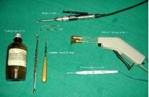
Figure 1: Simple tools used as adjuvants for extended curettage.

ijmrr_1335_2(1).JPG

ijmrr_1335_2(2).JPGFigure 2: Xrays showing a) ABC of talus b) post-op showing osteotomised fibula for talus exposure c) at 6months d) at 2years complete healing including osteotomised fibula.

ijmrr_1335_3(1).JPG

ijmrr_1335_3(2).JPGFigure 3: Xrays showing a) GCT of upper end tibia juxta-articular b) after curettage and bone grafting c) at 6months d) at 2years complete graft integration and maintenance of articular surface integrity.

ijmrr_1335_4(1).JPG

ijmrr_1335_4(2).JPGFigure 4: Xrays showing a) ABC of first metacarpal b) postoperative c) at 6months d) at 18months complete bone healing.

ijmrr_1335_5(1).JPG

ijmrr_1335_5(2).JPGFigure 5: Xrays showing a) fibrous dysplasia neck of the femur with fracture(x) b) one month after curettage, bone graft and screw fixation c) at 6months d) at 2years complete healing and graft integration.

In our study, we observed excellent clinical, radiological and functional results. We observed no post-surgical morbidity. Complications from the procedures were negligible in the form of two cases of mild superficial infection that was resolved by short term oral antibiotics. A prolonged period of local pain was also observed in three patients that were responsive to oral analgesics and resolved as the bone healing progressed. (Figure 2 & 3) (Table8) No recurrence, fracture, or infection was observed during the follow-up period.


A review of serial radiographs revealed that while smaller cysts filled up completely, the larger ones healed by initial thickening of the cortex followed by the development of septae within the defect. The earliest radiological evidence of bone formation was observed at 3months. The cavity wall became increasingly thick, sclerosed, and radio-opaque, even if complete bone filling of all cavities was not achieved. The healing of the bone progressed uneventfully and on expected lines resulting in fully functional limbs in all our patients. There was no disturbance in activities of daily living and the quality of life in our series. (Figure 4 & 5) While our study using simple protocol has significantly superior results (p<0.05) when compared to historical literature, there are certain limitations of this study that the series size was modest and therefore all histological types of benign bone tumors were not adequately represented to offer a robust inference of the treatment protocol used. Different types of benign bone tumors that have different biological behavior were treated by the same protocol.

Conclusion

To conclude, in this study we observed that careful selection of cases and executing extensive and meticulous curettage followed by extension of the cavity wall with a high-speed burr, treatment of the cavity wall by cauterizing on spray mode, using hydrogen peroxide and pulsatile lavage to clear the field of debris and adherent tumor tissue gives excellent results and prevents recurrences. It is very effective in low and moderate uncomplicated benign bone tumors and tumour-like lesions. We believe that this protocol can be kept as a first-line treatment option in carefully selected cases.  However, it is felt that there is a need for larger prospective randomized studies with longer follow up to evaluate the real effect of this simple treatment protocol in different histological types of benign bone tumors and tumor like lesions.

What does this study add to existing knowledge?

This study highlights that the cheap and easily available modalities can provide results at par with costly, machine intensive, or complicated methods used traditionally when used meticulously in the carefully selected patient group. This can help in bringing such treatment within the reach of a higher

proportion of the population in economically or geographically disadvantaged areas.

Contribution by authors: All authors were involved in the patient selection and management process and contributed to the preparation of this manuscript.

Reference

01. Heck RK Jr, Toy PC. General principles of tumors. In: Azar FM, Beaty JH, editors. Campbell’s operative orthopaedics. 14th ed. Philadelphia: Elsevier; 2021. p. 904-6 [Crossref][PubMed][Google Scholar]

02. McCaskie AW, Richardson JB, Gregg PJ. Further uses of polymethylmethacrylate in orthopaedic surgery. J R Coll Surg Edinb. 1998 Feb;43(1):37-9. [Crossref][PubMed][Google Scholar]

03. Wu MP, Ou CS, Chen SL, Yen EY, Rowbotham R. Complications and recommended practices for electrosurgery in laparoscopy. Am J Surg. 2000 Jan;179(1):67-73. doi: 10.1016/s0002-9610(99)00267-6 [Crossref][PubMed][Google Scholar]

04. Siddaiah-Subramanya M, Tiang KW, Nyandowe M. Complications, Implications, and Prevention of Electrosurgical Injuries: Corner Stone of Diathermy Use for Junior Surgical Trainees. Surg J (N Y). 2017 Sep 14;3(4):e148-e153. doi: 10.1055/s-0037-1606547 [Crossref][PubMed][Google Scholar]

05. Lewis VO, Wei A, Mendoza T, Primus F, Peabody T, Simon MA. Argon beam coagulation as an adjuvant for local control of giant cell tumor. Clin Orthop Relat Res. 2007 Jan;454:192-7. doi: 10.1097/01.blo.0000238784.98606.d4 [Crossref][PubMed][Google Scholar]

06. Nicholson NC, Ramp WK, Kneisl JS, Kaysinger KK. Hydrogen peroxide inhibits giant cell tumor and osteoblast metabolism in vitro. Clin Orthop Relat Res. 1998 Feb;(347):250-60. [Crossref][PubMed][Google Scholar]

07. Campanacci M, Baldini N, Boriani S, Sudanese A. Giant-cell tumor of bone. J Bone Joint Surg Am. 1987 Jan;69(1):106-14. [Crossref][PubMed][Google Scholar]

08. Bassiony AA. Giant cell tumour of the distal radius: wide resection and reconstruction by


non-vascularised proximal fibular autograft. Ann Acad Med Singap. 2009 Oct;38(10):900-4. [Crossref][PubMed][Google Scholar]

09. Enneking WF, Dunham W, Gebhardt MC, Malawar M, Pritchard DJ. A system for the functional evaluation of reconstructive procedures after surgical treatment of tumors of the musculoskeletal system. Clin Orthop Relat Res. 1993 Jan;(286):241-6. [Crossref][PubMed][Google Scholar]

10. Campanacci M, Capanna R, Picci P. Unicameral and aneurysmal bone cysts. Clin Orthop Relat Res. 1986 Mar;(204):25-36. [Crossref][PubMed][Google Scholar]

11. McDonald DJ, Sim FH, McLeod RA, Dahlin DC. Giant-cell tumor of bone. J Bone Joint Surg Am. 1986 Feb;68(2):235-42. [Crossref][PubMed][Google Scholar]

12. Carrasco CH, Murray JA. Giant cell tumors. Orthop Clin North Am. 1989 Jul;20(3):395-405. [Crossref][PubMed][Google Scholar]

13. Turcotte RE. Giant cell tumor of bone. Orthop Clin North Am. 2006 Jan;37(1):35-51. doi: 10.1016/j.ocl.2005.08.005 [Crossref][PubMed][Google Scholar]

14. Schreuder HW, Veth RP, Pruszczynski M, Lemmens JA, Koops HS, Molenaar WM. Aneurysmal bone cysts treated by curettage, cryotherapy and bone grafting. J Bone Joint Surg Br. 1997 Jan;79(1):20-5. doi: 10.1302/0301-620x.79b1.7097 [Crossref][PubMed][Google Scholar]

15. Marcove RC, Lyden JP, Huvos AG, Bullough PB. Giant-cell tumors treated by cryosurgery. A report of twenty-five cases. J Bone Joint Surg Am. 1973 Dec;55(8):1633-44 [Crossref][PubMed][Google Scholar]

16. Khalil el SA, Younis A, Aziz SA, El Shahawy M. Surgical management for giant cell tumor of bones. J Egypt Natl Canc Inst. 2004 Sep;16(3):145-52. [Crossref][PubMed][Google Scholar]

17. Chen C, Garlich J, Vincent K, Brien E. Postoperative complications with cryotherapy in bone tumors. J Bone Oncol. 2017 Apr 6;7:13-17. doi: 10.1016/j.jbo.2017.04.002 [Crossref][PubMed][Google Scholar]

18. Gitelis S, Mallin BA, Piasecki P, Turner F.

Intralesional excision compared with en bloc resection for giant-cell tumors of bone. J Bone Joint Surg Am. 1993 Nov;75(11):1648-55. doi: 10.2106/00004623-199311000-00009 [Crossref][PubMed][Google Scholar]

19. Stewart Mj, Richardson Tr. Giant cell tumor of bone. J Bone Joint Surg Am. 1952 Apr;34-A(2):272-86. [Crossref][PubMed][Google Scholar]

20. Provenzano MJ, Murphy KP, Riley LH 3rd. Bone cements: review of their physiochemical and biochemical properties in percutaneous vertebroplasty. AJNR Am J Neuroradiol. 2004 Aug;25(7):1286-90. [Crossref][PubMed][Google Scholar]

21. Eriksson RA, Albrektsson T, Magnusson B. Assessment of bone viability after heat trauma. A histological, histochemical and vital microscopic study in the rabbit. Scand J Plast Reconstr Surg. 1984;18(3):261-8. doi: 10.3109/02844318409052849 [Crossref][PubMed][Google Scholar]

22. Deramond H, Wright NT, Belkoff SM. Temperature elevation caused by bone cement polymerization during vertebroplasty. Bone. 1999 Aug;25(2 Suppl):17S-21S. doi: 10.1016/s8756-3282(99)00127-1 [Crossref][PubMed][Google Scholar]

23. Haas SS, Brauer GM, Dickson G. A characterization of polymethylmethacrylate bone cement. J Bone Joint Surg Am. 1975 Apr;57(3):380-91. [Crossref][PubMed][Google Scholar]

24. Sobti A, Agrawal P, Agarwala S, Agarwal M. Giant Cell Tumor of Bone - An Overview. Arch Bone Jt Surg. 2016 Jan;4(1):2-9. [Crossref][PubMed][Google Scholar]

25. Lim YW, Tan MH. Treatment of benign giant cell tumours of bone in Singapore. Ann Acad Med Singap. 2005 Apr;34(3):235-7. [Crossref][PubMed][Google Scholar]

26. Deventer N, Deventer N, Gosheger G, de Vaal M, Budny T, Laufer A, et al. Chondroblastoma: Is intralesional curettage with the use of adjuvants a sufficient way of therapy? J Bone Oncol. 2020 Dec 5;26:100342. doi: 10. 1016/j.jbo.2020.100342 [Crossref][PubMed][Google Scholar]

27. Marsh BW, Bonfiglio M, Brady LP, Enneking


WF. Benign osteoblastoma: range of manifestations. J Bone Joint Surg Am. 1975 Jan;57(1):1-9. [Crossref][PubMed][Google Scholar]

28. Blackley HR, Wunder JS, Davis AM, White LM, Kandel R, Bell RS. Treatment of giant-cell tumors of long bones with curettage and bone-grafting. J Bone Joint Surg Am. 1999 Jun;81(6):811-20. doi: 10.2106/00004623-199906000-00008 [Crossref][PubMed][Google Scholar]

29. Dürr HR, Maier M, Jansson V, Baur A, Refior HJ. Phenol as an adjuvant for local control in the treatment of giant cell tumour of the bone. Eur J Surg Oncol. 1999 Dec;25(6):610-8. doi: 10.1053/ejso.1999.0716 [Crossref][PubMed][Google Scholar]

30. Zhen W, Yaotian H, Songjian L, Ge L, Qingliang W. Giant-cell tumour of bone. The long-term results of treatment by curettage and bone graft. J Bone Joint Surg Br. 2004 Mar;86(2):212-6. doi: 10.1302/0301-620x.86b2.14362 [Crossref][PubMed][Google Scholar]

31. Suneja R, Grimer RJ, Belthur M, Jeys L, Carter SR, Tillman RM, et al. Chondroblastoma of bone: long-term results and functional outcome after intralesional curettage. J Bone Joint Surg Br. 2005 Jul;87(7):974-8. doi: 10.1302/0301-620X.87B7.16009 [Crossref][PubMed][Google Scholar]

32. Arbeitsgemeinschaft Knochentumoren, Becker WT, Dohle J, Bernd L, Braun A, Cserhati M, et al. Local recurrence of giant cell tumor of bone after intralesional treatment with and without adjuvant therapy. J Bone Joint Surg Am. 2008 May;90(5):1060-7. doi: 10.2106/JBJS.D.02771 [Crossref][PubMed][Google Scholar]

33. O'Donnell RJ, Springfield DS, Motwani HK, Ready JE, Gebhardt MC, Mankin HJ. Recurrence of giant-cell tumors of the long bones after curettage and packing with cement. J Bone Joint Surg Am. 1994 Dec;76(12):1827-33. doi: 10.2106/00004623-199412000-00009 [Crossref][PubMed][Google Scholar]