E-ISSN:2320-8686
P-ISSN:2321-127X

Research Article

Arthritis

International Journal of Medical Research and Review

2021 Volume 5 Number 9 September-October
Publisherwww.medresearch.in

Psoriatic Arthritis in the Eastern Part of Uttar Pradesh

Kumar Bhaskar K.1, Kumar Rai R.2, Mishra A.3, Chawla K.4, Kumar Shukla R.5*
DOI: https://doi.org/10.17511/ijmrr.2021.i05.06

1 Kamlesh Kumar Bhaskar, Consultant, Department of Skin & V.D., District Hospital Gazipur, Gazipur, Uttar Pradesh, India.

2 Rajesh Kumar Rai, Associate Professor, Department of Forensic Medicine, Moti Lal Nehru Medical College, Prayagraj, Uttar Pradesh, India.

3 Archana Mishra, Assistant Professor, Department of Biochemistry, Moti Lal Nehru Medical College, Prayagraj, Uttar Pradesh, India.

4 Kavita Chawla, Professor, Department of Physiology, Moti Lal Nehru Medical College, Prayagraj, Uttar Pradesh, India.

5* Rakesh Kumar Shukla, Assistant Professor, Department of Anatomy, Autonomous State Medical College and Hospital, Mirzapur, Uttar Pradesh, India.

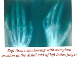
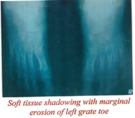
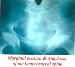
Aim: This original research article aims to study Psoriatic Arthritis in the Eastern Part of Uttar Pradesh. Material & Method: Two hundred twenty-eight patients of psoriasis regardless of age, sex, religion, occupation, attending the skin, and V. D. outpatients Department, B.R.D. Medical College, Gorakhpur for were taken because of the subject of this study. The bulk of patients belonged to the Eastern U.P. and adjoining areas of Bihar and Nepal. The clinical criteria for diagnosis of psoriasis were the presence of Erythematous and papulosquamous lesions with loosely adherent silvery-white scales. The auspitz's sign was demonstrated all told the cases. The detailed clinical history and examination were recorded with Tests for Rheumatoid factor and serum uric acid, Radiographs of both hands and feet, Radiographs of the lumbosacral Spine and both sacroiliac joints and Radiographs of affected joints (if any). Each patient was categorised into mild to severe psoriasis. Result: The total number of psoriatic arthritis cases was found, 26 of which male were 17 and female were 9. The maximum number of Arthritis cases, 18.5%, were seen with severe disease (PASI score > 15) with higher occurrence in males. The maximum number of joints involved distal interphalangeal joint was 69.2 3%, and the minimum number of joints involved was significant joint 7.69%. Conclusion: Psoriasis care is unbalanced, with men being more likely to undergo specialist treatment than women, causing higher distal interphalangeal (D.I.P.) joint arthropathy.

Keywords: Psoriasis, Psoriatic Arthritis, PASI score, PsA Radiology

Corresponding Author How to Cite this Article To Browse
Rakesh Kumar Shukla, Assistant Professor, Department of Anatomy, Autonomous State Medical College and Hospital, Mirzapur, Uttar Pradesh, India.
Email:
Kamlesh Kumar Bhaskar, Rajesh Kumar Rai, Archana Mishra, Kavita Chawla, Rakesh Kumar Shukla, Psoriatic Arthritis in the Eastern Part of Uttar Pradesh. Int J Med Res Rev. 2021;5(9):315-321.
Available From
https://ijmrr.medresearch.in/index.php/ijmrr/article/view/1324

Manuscript Received Review Round 1 Review Round 2 Review Round 3 Accepted
2021-07-26 2021-08-06 2021-08-17 2021-08-24 2021-09-03
Conflict of Interest Funding Ethical Approval Plagiarism X-checker Note
No Nil Yes 5%

© 2021 by Kamlesh Kumar Bhaskar, Rajesh Kumar Rai, Archana Mishra, Kavita Chawla, Rakesh Kumar Shukla and Published by Siddharth Health Research and Social Welfare Society. This is an Open Access article licensed under a Creative Commons Attribution 4.0 International License https://creativecommons.org/licenses/by/4.0/ unported [CC BY 4.0].

Introduction

Psoriasis would be a typical, genetically determined chronic, relapsing, inflammatory disorder of the skin characterised by red, scaly, sharply demarcated plaques that typically exhibit silvery-white scales.

Psoriasis is one of the significant common chronic inflammatory diseases of the skin, affecting 2–3% of the population [1]. The authors defined PsA as " inflammatory arthritis associated with psoriasis and negative for serum autoantibody." Some scientists refused to accept this definition, believing that the condition can coexist with two diseases: Arthritis (rheumatoid) and psoriasis. Psoriatic arthritis is a disease during which the cutaneous manifestation of psoriasis coexists with arthritis, usually within the absence of autoantibody.

The estimated prevalence of atrophic arthritis among patients with psoriasis is 4–42% (typically 7–10%) [2]. in additional recent studies, the prevalence of PsA is more often reported to be approximately 30% among patients with psoriasis, giving an overall prevalence of 0.3–1.0% within the general population [3-5]. Different clinical classifications are proposed for PsA since the initial subtypes were described in 1973; however, the foremost recent was developed by a world multicentre study leading to the CASPAR criteria and has now been widely adopted [6].

Psoriatic Arthritis (PsA) could be chronic inflammatory arthritis of unknown etiology, and currently, the cellular and molecular interactions that dictate its pathogenesis remain elusive. A task of the interleukin-23 (IL-23)/IL-23R (IL-23 receptor) interaction within the development of psoriasis and PsA is well established. As IL-23 regulates the differentiation and activation of innate and adaptive immunity, it pertains to complex pathophysiology involving a plethora of effectors and transducers. The positive effect of treatment in PsA depends on its early initiation before progressive joint destruction occurs. Modern diagnostic imaging techniques play an outsized role, especially in mildly symptomatic cases.

This study was planned at the Department of Skin and V.D., BRD Medical College Gorakhpur because no study was available on this topic in the eastern part of Uttar Pradesh and this medical college located in the east part of Uttar Pradesh with a sufficient number of cases, reported in the O.P.D. of the department and has adequate infrastructure.

Material and Methods

This study was conducted at the O.P.D. of the Department of the skin and V. D. B.R.D. Medical College, Gorakhpur. The total duration of the study was 16 months, and this study was a Cohort study (prospective and observational). ; A systematic sampling method was followed for sample collections; the bulk of patients belonged to the Eastern U.P. and adjoining areas of Bihar and Nepal. The sample size was calculated using the online software available at www. clincalc.com; a total of two hundred twenty-eight psoriasis cases were identified for study.

Inclusion criteria for study followed:

Psoriasis regardless of age, sex, religion, occupation, medical conditions.

Exclusion criteria for the study: Psoriasis cases from Geographically other than North India, Unable to follow up, and any other dermatological disorder associated. Ethical consideration was obtained from the ethical committee of BRD Medical College Gorakhpur. Every patient was informed regarding the study, and the consent of each patient was submitted to the concerned authority. The clinical criteria for diagnosis of psoriasis were the presence of Erythematous and papulosquamous lesions with loosely adherent silvery-white scales. The auspitz's sign was demonstrated. The patients in whom joint involvement the diagnosis was made based totally on history, physical examination, the same old absence of R.F. (Rheumatoid factor), and radiological features.

The physical examination excludes assessment of number, location, as distribution of joints involved, together with the presence of psoriatic skin lesion, key signs and symptoms indicative of rheumatism include asymmetrical joint involvement, Enthesitis, dactylitis, distal inter phalange (D.I.P.), and proximal inter phalange (P.I.P.) involvement and spinal inflammation. The detailed clinical history and examination were recorded. Each patient was categorised into mild to severe psoriasis on PASI Score. A PASI score is a tool used to measure the severity and extent of psoriasis. PASI is an acronym for Psoriasis Area and Severity Index. A representative area of psoriasis is selected for each body region. The intensity of redness, thickness, and scaling of the psoriasis is assessed as none (0), mild (1), moderate (2), severe (3), or very severe (4).


The percentage area affected by psoriasis is evaluated in the four regions of the body. In each region, the area is expressed as nil (0), 1-9% (score 1), 10-29% (score 2), 30-49% (score 3), 50-69% (score 4), 70-89% (score 5) or 90-100% (score 6).

  • Head and neck
  • Upper limbs
  • Trunk
  • Lower limbs

Each patient has undergone the following investigation.

  1. Routine blood tests
  2. Liver function test, kidney function test (if required)
  3. Skin biopsy
  4. Tests for Rheumatoid factor and serum uric acid.
  5. Radiographs of both hands and feet.
  6. Radiographs of the lumbosacral Spine and both sacroiliac joints.
  7. Radiographs of affected joints (if any).

ijmrr_1324_01.jpg

ijmrr_1324_02.jpg

ijmrr_1324_03.jpg

Observation

This study was conducted on 228 patients with psoriasis attending the Dermatology outpatient department of Nehru Hospital, B.R.D. Medical College, Gorakhpur, from July 2005 to October 2006.

Table 1: Age and Sex Distribution of the subjects

Age (Years) Male Female Total
No % No % No %
0-10
11-20
21-30
31-40
41-50
51-60
61-70
71-80
06
18
36
34
20
16
06
02
02.63
07.89
15.78
14.51
08.77
07.01
04.35
00.87
06
28
24
18
12
02
02.63
12.28
10.52
07.89
05.26
00.87
12
46
60
52
32
18
06
02
05.26
20.16
26.31
22.80
14.03
07.89
04.35
00.87
Total 138 100.0 90 100.0 228 100.0

The total numbers of cases studied were 228, of which were 138, and females were 90; the maximum numbers of patients were within the cohort of two 1-30 years. In males, most of the cases were within 21-30 years, while in females, 11 -20years (Table 1).

Table 2: Age and Sex Distribution of Psoriatic Arthritis

Age (Years) Male Female Total
No % No % No %
0-10
11-20
21-30
31-40
-
01
02
02
-
05.88
11.76
11.76
-
01
01
03
-
11.11
11.11
33.33
-
02
03
05
-
07.69
11.53
19.23

41-50
51-60
61-70
71-80
05
03
03
01
29.41
17.64
17.64
05.80
02
02
-
-
22.22
22.22
-
-
07
05
03
01
26.92
19.23
11.53
07.69
Total 17 100.0 9 100.0 26 100.0

Table 2 shows that the total number of psoriatic arthritis cases was found 26, in which males were 17 and females were 9. The maximum number of cases was in the age group 41-50 in male, while in female 31- 40 years.


Table 3: Correlation of Psoriatic Arthritis with Psoriasis

The severity of Psoriasis (PASI Score) Total Psoriatic cases Total No of Psoriatic cases Total No of Arthritis cases % of arthritis
Psoriasis in Male Arthritis in male Psoriasis in female Arthritis in female
0-3 mild
4-15 moderate
> 15 severe
32
63
43
02
07
08
20
41
29
01
03
05
52
104
72
02
11
13
03.84
11.57
18.05
Total 138 17 90 09 228 26  

Table 3 shows a maximum number of arthritis cases of 18.05% were seen with severe disease (PASI score > 15) in both male and female groups. Minimum numbers of arthritis cases 3.89 % were seen with mild disease (PASI score 0-3).

Table 4: Joint Involvement

Joint Involvement No of Patients Percentage (%)
Distal inter phalangeal
Proximal inter phalangeal
Sacroiliac joint
Spine
Major joint (Knee elbow)
18
03
04
04
02
69.23
11.53
15.38
15.38
07.69

Table 4 shows the maximum number of joints involved distal interphalangeal joint 69.2 3% and the minimum number of joints involved it was significant joint 7.69%.

Table 5: Radiological changes in upper and lower limb

  No of Patients Percentage (%)
Marginal erosion
Increase joint space
Periosteal new bone formation
Calcaneum spur
Deformity (Pencil in a cup)
06
01
05
01
02
23.17
03.84
19.23
03.84
07.69

Table 5 shows maximum radiological changes were seen in marginal erosion of distal interphalangeal joint 23.1 7%, and calcaneum spur was seen in 3.84 %.

Table 6: Radiological changes of Sacroiliac joint and Spine

  No of Patients Percentage (%)
Asymmetrical Joint Involvement
Erosion
Hazy joint margin
06
04
05
23.07
15.38
19.23
Narrowing of joint space
Ankylosis
03
02
11.53
07.69

Table 6 shows that asymmetrical joint involvement was 23.07%. Where marginal erosions were seen in 15.38 % and ankylosis was seen in 7.69% cases.

Discussion

Spondyloarthritis disorders are generally diagnosed more often in men than in women [7]. In relevancy the foremost representative quite these disorders, autoimmune disorder, three male cases are documented for every female patient [7]. PSA appears to be more frequent in men than women, particularly in its axial presentations [8].

Previous studies have shown that specialist treatment in psoriasis care is unbalanced, with men being more likely to undergo expert treatment than women [9]. Within the PsoReg, approximately 60% of registered patients are men. Most other European Registries for systemic psoriasis treatment show a more considerable dominance of enlisted men: Denmark 66%, Germany 60%, Italy 67%, Netherlands 68%, and Spain 63% [10]. In our present study, the total number of rheumatism cases was found, 26 of which male were 17 and female were 9. the maximum number of cases was within the cohort 41-50 in male, while in female 31- 40 years.

This support the previous study, that male are more in affected group, but little is assumed of the differential clinical expression of PsA between males and females. The explanation for these differences isn't clear, and also the participation of multifactorial parameters cannot be discarded.


Distal interphalangeal (D.I.P.) joint arthropathy is characteristic of both rheumatism (PsA) and osteoarthritis (O.A.), PsA is also distinguished from O.A. on the premise of more severe inflammation within the collateral ligaments and also the extensor tendons and more severe changes at the corresponding D.I.P. joint entheseal insertions. Swelling related to D.I.P. PsA usually affects the whole finger or toe. It's asymmetric, meaning it affects only one side of the body, which means that if all the joints in one hand are affected, the opposite hand might not be affected in the slightest degree, or four or fewer digits are involved.

However, research shows PsA can even be symmetrical, involving the joints on either side of the body.2 Moreover, D.I.P. PsA occurs more frequently within the toes [11]. Previously many authors [12,13] reported Distal interphalangeal (D.I.P.) joint arthropathy is highest among other joints. Our findings support the previous study because the maximum number of joints involved distal ginglymus was 69.2 3%, and also the minimum number of joints involved had been significant joint 7.69%. D.I.P. rheumatism also causes Enthesitis, inflammation at the places where ligaments and tendons meet at the bones. The kind of swelling and inflammation can erode the joints and result in deformity and loss of function.

Bone erosions were smitten by age and disease duration of PsA. Erosions reflect accumulating mechanical and inflammatory damage within the joints. In accordance, erosive damage was most significant in patients with PsA. No difference was found between healthy controls and patients with PSO. a noteworthy finding was that with an increase in age, bone erosions failed to only increase in patients with PsA, but also in patients with PSO and healthy controls. While individuals without inflammatory joint disease (healthy controls and patients with psoriasis) ;presumably accrue damage because of mechanical triggers, in patients with PsA the extra inflammatory trigger appears to hurry up erosions.

Hence, the common 20 to 40-year old patient with PsA already exhibits a burden of bone erosions, which equals that of a private who is over 60 years old and doesn't have inflammatory joint disease. These findings also indicate that judgement on the burden of erosive bone damage in private with PsA must consider age as a significant influencing factor.

The influence of inflammation as an enhancer of the progression of bone erosions is additionally supported by the notion that the duration of PsA is related to the burden of bone erosions [14].

Early erosive changes are quite common in PsA – they develop in 15–47% of patients within the first two years of disease [15]. Early erosions, like in R.A., are marginal and well-defined. Still, because the disease progresses, they become irregular and ill-defined thanks to the periosteal bone formation adjacent to erosions, which can make erosion of a speculated appearance, particularly towards its margin [16,17]. In our study maximum, radiological changes were seen in the marginal decline of distal hinge joint 23.1 7%, and calcaneum spur was seen in 3.84 %.

Asymmetric and bilateral involvement of the sacroiliac joints is typical, yet unilateral may occur moreover. Both synovial and syndesmotic parts are additionally engaged (like in autoimmune disorder and remaining spondyloarthritis). The iliac side is initially involved, presumably thanks to mechanical factors and anatomic features [18, 19]. Our finding shows that asymmetrical joint involvement was 23.07%. Where marginal erosions were seen in 15.38% and ankylosis was seen in 7.69% cases.

Conclusion

Psoriasis care is unbalanced, with men being more likely to undergo specialist treatment than women, causing higher distal interphalangeal (D.I.P.) joint arthropathy with early erosive changes in long bones of the limb with/without asymmetric and bilateral involvement of the sacroiliac joints.

By investigating the sex differences within the severity of psoriasis using the gold standard of severity measurement—the PASI score—and the distinct elements of the PASI score. Further research is required to substantiate this finding in numerous populations.

The outcome of this study

This is the first study under the above parameters in the eastern part of Uttar Pradesh, which shows 69.23% of Distal interphalangeal involvement, 23.07% asymmetrical joint involvement and 23.17% marginal erosion.


Authors contribution

Dr Kamlesh Kumar Bhaskar: Major contribution and performed all methods as a junior resident in this study.

Dr Rakesh Kumar Shukla: Major contribution and wrote the manuscript.

Dr. Rajesh Kumar Rai: Statistical analysis and data interpretation.

Dr Archana Mishra: Data Analysis and Review

Dr Kavita Chawla: Data Analysis and Review

Disclosure

The authors declare that there is no conflict of interest.

Reference

1. Wagner EF, Schonthaler HB, Guinea-Viniegra J, Tschachler E. Psoriasis: what we have learned from mouse models. Nat Rev Rheumatol. 2010 Dec;6(12):704-14. doi: 10.1038/nrrheum.2010.157 [Crossref][PubMed][Google Scholar]

2. Al-Heresh AM, Proctor J, Jones SM, Dixey J, Cox B, Welsh K, et al. Tumour necrosis factor-alpha polymorphism and the HLA-Cw*0602 allele in psoriatic arthritis. Rheumatology (Oxford). 2002 May;41(5):525-30. doi: 10.1093/rheumatology/41.5.525 [Crossref][PubMed][Google Scholar]

3. Green L, Meyers OL, Gordon W, Briggs B. Arthritis in psoriasis. Ann Rheum Dis. 1981 Aug;40(4):366-9. doi: 10.1136/ard.40.4.366 [Crossref][PubMed][Google Scholar]

4. Scarpa R, Oriente P, Pucino A, Torella M, Vignone L, Riccio A, Biondi Oriente C. Psoriatic arthritis in psoriatic patients. Br J Rheumatol. 1984 Nov;23(4):246-50. doi: 10.1093/rheumatology/23.4.246 [Crossref][PubMed][Google Scholar]

5. Salvarani C, Lo Scocco G, Macchioni P, Cremonesi T, Rossi F, Mantovani W, Battistel B, Bisighini G, Portioli I. Prevalence of psoriatic arthritis in Italian psoriatic patients. J Rheumatol. 1995 Aug;22(8):1499-503. [Crossref][PubMed][Google Scholar]

6. Taylor W, Gladman D, Helliwell P, Marchesoni A, Mease P, Mielants H; CASPAR Study Group. Classification criteria for psoriatic arthritis: development of new criteria from a large international study. Arthritis Rheum. 2006 Aug;54(8):2665-73. doi: 10.1002/art.21972 [Crossref][PubMed][Google Scholar]

7. Zochling J, Smith EU. Seronegative spondyloarthritis. Best Pract Res Clin Rheumatol. 2010 Dec;24(6):747-56. doi: 10.1016/j.berh.2011.02.002 [Crossref][PubMed][Google Scholar]

8. Queiro R, Sarasqueta C, Torre JC, Tinturé T, López-Lagunas I. Spectrum of psoriatic spondyloarthropathy in a cohort of 100 Spanish patients. Ann Rheum Dis. 2002 Sep;61(9):857-8. doi: 10.1136/ard.61.9.857 [Crossref][PubMed][Google Scholar]

9. Hotard RS, Feldman SR, Fleischer AB Jr. Sex-specific differences in the treatment of severe psoriasis. J Am Acad Dermatol. 2000 Apr;42(4):620-3. [Crossref][PubMed][Google Scholar]

10. Ormerod AD, Augustin M, Baker C, Chosidow O, Cohen AD, Dam TN, et al. Challenges for synthesising data in a network of registries for systemic psoriasis therapies. Dermatology. 2012;224(3):236-43. doi: 10.1159/000338572 [Crossref][PubMed][Google Scholar]

11. Gladman DD, Antoni C, Mease P, Clegg DO, Nash P. Psoriatic arthritis: epidemiology, clinical features, course, and outcome. Ann Rheum Dis. 2005 Mar;64 Suppl 2(Suppl 2):ii14-7. doi: 10.1136/ard.2004.032482 [Crossref][PubMed][Google Scholar]

12. Ash ZR, Tinazzi I, Gallego CC, Kwok C, Wilson C, Goodfield M, et al. Psoriasis patients with nail disease have a greater magnitude of underlying systemic subclinical enthesopathy than those with normal nails. Ann Rheum Dis. 2012 Apr;71(4):553-6. doi: 10.1136/annrheumdis-2011-200478 [Crossref][PubMed][Google Scholar]

13. Aydin SZ, Castillo-Gallego C, Ash ZR, Marzo-Ortega H, Emery P, Wakefield RJ, Wittmann M, McGonagle D. Ultrasonographic assessment of nail in psoriatic disease shows a link between onychopathy and distal interphalangeal joint extensor tendon enthesopathy. Dermatology. 2012;225(3):231-5. doi: 10.1159/000343607 [Crossref][PubMed][Google Scholar]


14. Simon D, Kleyer A, Faustini F, Englbrecht M, Haschka J, Berlin A, et al. Simultaneous quantification of bone erosions and enthesiophytes in the joints of patients with psoriasis or psoriatic arthritis - effects of age and disease duration. Arthritis Res Ther. 2018 Aug 31;20(1):203. doi: 10.1186/s13075-018-1691-z [Crossref][PubMed][Google Scholar]

15. Narváez J, Narváez JA, de Albert M, Gómez-Vaquero C, Nolla JM. Can magnetic resonance imaging of the hand and wrist differentiate between rheumatoid arthritis and psoriatic arthritis in the early stages of the disease?. Semin Arthritis Rheum. 2012 Dec;42(3):234-45. doi: 10.1016/j.semarthrit.2012.03.016 [Crossref][PubMed][Google Scholar]

16. Ory PA. Radiography in the assessment of musculoskeletal conditions. Best Pract Res Clin Rheumatol. 2003 Jun;17(3):495-512. doi: 10.1016/s1521-6942(03)00022-6 [Crossref][PubMed][Google Scholar]

17. Brown AK. How to interpret plain radiographs in clinical practice. Best Pract Res Clin Rheumatol. 2013 Apr;27(2):249-69. doi: 10.1016/j.berh.2013.03.004 [Crossref][PubMed][Google Scholar]

18. Paparo F, Revelli M, Semprini A, Camellino D, Garlaschi A, Cimmino MA, et al. Seronegative spondyloarthropathies: what radiologists should know. Radiol Med. 2014 Mar;119(3):156-63. doi: 10.1007/s11547-013-0316-5 [Crossref][PubMed][Google Scholar]

19. Coates LC, Hodgson R, Conaghan PG, Freeston JE. MRI and ultrasonography for diagnosis and monitoring of psoriatic arthritis. Best Pract Res Clin Rheumatol. 2012 Dec;26(6):805-22. doi: 10.1016/j.berh.2012.09.004 [Crossref][PubMed][Google Scholar]