E-ISSN:2320-8686
P-ISSN:2321-127X

Research Article

Aberrant

International Journal of Medical Research and Review

2020 Volume 9 Number 3 May-June
Publisherwww.medresearch.in

Aberrant promoter hypermethylation of RAR-β in endometrial carcinoma- an Indian study.

Shivanandappa N.1, N Swamy S.2, Kumar S S.3, Sheshadri S.4, Reddihalli Pallavi V.5, Gawari R.6*
DOI: https://doi.org/10.17511/ijmrr.2021.i03.05

1 Nagaratna Shivanandappa, Research Scholar, Department of Biochemistry, Kidwai Memorial Institute of Oncology, Bangalore, Karnataka, India.

2 Shalini N Swamy, Research Scholar, Department of Biochemistry, Kidwai Memorial Institute of Oncology, Bangalore, Karnataka, India.

3 Sandeep Kumar S, Senior Research Fellow, Department of Biochemistry, Kidwai Memorial Institute of Oncology, Bangalore, Karnataka, India.

4 Suma Sheshadri, Associate Professor, Department of Pathology, Kidwai Memorial Institute of Oncology, Bangalore, Karnataka, India.

5 Venkateshaiah Reddihalli Pallavi, Professor, Department of Gynaecology, Kidwai Memorial Institute of Oncology, Bangalore, Karnataka, India.

6* Ramesh Gawari, Professor, Department of Biochemistry, Kidwai Memorial Institute of Oncology, Bangalore, Karnataka, India.

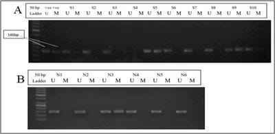
Endometrial cancer is the seventh most common cancer in women worldwide with an age-standardized rate of 8.4 per 100,000 women. Epigenetic alterations such as promoter hypermethylation of TSGs are known to be early events in carcinogenesis. The aim of the present study, we assessed the aberrant promoter hypermethylation pattern of RAR-β in 78 endometrial cancer samples. Methods: DNA was isolated from endometrial carcinoma samples and normal tissues and aberrant promoter hypermethylation was assessed using nested and methylation-specific PCR. The Chi-square test was used for statistical analysis and a p-value<0.05 was considered to be statistically significant. Results: 40 of the 78 (51.28%) endometrial carcinoma samples showed aberrant hypermethylation of the RAR-β gene. Methylation status in each histological subtype, grade and stage of the disease was also assessed. Conclusion: Aberrant hypermethylation is an important early epigenetic alteration that occurs during tumorigenesis. The Data shown here reports that promoter hypermethylation of RAR-β occurs in endometrial carcinoma and therefore could be used as a potential marker for early diagnosis and prognosis of the disease.

Keywords: DNA Hypermethylation, RAR-β, Endometrial cancer, epigenetic alterations, DNA hypermethylation, Methylation-specific PCR

Corresponding Author How to Cite this Article To Browse
Ramesh Gawari, Professor, Department of Biochemistry, Kidwai Memorial Institute of Oncology, Bangalore, Karnataka, India.
Email:
Shivanandappa N, Swamy SN, Kumar S S, Sheshadri S, Venkateshaiah Reddihalli Pallavi, Ramesh Gawari, Aberrant promoter hypermethylation of RAR-β in endometrial carcinoma- an Indian study.. Int J Med Res Rev. 2020;9(3):160-166.
Available From
https://ijmrr.medresearch.in/index.php/ijmrr/article/view/1294

Manuscript Received Review Round 1 Review Round 2 Review Round 3 Accepted
2020-05-16 2020-05-26 2020-06-08 2020-06-18 2020-06-22
Conflict of Interest Funding Ethical Approval Plagiarism X-checker Note
No Nil Yes 7%

© 2020 by Nagaratna Shivanandappa, Shalini N Swamy, Sandeep Kumar S, Suma Sheshadri, Venkateshaiah Reddihalli Pallavi, Ramesh Gawari and Published by Siddharth Health Research and Social Welfare Society. This is an Open Access article licensed under a Creative Commons Attribution 4.0 International License https://creativecommons.org/licenses/by/4.0/ unported [CC BY 4.0].

Introduction

Endometrial cancer (EC) is the seventh most common cancer in women worldwide with an age-standardized incidence rate (ASR) of 8.4 per 100,000 women. According to the 2018 Indian statistics, 13328 women were diagnosed with EC and an estimated 5010 women succumbed to the disease accounting for an ASR of 2.3 per 100,000 women [1-3]. Though most of the patients are diagnosed in the early stages when the disease is still confined to the primary site, about 30% of the patients are diagnosed in late stages where the disease would have disseminated from the uterus [4]. Various genetic and epigenetic alterations are known to be involved in the initiation and progression of EC. Aberrant promoter hypermethylation of Tumor suppressor genes (TSGs) is known to be an early event in many cancers including EC. Promoter hypermethylation is associated with transcriptional silencing of several tumour suppressor genes [5]. Retinoids are known to play a role in tumor suppression upon interaction with retinoic acid receptors. The retinoic receptors are nuclear receptors and include three retinoic acid receptors (RARs) - RAR-α, RAR-β, and RAR-γ [6,7]. Suppression of expression and function of RAR-β, have been reported in various cancers [8-10]. One of the mechanisms of suppressing the expression of RAR-β is by the epigenetic mechanism of promoter hypermethylation where a methyl group is added to deoxycytidine residues in the CpG islands of its promoter. This methylation of all or a few of the cytosine in the promoter region renders the closure of the chromatin structure eventually resulting in transcriptional silencing of the gene [11-13]. In the present study, we have attempted to analyze the methylation pattern of the RAR-β gene in 78 endometrial cancers of patients of the Indian cohort and associated the methylation patterns with various clinicopathological parameters.

Methods

Sample collection: 78 histologically confirmed endometrial cancer samples were collected post-surgery from patients undergoing surgery for endometrial cancer. All the samples were immediately stored at -800C until further processing.

Inclusion and exclusion criteria: Primary chemo naïve endometrial carcinoma patient samples were included in the study.

Patients with secondary metastasis to the endometrium and chemotherapy-treated samples were excluded from the study.

DNA extraction: Genomic DNA was isolated from all the samples using DNA easy mini kit (Qiagen, USA) following the manufacturer's protocol. The DNA concentration was estimated after extraction using Eppendorf Biospectrophotometer Kinetics™. 1μg of the eluted DNA samples were used for bisulfite modification.

Bisulfite modification: Genomic DNA was subjected to sodium bisulfite modification using the EZ DNA Methyl Lightening kit TM (Zymo Research D5031, CA, USA) following the manufacturer's procedure. The bisulfite modified DNA was aliquoted and used for methylation analysis.

Methylation Specific PCR: To assess the methylation status of the RAR-β promoter, methylation-specific PCR was used and was performed in two stages-first a nested PCR followed by Methylation-specific PCR. For the nested PCR, 2μl of sodium bisulfite modified DNA was used as a template. The PCR product from the first step nested PCR was diluted 10-folds and used as a template for the second step to specifically amplify the methylated and the unmethylated regions of the RAR-β promoter. CpG Methylated HeLa Genomic DNA (New England Biolabs, USA) was used as a positive control for methylated allele and DNA obtained from the normal ovary samples served as a negative control. The Primer sequences (table-1) and the cycling conditions for MSP and USP are summarized in table-2. The PCR products of methylation-specific and unmethylation specific PCR were loaded on a 2.5% Agarose gel and visualized by Ethidium Bromide Staining. The gel image was captured using Syngene G: Box gel Documentation system.

Statistical analysis: Chi-square test was used to analyze the statistical association of methylation with clinicopathological parameters. A p-value of <0.05 was considered to be statistically significant.

Ethical approval: The study was approved by the Institutional Scientific Review board and medical ethics committee and consent was obtained from patients before sample collection.

Results

RAR-β methylation was assessed in 78 endometrial cancer samples of 5 subtypes- endometrioid


(n=60), serous (n=06), mucinous (n=02), clear cell (n=02) and poorly differentiated carcinoma (n=08). Six normal endometrial samples were analyzed for RAR-β methylation as a control cohort. 40 of the 78 samples analyzed (51.28%) showed methylation of the RAR-β gene. Among the subtypes, 50 % of the Endometroid, mucinous, clear cell, and poorly differentiated samples showed methylation for the RAR-β gene and 66.6% of the samples of the serous subtype showed methylation for the gene. About 50% of the patient in Stage I and II and about 60% of the patients of stage III and IV showed methylation for the gene respectively. 20/44, 11/19 and 9/15 samples of grades 1,2,3 showed methylation respectively.

68 of the 78 patients (87.18%) included in the study posted menopausal women and 52 % of these postmenopausal patients showed methylation for the gene.

The results of the same are summarized in table 3. Though a good percentage of methylation was observed in various subtypes and subgroups of the clinicopathological parameters, there was no statistically significant association of the methylation status with these clinicopathological parameters.

Table 1: Primer sequence for nested and methylation specific PCR

Gene Forward (5´-3´) Reverse (5´-3´) Annealing temperature Product Size
RAR-β Nested AAGTGAGTTGTTTAGAGGTAGGAGGG CCTATAATTAATCCAAATAATCATTTACC 520 C 276
RAR-β MSP CCTATAATTAATCCAAATAATCATTTACC CCTATAATTAATCCAAATAATCATTTACC 600 C 146
RAR-β USP CCTATAATTAATCCAAATAATCATTTACC CTCAACCAATCCAACCAAAACA 610 C 146

Table 2: PCR cycling conditions for nested and Methylation-specific PCR.

Gene Initial Denaturation Cycling stage Final extension
Denaturation Annealing Extension
RAR-β Nested 950 C 950 C 520 C 720 C 720 C
10 mins 30 seconds 30 seconds 30 seconds 7 mins
RAR-β MSP/USP 950 C 950 C 600 C/610C (MSP/USP) 720 C 720 C
10 mins 30 seconds 30 seconds 30 seconds 7 mins

Table 3: Association of RAR-β methylation with clinicopathological parameters.

Clinicopathological Parameters N RAR-β methylation
Endometrial tumors 78  
Type of tumor Endometriod 60 30 (50%)
Serous 06 04 (66.6%)
Mucinous 02 01(50%)
Clear cell 02 01(50%)
Poorly differentiated 08 04(50%)
p-value   0.961293
FIGO stage I 64 32 (50%)
II 09 5 (55.5%)
III 02 1(50%)
IV 03 2 (66.6%)
p-value   0.91598
Histological grade 1 44 20(45.45%)
2 19 11(57.89)
3 15 9(60%)
p-value   0.499836
Menopausal status Premenopausal 10 4(40%)
Postmenopausal 68 36(52.94%)
p-value   0.444594
Invasion <50% 52 28(53.84%)
>50% 26 12(46.15%)

  p Value 0.521702
II. Normal control 06 01

One of the six normal endometrial samples analyzed showed positive for both unmethylated and unmethylated alleles.

ijmrr_1294_01.jpg

Figure 1: (A) Representative Agarose gel image of the RAR-β methylation pattern observed in the endometrial carcinoma samples and (B) in normal endometrial samples.

(S: Endometrial carcinoma sample; N: Normal; U: Unmethylated; M: Methylated allele)

Discussion

Cancers are known to evolve in a stepwise manner accumulating several genetic and epigenetic alterations [14,15]. One of the most well studied epigenetic alterations in the promoter methylation which plays significant roles in both normal and cancer cells. In normal cells, it is involved in processes such as chromatin organization, X-chromosome inactivation and genetic imprinting. On the contrary, in cancer cells aberrant hypermethylation is observed in the CpG islands of gene promoters which are involved in the regulation of cell cycle and apoptosis. This type of aberrant promoter hypermethylation leads to transcriptional gene silencing of several important tumour suppressor genes. Reports have also shown that the methylation patterns are gene-specific and cancer-specific and therefore specific methylation patterns may provide insights in unravelling specific molecular signatures of cancer [14-17].

The RAR-β gene is known to exhibit its tumour suppressor function by having significant roles in several important cellular pathways such as the apoptotic pathway and is known to inhibit cellular proliferation [18]. Aberrant hypermethylation of RAR-β has been studied across various cancer types.

Letetia C Jones et al reported 89% hypermethylation of RAR-β in patients with Myelofibrosis with Myeloid Metaplasia [19]. Yousseff EM et al have reported a 67% methylation frequency of RAR-β in head and neck cancer [20]. Other reports have shown that loss of RAR- β correlates with the progression of malignancy in tissues and cells of the Head and neck, breast, lung, esophagus, pancreas, cervix and prostate [21]. Around 40% methylation of RAR- β was reported by three independent studies in NSCLC [22-25]. Methylation of 23.61% was reported in ovarian carcinoma and 10% methylation was reported in breast cancer [26,27]. However, studies in other cancers have reported much higher frequencies of RAR-β methylation such as, cervical cancer (40%) and ovarian cancer (31%) [28,29].

44% methylation was reported for RAR-β in endometrial cancer of the endometrioid subtype [30]. A 75% and 92.2% methylation was reported in the endometrial hyperplasia and carcinoma respectively [31]. suggesting that the hypermethylation of RAR-β is an early epigenetic alteration in the endometrial carcinogenesis. Two independent studies have reported a methylation frequency as low as 2.3% and 1% methylation (in Endometroid and serous subtype) for the RAR-β gene [32,33]. These findings suggest that epigenetic silencing of the RAR-β gene promoter through promoter hypermethylation is a common event in the initiation and progression of several cancers and thus can be used as a marker for diagnosing the disease at the early stages. The methylation analysis also has the benefit of serving as a molecular marker for the prognosis of cancers. In addition methylation patterns can also be detected in circulating cell-free DNA of cancer patients thereby making it a promising minimally invasive marker of diagnosis and prognosis.

Conclusion

Promoter hypermethylation of tumor suppressor genes is an early event in cancer. Analysis of promoter hypermethylation with a larger cohort of samples will aid in the development of a methylation-based biomarker for endometrial cancer which could potentially aid in the diagnosis and management of the disease along with existing clinical modalities.


What this study adds to the existing knowledge

Analysis of promoter hypermethylation pattern of tumor suppressor gene panels in larger sample cohorts will provide a deeper understanding of the underlying epigenetic alteration that contributes to endometrial carcinogenesis. These hypermethylation patterns can aid in the development of methylation-based biomarker assays that could aid in better diagnosis and management of the disease.

Author Contributions

Nagaratna Shivanandappa: designed the study, Experimentation and data collection, manuscript writing, statistical analysis and manuscript editing Shalini N Swamy: experimental design, manuscript writing and editing, statistical analysis, Sandeep Kumar S: manuscript writing and editing, statistical analysis, Suma Sheshadri: data collection and sample screening, Pallavi V R: data collection and sample selection, Ramesh Gawari: designed the study, manuscript writing, editing, statistical analysis.

Reference

01. Banno K, Kisu I, Yanokura M, Masuda K, Kobayashi Y, Ueki A, et al. Endometrial Cancer and Hypermethylation- Regulation of DNA and MicroRNA by Epigenetics. Biochem Res Int. 2012;738274. doi: 10.1155/2012/738274 [Crossref][PubMed][Google Scholar]

02. World Health Organization. "Global cancer observatory. " Cancer today. 2019. Available from: [Article][Crossref][PubMed][Google Scholar]

03. Balasubramaniam G, Sushama S, Rasika B, Mahantshetty U. Hospital-based study of endometrial cancer survival in Mumbai, India. Asian Pac J Cancer Prev. 2013;14(2)977-80. doi: 10.7314/apjcp.2013.14.2.977 [Crossref][PubMed][Google Scholar]

04. Fan Y, Wang Y, Fu S, Yang L, Lin S, Fan Q, et al. The diagnostic role of DNA methylation in sporadic endometrial cancer- a systematic review and meta-analysis. Oncotarget. 2017 Dec 20;9(9)8642-8652. doi: 10.18632/oncotarget.23480 [Crossref][PubMed][Google Scholar]

05. S SK, Swamy SN, Premalatha CS, Pallavi VR, Gawari R. Aberrant Promoter Hypermethylation of RASSF1a and BRCA1 in Circulating Cell-Free Tumor DNA Serves as a Biomarker of Ovarian Carcinoma. Asian Pac J Cancer Prev. 2019 Oct 1;20(10)3001-3005. doi: 10.31557/APJCP.2019.20.10.3001 [Crossref][PubMed][Google Scholar]

06. Sun SY, Lotan R. Retinoids and their receptors in cancer development and chemoprevention. Crit Rev Oncol Hematol. 2002 Jan;41(1)41-55. doi: 10.1016/s1040-8428(01)00144-5 [Crossref][PubMed][Google Scholar]

07. Altucci L, Gronemeyer H. Decryption of the retinoid death code in leukemia. J Clin Immunol. 2002;22(3)117-23. doi: 10.1023/a:1015490627613 [Crossref][PubMed][Google Scholar]

08. Xu XC, Lotan R. Aberrant expression and function of retinoid receptors in cancer. In Retinoids, Springer, Berlin, Heidelberg. 1999;323-343). [Crossref][PubMed][Google Scholar]

09. Piedrafita FJ. The biochemical and molecular basis of vitamin A and retinoid action. Retinoids. 1999;153-84. [Crossref][PubMed][Google Scholar]

10. Zou CP, Clifford JL, Xu XC, Sacks PG, Chambon P, Hong WK, Lotan R. Modulation by retinoic acid (RA) of squamous cell differentiation, cellular RA-binding proteins, and nuclear RA receptors in human head and neck squamous cell carcinoma cell lines. Cancer Res. 1994 Oct 15;54(20)5479-87. [Crossref][PubMed][Google Scholar]

11. Jones PL, Wolffe AP. Relationships between chromatin organization and DNA methylation in determining gene expression. Semin Cancer Biol. 1999 Oct;9(5)339-47. doi: 10.1006/scbi.1999.0134 [Crossref][PubMed][Google Scholar]

12. Baylin SB, Esteller M, Rountree MR, Bachman KE, Schuebel K, Herman JG. Aberrant patterns of DNA methylation, chromatin formation and gene expression in cancer. Hum Mol Genet. 2001 Apr;10(7)687-92. doi: 10.1093/hmg/10.7.687 [Crossref][PubMed][Google Scholar]

13. Ahuja N, Issa JP. Aging, methylation and cancer. Histol Histopathol. 2000;15(3)835-42. doi: 10.146 70/HH-15.835 [Crossref][PubMed][Google Scholar]

14. Baylin SB, Herman JG. DNA hypermethylation in tumorigenesis- epigenetics joins genetics. Trends Genet. 2000 Apr;16(4)168-74. doi: 10.1016/s0168-9525(99)01971-x [Crossref][PubMed][Google Scholar]


15. Jones PA, Baylin SB. The fundamental role of epigenetic events in cancer. Nat Rev Genet. 2002 Jun;3(6)415-28. doi: 10.1038/nrg816 [Crossref][PubMed][Google Scholar]

16. Costello JF, Frühwald MC, Smiraglia DJ, Rush LJ, Robertson GP, Gao X, et al. Aberrant CpG-island methylation has non-random and tumour-type-specific patterns. Nat Genet. 2000 Feb;24(2)132-8. doi: 10.1038/72785 [Crossref][PubMed][Google Scholar]

17. Yang HJ, Liu VW, Wang Y, Tsang PC, Ngan HY. Differential DNA methylation profiles in gynecological cancers and correlation with clinico-pathological data. BMC Cancer. 2006 Aug 23;6;212. doi: 10.1186/1471-2407-6-212 [Crossref][PubMed][Google Scholar]

18. Tao MH, Freudenheim JL. DNA methylation in endometrial cancer. Epigenetics. 2010 Aug 16;5(6)491-8. doi: 10.4161/epi.5.6.12431 [Crossref][PubMed][Google Scholar]

19. Jones LC, Tefferi A, Idos GE, Kumagai T, Hofmann WK, Koeffler HP. RARbeta2 is a candidate tumor suppressor gene in myelofibrosis with myeloid metaplasia. Oncogene. 2004 Oct 14;23(47)7846-53. doi: 10.1038/sj.onc.1207510 [Crossref][PubMed][Google Scholar]

20. Jones LC, Tefferi A, Idos GE, Kumagai T, Hofmann WK, Koeffler HP. RARbeta2 is a candidate tumor suppressor gene in myelofibrosis with myeloid metaplasia. Oncogene. 2004 Oct 14;23(47)7846-53. doi: 10.1038/sj.onc.1207510 [Crossref][PubMed][Google Scholar]

21. Xu XC. Tumor-suppressive activity of retinoic acid receptor-beta in cancer. Cancer Lett. 2007 Aug 8;253(1)14-24. doi: 10.1016/j.canlet.2006.11.019 [Crossref][PubMed][Google Scholar]

22. Zöchbauer-Müller S, Fong KM, Virmani AK, Geradts J, Gazdar AF, Minna JD. Aberrant promoter methylation of multiple genes in non-small cell lung cancers. Cancer Res. 2001 Jan 1;61(1)249-55. [Crossref][PubMed][Google Scholar]

23. Kim JS, Lee H, Kim H, Shim YM, Han J, Park J, et al. Promoter methylation of retinoic acid receptor beta 2 and the development of second primary lung cancers in non-small-cell lung cancer. J Clin Oncol. 2004 Sep 1;22(17)3443-50. doi: 10.1200/JCO.2004.11.135 [Crossref][PubMed][Google Scholar]

24. Esteller M, Sanchez-Cespedes M, Rosell R, Sidransky D, Baylin SB, Herman JG. Detection of aberrant promoter hypermethylation of tumor suppressor genes in serum DNA from non-small cell lung cancer patients. Cancer Res. 1999 Jan 1;59(1):67-70. [Crossref][PubMed][Google Scholar]

25. Virmani AK, Rathi A, Zöchbauer-Müller S, Sacchi N, Fukuyama Y, Bryant D, et al. Promoter methylation and silencing of the retinoic acid receptor-beta gene in lung carcinomas. J Natl Cancer Inst. 2000;92(16)1303-7. doi: 10.1093/jn ci/92.16.1303 [Crossref][PubMed][Google Scholar]

26. Ramesh G, Kumar S S, Swamy S N, Premalata C S, Pallavi V R. A Study On The Aberrant Promoter Hypermethylation Of Rar-Β Gene In The Plasma Of Epithelial Ovarian Carcinoma Patients. J Evidence Based Med & Hlthcare. 2015;May 25;2(21)2349-2570. [Crossref][PubMed][Google Scholar]

27. Shukla S, Mirza S, Sharma G, Parshad R, Gupta SD, Ralhan R. Detection of RASSF1A and RAR beta hypermethylation in serum DNA from breast cancer patients. Epigenetics. 2006 Apr-Jun;1(2)88-93. doi: 10.4161/epi.1.2.2679 [Crossref][PubMed][Google Scholar]

28. Ivanova T, Petrenko A, Gritsko T, Vinokourova S, Eshilev E, Kobzeva V, et al. Methylation and silencing of the retinoic acid receptor-beta 2 gene in cervical cancer. BMC Cancer. 2002;2;4. doi: 10.1186 /1471-2407-2-4[Crossref][PubMed][Google Scholar]

29. Bhagat R, Kumar SS, Vaderhobli S, Premalata CS, Pallavi VR, Ramesh G, et al. Epigenetic alteration of p16 and retinoic acid receptor beta genes in the development of epithelial ovarian carcinoma. Tumour Biol. 2014 Sep;35(9)9069-78. doi: 10.1007/s13277-014-2136-1 [Crossref][PubMed][Google Scholar]

30. Arafa M, Kridelka F, Mathias V, Vanbellinghen JF, Renard I, Foidart JM, et al. High frequency of RASSF1A and RARb2 gene promoter methylation in morphologically normal endometrium adjacent to endometrioid adenocarcinoma. Histopathology. 2008;53(5)525-32. doi: 10.1111/j.1365-2559. 2008.03147.x [Crossref][PubMed][Google Scholar]

31. Li R, Saito T, Tanaka R, Satohisa S, Adachi K, Horie M, et al. Hypermethylation in promoter region of retinoic acid receptor-beta gene and immunohistochemical findings on retinoic acid receptors in carcinogenesis of endometrium. Cancer Lett. 2005;219(1)33-40. doi: 10.1016/j.canlet. 2004.06.044 [Crossref][PubMed][Google Scholar]


32. Banno K, Yanokura M, Susumu N, Kawaguchi M, Hirao N, Hirasawa A, et al. Relationship of the aberrant DNA hypermethylation of cancer-related genes with carcinogenesis of endometrial cancer. Oncol Rep. 2006 Dec;16(6)1189-96. [Crossref][PubMed][Google Scholar]

33. Seeber LM, Zweemer RP, Marchionni L, Massuger LF, Smit VT, van Baal WM, et al. Methylation profiles of endometrioid and serous endometrial cancers. Endocr Relat Cancer. 2010;17(3)663-73. doi: 10.1677/ERC-10-0014 [Crossref][PubMed][Google Scholar]