E-ISSN:2320-8686
P-ISSN:2321-127X

Case Report

Breast

International Journal of Medical Research and Review

2020 Volume 8 Number 5 September-October
Publisherwww.medresearch.in

Papillary carcinoma of the male breast in a comparatively young patient: A case report of a rare entity

Prashant D.1, Prashant V.2*
DOI: https://doi.org/10.17511/ijmrr.2020.i05.05

1 Dolly Prashant, Associate Professor, Department of Pathology, Government Dental College, Raipur, Chhattisgarh, India.

2* Viplav Prashant, Assistant Professor, Department of Biochemistry, Government Dental College, Raipur, Chhattisgarh, India.

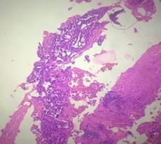
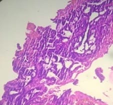
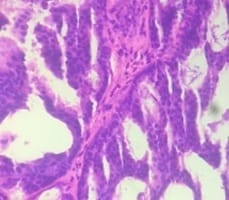
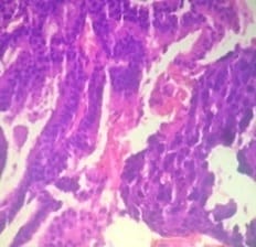
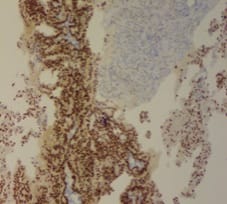
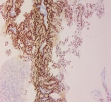
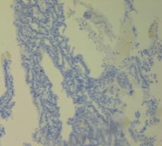
Breast carcinoma in men is rare and intra-cystic papillary carcinoma of male breast is an extremely rare entity. Papillary lesions of male breast comprise of the spectrum of lesions ranging from benign intraductal papilloma to intraductal papillary carcinoma and invasive papillary carcinoma. It occurs most commonly in the seventh decade of life and accounts for <1% of all breast malignancies. The present case report is of a comparatively younger, 37 years old male who presented with a mass on lower quadrant (retro-areolar region) of the left breast.

Keywords: Male breast, Papillary lesions, Intra-cystic papillary carcinoma, Invasive papillary carcinoma

Corresponding Author How to Cite this Article To Browse
Viplav Prashant, Assistant Professor, Department of Biochemistry, Government Dental College, Raipur, Chhattisgarh, India.
Email:
Prashant D, Prashant V. Papillary carcinoma of the male breast in a comparatively young patient: A case report of a rare entity. Int J Med Res Rev. 2020;8(5):369-374.
Available From
https://ijmrr.medresearch.in/index.php/ijmrr/article/view/1214

Manuscript Received Review Round 1 Review Round 2 Review Round 3 Accepted
2020-09-03 2020-10-08 2020-10-15 2020-10-30
Conflict of Interest Funding Ethical Approval Plagiarism X-checker Note
No Nil Yes 6%

© 2020 by Dolly Prashant, Viplav Prashant and Published by Siddharth Health Research and Social Welfare Society. This is an Open Access article licensed under a Creative Commons Attribution 4.0 International License https://creativecommons.org/licenses/by/4.0/ unported [CC BY 4.0].

Introduction

Breast masses include benign as well as malignant lesions and occur less frequently in male as compared to female. Malignant neoplasms account for 0.6 % of all breast carcinomas and less than 1% of all malignant lesions in men [1-2]. Papillary lesions are far more uncommon in men than women. The exact aetiology of male breast cancer is unknown. However, gynecomastia, imbalance in oestrogen-testosterone ratio, radiation exposure, obesity, BRCA2 mutations have all been implicated as risk factors [3-6]. It has been observed that intra-cystic papillary carcinoma (IPC) in particular may present more often in men with gynecomastia [7,8]. IPC is rare in male breast cancer, reported comprising 2.6-5% of male breast cancers [9,10]. Here, the present study reports a case of intra-cystic papillary carcinoma of the breast in a 37 years old male patient.

Case Report

A 37-year-old male presented with a mass (lump) over the retro-areolar region of left breast which according to the patient was of small size initially but rapidly increased in size within 15-20 days. On examination, the mass was of variegated consistency i.e., soft to cystic at one place and hard in consistency at another. Also, it was non-tender and free from underlying structures with no nipple retraction. However, the bloody discharge was present (Figure 1).

ijmrr_1214_01.jpg

Fig-1: Clinical appearance of the lesion.

Cytology of the lesion revealed that the lesion was suspicious of malignancy (grade c4). Mammography of the lesion revealed a large solid cystic mass lesion with predominant solid component measuring approximately 7.1 x 6.2 cm in size. This mass showed significant internal vascularity, with suspicion of involvement of underlying musculature. No lymph node involvement was seen. The lesion was highly suspicious of neoplastic aetiology. Histopathological examination and CECT were advised for confirmation of the same.

A core biopsy of the lesion was done which revealed tumour cells arranged in multi-layered papillae with foci of the cribriform and solid pattern. Mitotic figures were also noted (Figure 2a-2e).

ijmrr_1214_02.jpg

Fig-2a: H and E (4x).

ijmrr_1214_03.jpg

Fig-2b: H and E (10x).


ijmrr_1214_04.jpg

Fig-2c: H and E (40x).

ijmrr_1214_05.jpg

Fig-2d: H and E (40x).

ijmrr_1214_06.jpg

Fig-2e: H and E (40x).

Figure 2a-2e: Tumour cells arranged in multi-layered papillae with foci of the cribriform and solid pattern. Mitotic figures are also noted.

IHC: The immunohistochemistry of the same tissue block was done for confirmation and further evaluation of the tumour.

It showed the strong positive intensity of staining for ER (EP-1) receptors as well as PR (EP-2) receptors with nearly 80% of cells nuclear staining in the invasive component of the tumour.

However, Her-2/neu (EP-3) was negative whereas P-63 showed non-specific staining (Figure 3-6).

ijmrr_1214_07.jpg

Fig-3: Immunohistochemistry: ER-positive.

ijmrr_1214_08.jpg

Fig-4: Immunohistochemistry: PR positive.


ijmrr_1214_09.jpg

Fig-5: Immunohistochemistry: Her-2/neu negative.

ijmrr_1214_10.jpg

Fig-6: Immunohistochemistry: P-63 non-specific.

Based on these findings, a final diagnosis of intra-cystic papillary carcinoma was made. The patient had received one cycle of chemotherapy but did not turn up for further follow-up.

Discussion

Invasive Papillary Carcinoma of the breast is a rare tumour and constitutes fewer than 1% in most symptomatic series. Intra-cystic Papillary Carcinoma (IPC) is a variant of papillary carcinoma and accounts for 0.5-1% of breast cancers. The prevalence of male breast cancer increases with age [2]. Male patients are usually diagnosed in their 70s and 80s [6]. Approximately 15-20% of male patients with breast cancer have a positive family history [1].

The most common clinical finding in 75-90% of the patients is a painless mass, which is centrally located in 70-90% of the cases. IPCs can be asymptomatic or present with a palpable breast lump or bloody nipple discharge. Preoperative diagnosis of IPC can be difficult and definitive diagnosis is usually made after excisional biopsy [10].

IPC is usually located in the retro-areolar or the central area. It arises in the larger, more centrally placed ducts, and the tumour development and its secretion cause the cystic dilatation [4]. Ultrasonography of the IPC lesion typically reveals a hypo-echoic area (which represents the cyst) with soft tissue echoes projecting from the wall of the cyst (intra-cystic tumour).

The aetiology of male breast cancer remains poorly understood, but an imbalance in the oestrogen-testosterone ratio has been implicated [5]. The other etiological factors include obesity, cirrhosis, radiation exposure, drugs, head trauma (by increasing the prolactin production), local chest trauma, and smoking [1-3].

Increased risk has been seen in patients with an undescended testis, congenital inguinal hernia, orchidectomy, orchitis, testicular injury and infertility. BRCA1 and BRCA2 can cause breast cancer in females, but only a BRCA2 mutation can confer a significant risk in men [1]. Tsuda et al reported that a loss of heterozygosity on chromosome l6q was a useful marker for IPC since intraductal papilloma showed no loss of heterozygosity [11].

The papillary lesions in the breast include benign papilloma, the papillary variants of Ductal Carcinoma in Situ (DCIS), and invasive papillary carcinoma and comprises of a wide spectrum in terms of their clinical presentation, behaviour, morphologic features and malignant potential. IPC can be present as a pure form or associated with ductal carcinoma in situ or ductal carcinoma invasive around the tumour [3,9].

Recent studies have suggested that IPC should be considered as a low-risk invasive carcinoma [12]. Kraus and Neubecker established criteria to distinguish the benign papilloma from the papillary carcinomas [7]. Subsequently, the papillary carcinomas can be divided into invasive and non-invasive types. The further subdivision of papillary carcinomas into 2 types was made by Carter et al [8].


IPCs do not appear to have a basal myoepithelial cell (MEC) layer, and MEC markers, such as p63, actin, and calponin can be useful in differentiating between IPCs from papilloma [15,16]. Hill et al. suggested a spectrum of progression from an in-situ disease to an invasive disease by using myoepithelial cell staining which is considered as “gold standard” method, as it has relatively high sensitivity and denotes the invasiveness of the tumour cells in malignant papillary breast lesions [7,9]. Lobular carcinoma is much less common in men than in women and it represents only 1% of all the cases [4]. The rarer subtypes such as the medullary, tubular, mucinous and the squamous carcinomas have all been reported in men. The tumours with high nuclear grades, histologic grades and large surface areas are more likely to metastasize to the lymph nodes or to recur locally [6,7]. The frequency of lymph node involvement, local recurrence and distant recurrence is 0% to 11%, 3% to 70% and 0% to 4% respectively [11-13].

The carcinomas of the male breast have higher rates of oestrogen and progesterone receptor positivity than are found in women, but similar percentages may be expressed for Her-2 and p53 in men and women [1]. Our case was Her-2 negative. The standard treatment of IPC should be based on the associated pathology [10]. There are no definite guidelines for the treatment of IPC, however, a radical surgical excision with a clear resection of margin is the important mainstay of the treatment.

Mastectomy is usually unessential unless it is required because IPC has an excellent prognosis, a low local recurrence, and a rare distant metastasis [9]. Long-term follow-up is necessary after the surgical treatment. Clinical assessment, however, could not be done in our case as the patient absconded after the first cycle of chemotherapy. There has been no clear indication for adjuvant endocrine therapy, even among the patients with oestrogen receptor-positive tumours [17]. Lefkowitz et al reported a 10-year disease-free survival rate of 91% [14].

Conclusion

To conclude, invasive papillary breast carcinoma of males is an uncommon disease with a peak incidence in the seventh decade of life, however, it is extremely rare in comparatively younger patients of the third to the fourth decade.

A triple assessment, i.e., clinical examination, radiological investigation and histological assessment with a high level of clinical suspicion is necessary to diagnose IPC in men due to its rarity. Risk factors for male breast cancer include genetic and hormonal abnormalities. Furthermore, genetic testing and risk-reducing mastectomy should also be considered in cases with a strong family history for male breast cancer. The use of adjuvant therapy with hormonal therapy and radiotherapy remains unclear. Because of its good prognostic value, it is essential to diagnose the disease meticulously at an early stage.

Reference

  1. Siegel R, Ward E, Brawley O, Jemal A. Cancer statistics, 2011- the impact of eliminating socioeconomic and racial disparities on premature cancer deaths. CA Cancer J Clin. 2011;61(4)212-236.
  2. White J, Kearins O, Dodwell D, Horgan K, Hanby AM, Speirs V. Male breast carcinoma- increased awareness needed. Breast Cancer Res. 2011;13(5)219.
  3. Sasco AJ, Lowenfels AB, Pasker-de Jong P. Review article- epidemiology of male breast cancer, A meta-analysis of published case-control studies and discussion of selected aetiological factors. Int J Cancer. 1993;53(4)538-549.
  4. Shah P, Robbani I, Shah O. Clinicopathological study of male breast carcinoma- 24 years of experience. Ann Saudi Med. 2009;29(4)288-293.
  5. Kwiatkowska E, Teresiak M, Filas V, Karczewska A, Breborowicz D, Mackiewicz A. BRCA2 mutations and androgen receptor expression as independent predictors of outcome of male breast cancer patients. Clin Cancer Res. 2003;9(12)4452-4459.

  1. Ruddy KJ, Winer EP. Male breast cancer- risk factors, biology, diagnosis, treatment, and survivorship. Ann Oncol. 2013;24(6)1434-1443.
  2. Kraus FT, Neubecker RD. The differential diagnosis of papillary tumors of the breast. Cancer. 1962;15;444-455.
  3. Fisher ER, Palekar AS, Redmond C, Barton B, Fisher B. Pathologic findings from the National Surgical Adjuvant Breast Project (protocol no-4) VI, Invasive papillary cancer. Am J Clin Pathol. 1980;73(3)313-322.
  4. Romics L, O’Brien ME, Relihan N, O’Connell F, Redmond HP. Intracystic papillary carcinoma in a male as a rare presentation of breast cancer- a case report and literature review. J Med Case Rep. 2009;3(13)1-4.
  5. Solorzano CC, Middleton LP, Hunt KK, Mirza N, Meric F, Kuerer HM, et al. Treatment and outcome of patients with intracystic papillary carcinoma of the breast. Am J Surg. 2002; 184(4)364-8.
  6. Tsuda H, Uei Y, Fukutomi T, Hirohashi S. Different incidence of loss of heterozygosity on chromosome 16q between intraductal papilloma and intracystic papillary carcinoma of the breast. Jpn J Cancer Res. 1994;85(10)992-996.
  7. Lee SK, Choi JH, Lim HI, Kim WW, Kim S, Cho EY, et al. Experience with Intracystic Papillary Carcinoma of the Breast at a Single Institute in Korea. J Breast Cancer. 2009;12(4)338-343.
  8. Pant I, Joshi SC. Invasive papillary carcinoma of the male breast- Report of a rare case and review of the literature. J Can Res Ther. 2009;5(3)216-218.
  1. Lefkowitz M, Usar CM, Lefkowitz W, Wargotz ES. Intraductal (intracystic) papillary carcinoma of the breast and its variants- A clinicopathological study of 77 cases. Hum Pathol. 1994;25(8)802-609.
  2. Hill CB, Yeh IT. Myoepithelial cell staining patterns of papillary breast lesions: from intraductal papillomas to invasive papillary carcinomas. Am J Clin Pathol. 2005;123(1)36-44.
  3. Collins LC, Carlo VP, Hwang H, Barry TS, Gown AM, Schnitt SJ. Intracystic papillary carcinomas of the breast- a reevaluation using a panel of myoepithelial cell markers. Am J Surg Pathol. 2006;30(8)1002-1007.
  4. Dixon JM, Page DL, Anderson TJ, Lee D, Elton RA, Stewart HJ. Long term survivors of breast cancer. Br J Surg. 1985;72(6)445-448.