E-ISSN:2320-8686
P-ISSN:2321-127X

Case Report

Supraventricular Tachycardia

International Journal of Medical Research and Review

2023 Volume 11 Number 1 January-February
Publisherwww.medresearch.in

A Case of Refractory Supraventricular Tachycardia in a Newborn

P.Wali P.1*, Khan A.2, Parakh H.3
DOI: https://doi.org/10.17511/ijmrr.2023.i01.04

1* Pradnya. P.Wali, MBBS, Dch,DNB Pediatrics, Fellowship in Neonatology, Shalini Hospital, Hyderabad, Telangana, India.

2 Aswad Khan, DNB Paediatrics, Fellowship in Neonatology, Consultant Neonatologist, Shalini Hospital, Hyderabad, Telangana, India.

3 Hemant Parakh, MD Paediatrics, DM Neonatology, Senior Consultant Neonatologist, Shalini Hospital, Hyderabad, Telangana, India.

Supraventricular Tachycardia (SVT) is the most common neonatal dysrhythmia with the incidence being 1 in 100 for children of all ages and 1 in 250 for neonates. The origin of supraventricular tachycardia is either above the bifurcation of the bundle of His or it has mechanisms dependent on the bundle of His. Newborns may present with irritability, poor feeding and tachypnea. The most important clinical sign of SVT is tachycardia sometimes associated with hypotension, heart failure, pallor, or decreased level of consciousness. Diagnosis is done with heart rate continuously remaining ≥ 220 beats per minute with a QRS < 0.08 seconds. Adenosine is the first-line abortive therapy of choice. Intractable SVTs are treated with amiodarone, esmolol, and procainamide. SVT with Circulatory collapse needs a synchronized DC cardioversion. Prognosis of SVT is generally excellent in the absence of structural heart disease.

Keywords: Neonatal tachyarrhythmia, Supraventricular tachycardia, Adenosine, Amiodarone, synchronized DC cardioversion

Corresponding Author How to Cite this Article To Browse
Pradnya. P.Wali, Consultant Neonatologis, Department of Neonatology, Shalini Hospital, Hyderabad, Telangana, India.
Email:
Pradnya. P.Wali, Aswad Khan, Hemant Parakh, A Case of Refractory Supraventricular Tachycardia in a Newborn. Int J Med Res Rev. 2023;11(1):22-27.
Available From
https://ijmrr.medresearch.in/index.php/ijmrr/article/view/1193

Manuscript Received Review Round 1 Review Round 2 Review Round 3 Accepted
2023-02-04 2023-02-07 2023-02-14 2023-02-21 2023-02-28
Conflict of Interest Funding Ethical Approval Plagiarism X-checker Note
Nil Nil Yes 17%

© 2023by Pradnya. P.Wali, Aswad Khan, Hemant Parakhand Published by Siddharth Health Research and Social Welfare Society. This is an Open Access article licensed under a Creative Commons Attribution 4.0 International License https://creativecommons.org/licenses/by/4.0/ unported [CC BY 4.0].

Introduction

Supraventricular tachycardia (SVT) is the most common arrhythmia in children with an estimated incidence of 1/250 among newborns and 1/10 among infants with congenital heart disease (CHD) [1-6]. Most of the SVTs in the neonate are re-entry atrioventricular SVTs utilizing an accessory pathway. The most important clinical sign of SVT is tachycardia sometimes associated with hypotension, heart failure, and signs of shock, pallor, or decreased level of consciousness. Adenosine is the first-line abortive therapy of choice for the majority of infants. Refractory SVT is defined as SVT which did not convert to sinus rhythm despite the administration of 2 doses of adenosine at or above the AHA-recommended doses and which often requires amiodarone, esmolol, and procainamide for its treatment. Hereby we present a newborn with recurrent intractable SVT requiring multiple doses of adenosine and high doses of amiodarone infusion for its reversal.

Case Report

A 19-day-old Term, AGA, Female baby weighing 3.5 kgs born out of spontaneous vaginal delivery and cried at birth was brought with complaints of hurried breathing and fast heart beating since 6 days. The baby was treated with oral digoxin at the local hospital and was referred to a higher centre for further management. At admission, the baby was alert, and active with mild respiratory distress.HR – 228/min, RR-64/ min, SPO2- 96% at room air, CFT < 3 seconds, Peripheral pulses well felt. BP- 63/40(48) mm Hg. Respiratory system – Mild tachypnea and mild subcostal retractions were present. B/L NVBS were present. CVS- Tachycardia present. HR-228-230/Min, regular. No murmur. P/A – soft.CNS- cry and activity good. Sensorium- normal. Investigations revealed: Hb- 16.6 gms/dl, TLC- 8100 /cu mm, platelet count 4.23 lakhs/cu mm. CRP- negative. Ionic calcium, sr. electrolytes, RFT and LFT were normal. ECG done showed narrow complex tachycardia s/o SVT. The baby was treated with oxygen, IVF and other supportive measures. SVT was treated with the rapid push of IV Adenosine with an initial dose of 0.1 mg/kg and was gradually increased up to 0.3 mg/kg as there was no reversal. The heart rate reversed back to a normal rhythm after 3 doses of adenosine. 2D Echo done was s/o mild dilated atria, normal

ventricles, No PAH and good biventricular function. Cardiologist Opinion was taken and the baby was started on oral propranolol.

Table 1: Chart Showing Events Occurring in The Baby and the Interventions from the Time Of Admission: 

TIME HR INTERVENTION EFFECT INVESTIGATION
At admission SVT Loading dose of Adenosine (0.1mg/kg). No reversal 2DECHO- Mild dilatation of atria, no PAH, good biventricular function.
3 doses of Adenosine(0.1-0.3mg/kg). Reversal of HR to a normal rhythm.  
Propranolol(1mg/kg/dose every 6th hour) started after the cardiologist's opinion.    
2 hrs of admission Tachycardia (rise in HR 190-200/min) Loading dose of Amiodarone followed by continuous infusion(5mcg/kg/min). HR 170-180/min for 12 hrs.  
12 hrs of admission HR started rising to 200/min Amiodarone infusion increased to 10mcg/kg/min. Hr decreased to 180-190/min.  
24 hrs of admission Recurrence of svt Adenosine 3 doses starting with 0.1mg/kg – 0.3mg/kg. Propranolol dose increased (1.5mg/kg/dose every 6th hour). Amiodarone infusion increased to 15mcg/kg/min. Reversal of SVT after 30 min. 2DECHO- small PFO(left to right shunt), mild to mod PAH, dilated atria, good biventricular function.
34 hrs of admission Recurrence of SVT 3 doses of Adenosine 0.3mg/kg. Propranolol increased(2mg/kg/dose every 6th hour). Amiodarone infusion increased to 20mcg/kg/min. Reversal of SVT. HR 120-160/min.  

ijmrr_1193_01.JPG
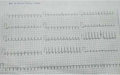
Figure 1: ECG showing Narrow Complex Tachycardia suggestive of SV


ijmrr_1193_02.JPG
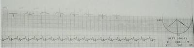
Figure 2: Resolution of SVT after Adenosine therapy.

Baby had one episode of seizure at admission which was treated with a loading dose of Levipil and the maintenance dose was continued. No further seizures were noted. The condition of the baby and the need for cardioversion in case of worsening of cardiac function given recurrent SVTs were explained to the attendees in detail.

The baby continued to maintain HR in sinus rhythm and HR reversed to the normal range(124-160/min) after 36 hours of admission after being treated with three doses of Adenosine and optimal doses of Amiodarone infusion and propranolol. Repeat septic screen on day 3 of admission was positive with CRP- 19.9 mg/dl. The blood culture was repeated and antibiotics were hiked up. Repeat Sr electrolytes, LFT, and Ionic calcium were within normal range. TSH done was 2.23 Uiu/ml. 48 hours blood c/s was sterile. Antibiotics were given for a total of 7 days. Baby gradually improved with treatment and maintained HR in sinus rhythm. The Amiodarone drip was slowly tapered and stopped. Oral amiodarone and propranolol was continued.Feeds were gradually upgraded to full feeds and DBF was started. The mother involved in feeding and rooming in was done. The baby continued to maintain HR in the normal range, and activity and feeding were good, hence baby was discharged in stable condition.

Discussion

Supraventricular Tachycardia (SVT) is the most common neonatal dysrhythmia with the incidence being 1 in 100 for children of all ages and 1 in 250 for neonates [1]. Origin of Supraventricular tachycardia is either from above the bifurcation of the bundle of His or it has mechanisms dependent on the bundle of His.

  • SVTs can be classified as either atrial or atrioventricular and may be further sub-classified into automatic or re-entry by its mechanism.
  • Most of the SVTs in the neonate are re-entry atrioventricular SVTs which utilize an accessory pathway (2).
  • Sustained/paroxysmal SVT may develop in utero and may lead to fetal hydrops.

Epidemiology: The true incidence of SVT in children is not known but is generally estimated as 1 in 250 in newborns. Approximately 50% of children will have the first episode of SVT in their first year of life [3,4]. The incidence peaks in early childhood (ages 6-9 years) and then again in adolescence [4,5]. Spontaneous resolution of SVT occurs in more than 90% of infants by 1 year of age. Most of the babies with SVT have a structurally normal heart but 9% to 32% of children with SVT have structural congenital heart disease. The most common association is with WPW syndrome and the Ebstein anomaly, and then less commonly with ventricular or atrial septal defects [4-6].

Genetics: Most cases of reentrant SVT are sporadic, with approximately 7% of patients having documented SVT in a first-degree relative [7].

Evaluation:

History: The clinical presentation of SVT is age and duration dependent. In infants, the heart rate is generally 220 to 320 beats/minute and in older children, it ranges from 160 to 280 beats/min [3]. Symptoms are usually nonspecific like poor feeding, irritability, vomiting, cyanosis, and pallid spells. If the symptoms go unnoticed for hours to days, the infant can present with symptoms of heart failure [8]. Congestive heart failure was noted in 19% of infants who had SVT for 24 to 36 hours and in 50% who had SVT for more than 48 hours [3].

Physical Examination: Most of the babies with SVT have a structurally normal heart and normal findings on the physical examination. If the tachycardia goes unrecognized for longer periods, infants may present with congestive cardiac failure.

Diagnosis: Recording a heart rhythm strip (24-hour ambulatory monitoring, event recorders, and ECGs) is key for correct diagnosis and treatment in symptomatic infants. Event recorders are used in children with symptoms more than once per month. ECG typically demonstrates Narrow complex tachycardia with no visible P waves and pseudo-R waves in V1-2. Most children with SVT do not have daily symptoms hence Holter monitoring is practically not possible.

Treatment: The treatment of SVT depends on the age of the patient, the duration and frequency


of the SVT and the presence of ventricular dysfunction. Infants with mild symptoms generally do not require treatment. Children with frequent or difficult-to-terminate SVTs require medical therapy or transcatheter ablation.

Medical Therapy: Medications used to treat SVT are of 3 types: 1) Abortive therapy 2) Acute therapies used to achieve rate control 3) Secondary prevention or “prophylactic” therapies used to prevent SVT recurrence [9,10].

1. Abortive Therapy: Adenosine is the abortive therapy of choice for the majority of infants for all types of SVT except Atrial flutter when cardioversion is favoured [11-13].

Intravenous Adenosine: Full resuscitation facilities must be available before starting Adenosine Therapy.

  • Monitor ECG continuously throughout the administration of adenosine.
  • Adenosine can be started at a dose of 0.1 mg/kg and can be increased up to 0.3 mg/kg.
  • Wait two minutes between doses and check the patient’s vital signs.
  • Adenosine should be given as a rapid bolus and in the intravenous line as proximal as possible. Adenosine should be quickly flushed with normal saline using a three-way stop cock.
  • After conversion to sinus rhythm ECG strip should be immediately observed for concealed pre-excitation, which may be revealed during the first few beats.
  • Sometimes rapid re-initiation of tachycardia may occur due to premature atrial contractions stimulated by adenosine which can be treated with repeat doses of adenosine.

Contraindications to Adenosine: Adenosine is contraindicated in a rare form of immune deficiency called Adenosine-deaminase deficiency.
2. Acute management therapies: Amiodarone, Esmolol, and Procainamide are often used to treat intractable SVT which cannot be aborted with adenosine or cardioversion or which recurs rapidly. These drugs are generally used to achieve rate control [14,15].
3. Secondary prevention or “prophylactic” therapies: are used to prevent SVT recurrence. Digoxin and beta-blockers are generally

considered first-line treatments for the secondary prevention of SVT [9,16,17].
Direct current cardioversion:

  • Defibrillation should be considered for hemodynamically unstable patients after intubation and initiation of ventilation.
  • For tachycardia with a regular well-defined QRS complex, the discharges synchronized with the QRS complex should be given to avoid ventricular fibrillation.
  • For ventricular fibrillation or polymorphic VT, an unsynchronized shock is necessary to avoid the device delaying the shock while trying to track the QRS complex.
  • SVTs usually require 1 joule per kg and ventricular arrhythmia 2-4 joules per kg.

In our baby, SVT was intractable and refractory. RefractorySVT is the one which does not convert to sinus rhythm despite the administration of 2 doses of adenosine at or above the AHA-recommended doses. The reason for a low response rate to adenosine in infants is generally due to: [1] inadequate delivery of adenosine to infants due to the slow rate of infusion by using a small intravenous catheter or intravenous access located far from the conduction system [2] relative resistance of infants’ atrioventricular node to adenosine; or [3] infant presenting later in the course of the illness, which might be associated with a low response to treatment secondary to lower cardiac output. These babies often require treatment with amiodarone, esmolol, and procainamide to treat intractable SVT. Our baby required a higher dose of amiodarone and propranolol for the reversal of SVT to a normal rhythm. According to a study Acute Management of Refractory and Unstable Pediatric Supraventricular Tachycardia, by Jonathan Lewis, Gaurav Arora, Dana L. Tudorascu, Robert W. Hickey, Richard A. Saladino, and Mioara D. Manole, it was seen that Refractory SVT occurred in 15% of all acute SVT episodes indicating success rates of adenosine being 60%-80% [19,20,21]. All cases with refractory SVT received adenosine at or above the AHA recommended doses[19].85%episodes of refractory SVT were treated with additional (≥3) doses of adenosine. Around 45% of babies with persistent SVT after 3 doses of adenosine were treated with one or more antiarrhythmic agents[19].


Prognosis: In the absence of structural heart disease or cardiomyopathy, the prognosis of SVT is generally excellent[23]. 3-4 % of WPW syndrome patients have the risk of sudden cardiac death. In stable patients with refractory SVT, medical management with multiple adenosine doses or additional antiarrhythmics is generally successful.

Conclusion

Supraventricular tachycardia is the commonest tachyarrhythmia in newborns with a good prognosis if detected early. Hence early diagnosis and prompt treatment are essential for better outcomes and to prevent complications in newborns. Refractory SVTs account for around 15% of all cases and those too if treated promptly give better results.

Reference

1.Garson AJ, Gillette PC, McNamara DG. Supraventricular tachycardia in children: clinical features, response to treatment, and long-term follow-up in 217 patients. J Pediatr. 1981; 98(6) : 875 -82.

2.Fox DJ, Tischenko A, Krahn AD, Skanes AC, Gula LJ, Yee RK, et al. Supraventricular tachycardia: diagnosis and management. Mayo Clin Proc.2008; 83(12) : 1400 -11.

3.Nadas ASDaeschner CWRoth ABlumenthal SL Paroxysmal tachycardia in infants and children: study of 41 cases. Pediatrics 1952;9 (2) 167- 181.

4.Perry JCGarson A Jr Supraventricular tachycardia due to Wolff-Parkinson-White syndrome in children: early disappearance and late recurrence. J Am Coll Cardiol 1990;16 (5) 1215- 1220.

5.Deal BJKeane JFGillette PCGarson A Jr Wolff-Parkinson-White syndrome and supraventricular tachycardia during infancy: management and follow-up. J Am Coll Cardiol 1985;5 (1) 130- 135.

6.Giardina ACEhlers KHEngle MA Wolff-Parkinson-White syndrome in infants and children: a long-term follow-up study. Br Heart J 1972;34 (8) 839- 846.

7.Garson ABricker JTMcNamara DG The Science and Practice of Pediatric Cardiology. Philadelphia, PA Lea & Febiger1990.

8.Garson A JrGillette PCMcNamara DG Supraventricular tachycardia in children: clinical

features, response to treatment, and long-term follow-up in 217 patients. J Pediatr 1981; 98 (6) 875- 882.

9.Wong KK, Potts JE, Etheridge SP, Sanatani S. Medications used to manage Supraventricular tachycardia in the infant a North American survey. Pediatr Cardiol. 2006;27:199–203.

10.Tavera MC, Bassareo PP, Neroni P, Follese C, Manca D, Montis S, et al. Supraventricular tachycardia in neonates: antiarrhythmic drug choice dilemma. J Matern Fetal Neonatal Med. 2010;23(Suppl 3):30–3.

11.Brugada J, Blom N, Sarquella-Brugada G, Blomstrom-Lundqvist C, Deanfield J, Janousek J, et al. Pharmacological and non-pharmacological therapy for arrhythmias in the pediatric population: EHRA and AEPC-Arrhythmia Working Group joint consensus statement. Europace. 2013;15:1337–82.

12.Texter KM, Kertesz NJ, Friedman RA, Fenrich AL., Jr Atrial flutter in infants. J Am Coll Cardiol. 2006;48:1040–6.

13.Van Gelder IC, Tuinenburg AE, Schoonderwoerd BS, Tieleman RG, Crijns HJ. Pharmacologic versus direct-current electrical cardioversion of atrial flutter and fibrillation. Am J Cardiol. 1999;84:147R–51R.

14.Chang PM, Silka MJ, Moromisato DY, Bar-Cohen Y. Amiodarone versus procainamide for the acute treatment of recurrent supraventricular tachycardia in pediatric patients. Circ Arrhythm Electrophysiol. 2010;3:134–40.

15.Manole MD, Saladino R. Emergency department management of the pediatric patient with supraventricular tachycardia. Pediatr Emerg Care. 2007;23:176–85.

16.Weindling SN, Saul JP, Walsh EP. Efficacy and risks of medical therapy for Supraventricular tachycardia in neonates and infants. Am Heart J. 1996;131:66–72.

17.Seslar SP, Garrison MM, Larison C, Salerno JC. A multi-institutional analysis of inpatient treatment for supraventricular tachycardia in newborns and infants. Pediatr Cardiol. 2013;34:408–14.

18.Jonathan Lewis, Gaurav Arora, Dana L. Tudorascu, Robert W. Hickey, Richard A. Saladino, and Mioara D. Manole.Acute Management of Refractory and Unstable Pediatric Supraventricular Tachycardia.J Pediatr 2017;181:177-82).


19.Clausen H, Theophilos T, Jackno K, Babl FE. Paediatric arrhythmias in the emergency department. Emerg Med J 2012;29:732-7.

20.Till J, Shinebourne EA, Rigby ML, Clarke B,Ward DE, Rowland E. Efficacy and safety of adenosine in the treatment of Supraventricular tachycardia in infants and children. Br Heart J 1989; 62:204-11.

21.Ralston MA, Knilans TK, Hannon DW, Daniels SR. Use of adenosine for diagnosis and treatment of tachyarrhythmia in pediatric patients. J Pediatr 1994; 124:139-43.

22.JACK C. Salerno, MD; Stephen P. Seslar, MD, PhDSupraventricular Tachycardia.Arch Pediatr Adolesc Med. 2009; 163(3):268-274. doi: 10.1001/archpediatrics.2008.547.

23.Munger TM Packer DL Hammill SC et al. A population study of the natural history of Wolff-Parkinson-White syndrome in Olmsted County, Minnesota, 1953-1989. Circulation 1993;87 (3) 866- 873.