E-ISSN:2320-8686
P-ISSN:2321-127X

Case Report

Topography

International Journal of Medical Research and Review

2020 Volume 8 Number 3 May-June
Publisherwww.medresearch.in

Isthmus agenesis with variant topography of levator glandulae thyroideae: A case report

Potaliya P.1*, Choudhary A.2, Ghatak S.3
DOI: https://doi.org/10.17511/ijmrr.2020.i03.10

1* Pushpa Potaliya, Assistant Professor, Department of Anatomy, All India Institute of Medical Sciences, Jodhpur, Rajasthan, India.

2 Anju Choudhary, Senior Resident, Department of Anatomy, All India Institute of Medical Sciences, Jodhpur, Rajasthan, India.

3 Surajit Ghatak, Professor and Head, Department of Anatomy, All India Institute of Medical Sciences, Jodhpur, Rajasthan, India.

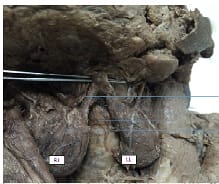
The thyroid gland is an imperative endocrine gland present in the neck known commonly for its morphological variations. During routine dissection agenesis of the isthmus of the thyroid gland was observed with levator glandulae thyroidea extending from both the lateral lobes with stout stalks of pyramidal lobes bilaterally. The Levator glandulae thyroidea is a fibro-musculo-glandular band. usually, present unilaterally or extending from isthmus connecting the pyramidal lobe of the thyroid gland to the hyoid bone. The site of the isthmus is often associated with anastomosis of vessels supplying thyroid gland but strikingly no such anastomosis was observed. The presence of such variant of levator glandulae thyroidea with agenesis of the isthmus is a rare finding and knowledge of such topographical anatomical variations is crucial for understanding associated pathologies related to the thyroid gland and modus operandi in their management.

Keywords: Anatomic variation, Thyroid gland, Anatomy, Isthmus, Agenesis

Corresponding Author How to Cite this Article To Browse
Pushpa Potaliya, Assistant Professor, Department of Anatomy, All India Institute of Medical Sciences, Jodhpur, Rajasthan, India.
Email:
Potaliya P, Choudhary A, Ghatak S. Isthmus agenesis with variant topography of levator glandulae thyroideae: A case report. Int J Med Res Rev. 2020;8(3):273-276.
Available From
https://ijmrr.medresearch.in/index.php/ijmrr/article/view/1192

Manuscript Received Review Round 1 Review Round 2 Review Round 3 Accepted
2020-05-20 2020-05-28 2020-06-03 2020-06-07
Conflict of Interest Funding Ethical Approval Plagiarism X-checker Note
No Nil Yes 9%

© 2020 by Pushpa Potaliya, Anju Choudhary, Surajit Ghatak and Published by Siddharth Health Research and Social Welfare Society. This is an Open Access article licensed under a Creative Commons Attribution 4.0 International License https://creativecommons.org/licenses/by/4.0/ unported [CC BY 4.0].

Introduction

Anatomical variations of the structures in the neck are of clinically significance and success of various surgical procedures and circumventing potential possibilities of iatrogenic lesions in the region entirely depends on precise knowledge [1,2]. The thyroid gland consists of two lateral lobes joined by a midline isthmus.

Isthmus lies at the level of second to fourth tracheal rings and is enveloped by infrahyoid muscles. Occasionally an extrapyramidal lobe is also present attached to the isthmus [3]. The upward-pointing apex of this pyramidal lobe has a fibromuscular band extending from here to hyoid bone [4]. It develops from remains of the thyroglossal duct, which seldom gets muscle fibers and eventually form this band.

Embryologically, the thyroid gland is the foremost to start developing amongst endocrine glands. In addition, it is frequently observed to be associated with congenital anomalies of varying degrees. They range from common ones as variations in lobes pyramidal lobe and presence of thyroglossal duct cyst to sporadic ones as partial or complete agenesis of lateral lobes, isthmus, or even whole gland [5,6].

The congenital anomalies during the development of the gland alter the usual morphology. As a result, it ends up leading to several clinical conditions and various thyroid disorders [7]. Literature reported isthmus agenesis to vary from a range of 5-10% and even confined to 8-10% [8,5]. It is challenging and indefinite to find the incidence of isthmus agenesis for the reason that the analysis predominantly based on populations presented with some or other thyroid diseases [8].

Case Report

During routine dissection in the Department of Anatomy at All India Institute of Medical Sciences Jodhpur, a 52-year-old male cadaver exhibited agenesis of the isthmus of the thyroid gland leading to its absence. Intact skin of anterior cervical region with no visible scar ruled out any possibility of surgical procedure carried out in the past.

The thyroid gland was found with two separate lateral lobes, with complete agenesis of the isthmus. The pyramidal lobe on the left side was large and prominent whereas on the right side lobe had a

stalk of small triangular kind of pyramidal lobe with glandular tissue from the tip of which was extending fibromuscular band of levator glandulae thyroideae on both the sides.

Any aberrant thyroid tissue was not found in the vicinity of dissection in the region. The two lateral lobes were discrete with no midline flanked intervening tissue (Figure 1).

ijmrr_1192_01.jpg

Fig-1: Agenesis of thyroid isthmus (Lt py and Rt py: Left and right pyramidal lobes; RL: Right lateral lobe; LL: Left lateral lobe; Levator glandulae thyroidea)

The dimensions of lateral lobes were as:

  1. Right side- Length: 5.6 cm, breadth: 3.4 cm
  2. Left side- Length: 6.3 cm, breadth: 3.7 cm

The slips of levator glandulae thyroideae from both sides extended medially. A left slip was wider (2 cm) whereas right-sided slip was just 1.4 cm in width. For running around five cm both intermingled and attached on the body of the hyoid bone.

Arterial supply was by branches of superior and inferior thyroid arteries for both the lobes with no anastomosis between two of them. The anterior branch of the superior thyroid artery anastomosed with the ascending branch of the inferior thyroid artery around the pyramidal lobe. The posterior border on both sides showed anastomoses between the posterior and inferior branches of both superior and inferior thyroid arteries.

Discussion

The midline juncture isthmus of the thyroid gland comprises of normal thyroid tissues, with dimensions 1.25 x 1.25 cm in length and breadth


[9,10]. Agenesis of the isthmus is a rare developmental entity. It also showed gender disparity with the predominance of the condition in males as compared to females [10]. In the present study, the agenesis is reported in a male cadaver.

According to Gruber (cited by Testut), the incidence of agenesis is about 5% [6]. Allan and authors in there study found it be in the range of 2-4% but according to Marshall, it is higher i.e. up to 15% [5,11]. In an Indian study, the incidence was moreover even found a little higher than this i.e. 16.66% [12].

Levator glandulae thyroidea is found to be 30% in a study on Indian diaspora [12]. Incidence of it was slightly higher up to 43.33% in the Bangladesh population [13]. According to Ranade et al the incidence reported was even more i.e. 49.5% [14]. In classical literature levator glandulae, thyroidea is seen to extend from the pyramidal lobe or moreover mostly from the left lobe or left side of the isthmus to be attached to the hyoid bone cranially [15]. In the present case, there is an unusual presentation of the levator glandulae thyroidae. It extended as a slip from the tip of both the lobes and eventually join to form a single band and attached to the body of the hyoid bone. The occurrence of isthmus agenesis was reported ranging from 3 to 10% [14,16]. Lack of data with the precise number was not found and it might be because generally occurrence is in non-symptomatic population and reported data are based mostly on cadaveric studies. In clinical setting agenesis of the isthmus is often seen to be associated with conditions as nodular goiter, inflammation, or primary carcinoma [8]. It is observed that the etiology of agenesis is moreover indistinct. Innategenetic factors and defects in development inintra uterine life has a pivotal role. Genetic mutations accountable for normal development can be another reason for isthmus agenesis. Principally it involves TITF1-2 genes and chromosome 22 [17,18].

In the early gestation phase on the 24th day, the thyroid starts developing. The lateral thyroid develops from neural crest cells, while the isthmus arises from the primitive pharynx. The development of thyroglossal duct arises from the endodermal layer in the pharyngeal floor at the level of second and third pharyngeal pouches. It grows downwards and anterior to the trachea to split into two lateral lobes [5,8,14,16]. It was also found that defective or imperfect fusion of the thyroglossal duct in the midline basically is responsible for ending into the

formation of two independent lobes with the absence of middle isthmus. Absence of isthmus described in lower animals like amphibians, birds, and among certain mammals [8]. This case report emphasizes the importance of such variations to consider during surgical interventions, as trans-thyroid tracheotomy etc. to avoid iatrogenic lesions and individuals not to be missed for differential diagnosis.

Reference

  1. Bhargav PR. Salient anatomical landmarks of the thyroid and their practical significance in thyroid surgery- a pictorial review of thyroid surgical anatomy (revisited). Ind J Surg. 2014;76(3)207-211.
  2. Bribriesco A, Patterson GA. Cricothyroid approach for emergency access to the airway. Thorac Surg Clin. 2018;28(3)435-440.
  3. Veerahanumaiah S, Dakshayani KR, Menasinkai SB. Morphological variations of the thyroid gland. Int J Res Med Sci. 2015;3(1)53-57.
  4. Hegcazy AA. Clinical embryology for medical students and postgraduate doctors. LAP ‘Lambert Academic Publishing, Berlin. 2014.
  5. Marshall CF. Variation in the form of the thyroid in man. J Anat Physiol. 1895;29(2)234-239.
  6. Testut L, Latarjet A. Anatomia Humana, Tomo IV- Ed. Barcelona, Salvat. 1978;402–404.
  7. Larochelle D, Arcand P, Belzile M, Gagnon N-B. Ectopic thyroid tissue - a review of the literature. J Otolaryngol. 1979;8(6)523-530.
  8. Pastor Vázquez JF, Gil Verona JA, De Paz Fernández FJ, Barbosa Cachorro M. Agenesis of the thyroid isthmus. Eur J Anat. 2006;10(2)83-84.
  9. Kosiak W, Swieton D, Piskunowicz M, Kujawa M. Thyroid isthmus thickness in prepubertal healthy children in an Iodine-sufficient region. Polish J Radiol. 2010;75(3)17-19.

  1. Dixit D, Shilpa MB, Harsh MP, Ravishankar MV. Agenesis of isthmus of thyroid gland in adult human cadavers- a case series. Cases J. 2009; 2;6640.
  2. Allan FD. An accessory or superficial inferior thyroid artery in a full term infant. Anatomy Rec. 1952;112(3)539-542.
  3. Joshi SD, Joshi SS, Daimi SR, Athavale SA. The thyroid gland and its variations- a cadaveric study. Folia Morpologica. 2010; 69(1)47-50.
  4. Sultana SZ, Khalil M, Khan MK, Shamim R, Parveen S, Ara ZG. Morphological study of levator glandulae thyroidea in Bangladeshi cadaver. Mymensingh Med J. 2009;18(2)179-183.
  5. Ranade AV, Rai R, Pai MM, Nayak SR, Prakash, Krisnamurthy A, et al. Anatomical variations of the thyroid gland- possible surgical implications. Singapore Med J. 2008;49(10)831-834.
  1. Standring S. In- Gray's Anatomy, 39th Edition. London- Elsevier Chruchill Livingstone. 561;2006.
  2. Won HS, Chung IH. Morphologic variations of the thyroid gland in Korean adults. Korean J Phys Anthropol. 2002;15(2)119-125.
  3. Devi Sankar K, Sharmila Bhanu P, Bapuji P. Agenesis of isthmus of thyroid gland with embryological and clinical basis. Narayana Med J. 2012;1(1)32-34.
  4. Gangbo F, Lacombe D, Alberti EM, Taine L, Saura R, Carles DN. Trisomy 22 with thyroid isthmus agenesis and absent gall bladder. Genetic Counseling. 2004;15(3)311-315.