E-ISSN:2320-8686
P-ISSN:2321-127X

Research Article

Radiotherapy

International Journal of Medical Research and Review

2020 Volume 8 Number 2 March-April
Publisherwww.medresearch.in

The necessity of replanning during the intensity-modulated radiotherapy (IMRT) for head and neck cancer, to ensure adequate coverage of target volume

Naveen B.1*, S. Narayanan G.2, Narayanan S.3
DOI: https://doi.org/10.17511/ijmrr.2020.i02.10

1* Naveen B., Assistant Professor, Department of Radiation Oncology, VTSM Peripheral Cancer Centre, Kalaburagi, Karnataka, India.

2 Geeta S. Narayanan, Professor and HOD, Department of Radiation Oncology, Vydehi Institute of Medical Sciences and Research Centre, Bengaluru, Karnataka, India.

3 Sowmya Narayanan, HOD, Department of Radiation Physics, Vydehi Institute of Medical Sciences and Research Centre, Bengaluru, Karnataka, India.

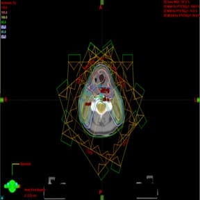
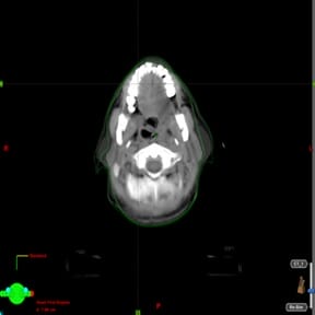
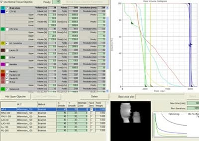
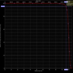
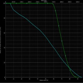
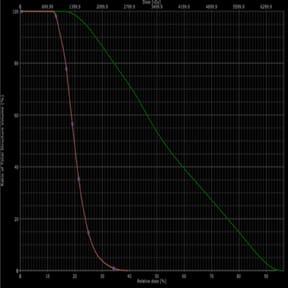
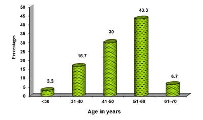
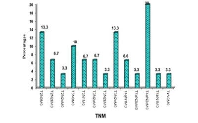
Introduction: In head and neck cancer the location, size, shape of disease, and normal anatomy change in 6-7 weeks radiotherapy. As a result, steep dose gradients move across target and critical structures resulting in underdose to target and overdose to critical structures. Aim: comparison of target coverage in initial IMRT plan and replan and to quantify dose changes to normal structures in two plans. Methods and Material: 30 patients with locally advanced head and neck cancer patients planned for curative radiotherapy were selected and treated with 3DCRT plan. For dosimetric comparison IMRT plan was created for pre-treatment and repeat CT, which was done after 40Gy. Statistical analysis used: Statistical methods (student’s paired t-test) were applied. Results: Both PTV coverage (V95 from 96.29±1.12 to 97.33±0.80) and dose (D95 from 66.64±0.87 to 67.57±0.74) increased in replanned CT. Both max and mean doses to the brainstem and spinal cord along with mean dose to parotid glands increased in replanned CT. Conclusions: Replanning is necessary during mid-treatment to accommodate anatomical and dosimetric changes during curative radiotherapy.

Keywords: Head and Neck cancer, IMRT, Replanning

Corresponding Author How to Cite this Article To Browse
Naveen B., Assistant Professor, Department of Radiation Oncology, VTSM Peripheral Cancer Centre, Kalaburagi, Karnataka, India.
Email:
Naveen B, Narayanan GS, Narayanan S. The necessity of replanning during the intensity-modulated radiotherapy (IMRT) for head and neck cancer, to ensure adequate coverage of target volume. Int J Med Res Rev. 2020;8(2):189-200.
Available From
https://ijmrr.medresearch.in/index.php/ijmrr/article/view/1178

Manuscript Received Review Round 1 Review Round 2 Review Round 3 Accepted
2020-04-10 2020-04-20 2020-04-25 2020-04-30
Conflict of Interest Funding Ethical Approval Plagiarism X-checker Note
No Nil Yes 5%

© 2020 by Naveen B., Geeta S. Narayanan, Sowmya Narayanan and Published by Siddharth Health Research and Social Welfare Society. This is an Open Access article licensed under a Creative Commons Attribution 4.0 International License https://creativecommons.org/licenses/by/4.0/ unported [CC BY 4.0].

Introduction

Radiotherapy (RT) plays an important role in the management of head and neck cancer. Intensity-modulated radiotherapy (IMRT) can conform the dose to target with complex shape and spare organs at risk (OAR), when compared to three-dimensional conformal radiotherapy (3DCRT). Location, shape, and size of disease and anatomy change over 6-7 weeks radiotherapy.

Much research has been reported on the sparing of the parotid gland with IMRT. This organ irresponsible for 60% to 65% of the saliva produced and xerostomia is a major acute and late side effect that can have a significant negative impact on a patient’s quality of life. Six months post-treatment, the stimulated salivary flow is reduced exponentially for each parotid gland at a rate of approximately 4% per Gy of mean parotid dose [1].

Eisbruch et al. [2] reported that the sparing of major salivary glands by IMRT increased late salivary flow rates, and improved xerostomia. They also noted that the sparing of minor salivary glands in the oral cavity was a significant independent predictor of xerostomia.

An analysis of dose, volume, and function relationships in the parotid glands after IMRT suggested that a mean parotid dose of 26 Gy was necessary for the substantial sparing of the gland.

Barker et al [3] conducted a pilot study to quantify the magnitude of the anatomical changes using an integrated CT linear accelerator system. They concluded that GTVs decreased throughout the course of RT, at a median rate of 1.8% per treatment day. On the last day of treatment, this corresponded to a median total relative loss of 69.5% of the initial GTV.

In addition, the center of mass of the GTV changed position with time, indicating that tumor loss was frequently asymmetric. At treatment completion, the median center of mass displacement was 3.3 mm. Parotid glands also decreased in volume by 0.6% per treatment day and shifted medially (median shift of 3.1 mm) with time. This medial displacement of the parotid glands correlated with the weight loss that occurred during treatment.

Hansen et al [4] performed a study on H and N cancer patients treated with IMRT. Planning CT scans were performed before treatment and after an average dose of 36 Gy. A mean reduction in the

parotid volume of 21.5% and 15.6% was observed for the left and right gland, respectively. Surprisingly, no changes were observed for the GTV.

Patients were treated with IMRT and had repeated CT imaging during the course of RT. For both therapeutic and prophylactic PTVs, the mean dose to 99% of the volume (D), the mean dose to 95% of volume (D), and the mean percentage of the volume receiving more than 93% of the prescribed dose, all decreased.

Moreover, the doses to OARs also increased, which is the percentage of the volume of the right parotid receiving more than 26 Gy (V) and the percentage of the volume of the mandible receiving more than 60 Gy.

In Robar’s study [5], 26Gy for each patient, the IMRT dose distribution was recalculated on each CT image set to determine the dosimetric consequences of anatomical modifications. For the left and right parotids, the mean doses increased by 2.6% ±4.3% and 0.2% ± 4.0%, respectively.

The volume that received 26 Gy increased by 3.5%± 5.2% for the left gland and 0.3%± 4.7% for the right gland.

Because of these changes, steep dose gradients with IMRT may move across target volume and critical structures resulting in underdose to the target volume and overdose to the critical structures. Hence, there is a necessity of replanning at mid-treatment.

Subjects and Methods

Source of Data: Patients presenting to the Department of radiation oncology with symptoms and signs of head and neck cancer with newly biopsy-proven squamous cell carcinoma. Stage III, IVA, and IVB and in whom curative radiotherapy with or without chemotherapy is planned are included for the study.

Duration of Study: Jan.2012 to Dec. 2012.

Sample Size: The Sample size has been estimated in consultation with a biostatistician. The sample size chosen is 30. This was estimated based on data obtained with the historical studies.

Method of Collection of Data (Including Sampling, Procedure, if any)

Thirty patients of head and neck cancer were included in this study.


    • A standardized data collection proforma was used for the study.
    • All the cases underwent a biopsy for confirmation of malignancy.
    • Clinical examination, Computer tomography (CT) or Magnetic Resonance Imaging (MRI) with contrast was carried out for staging.

    Inclusion criteria

    • Head and neck cancer patients considered suitable for curative treatment with radiotherapy with or without chemotherapy.
    • Positive biopsy showing squamous-cell carcinoma.
    • Staging according to AJCC (American joint cancer committee) stage III, IVA and IVB.

    Exclusion criteria

    • Metastatic disease.
    • Previous Radiation therapy to Head and neck cancer.

    All the patients included for the study underwent CT simulation and were treated with 3DCRT plan. IMRT plan was used for dosimetric comparison only. The IMRT plan was created on the pre-treatment planning study.

    Repeat CT simulation was done at the end of 40 Gy and fused with pre-treatment CT. The cone-beam CT images were not used because of the poor delineation of tumor volume. The coverage of PTV and doses to critical structures were compared for the repeat CT using the pretreatment IMRT plan.

    Steps

    Immobilization and Image Acquisition-Patients were immobilized on carbon fiber head and neck board with proper neck rest using the thermoplastic mask. For patients with a short neck, the shoulders were depressed by using shoulder retractors. Three fiducial markers were used as reference marks and to localize isocentre using room lasers. The anterior topo was obtained and the area to be scanned was localized. The scan slice thickness was 5 mm. The images were transferred to the treatment planning system.

    Contouring

    The following structures were contoured on pretreatment planning CT Using the Planning System Eclipse V8.1

    GTV1 – Gross disease at primary based on clinical,

    scopy and imaging evaluation

    CTVT1- 1 to 1.5 cm margin to GTV1 taking microscopic disease spread into account

    GTVN1- Gross involved nodes

    CTVN1 – 5mm margin to GTVN1

    PTV70Gy1 – CTVT1+CTVN1

    CTV60Gy1 – Uninvolved next echelon group of nodes

    CTV50Gy – Uninvolved group of nodes (In most of the cases represented level IV nodes and was not included in the IMRT plan. This volume was treated with separate direct anterior portal).

    Brain stem1

    Spinal cord1

    Right parotid1 (Both superficial and deep lobes)

    Left parotid 1(Both superficial and deep lobes)

    The following contours were drawn on repeat CT

    PTV70Gy2- PTV70Gy1 structure was copied and pasted on a repeat CT scan. The volume which was extending beyond the body contour (due to weight loss, shrinkage in nodal size) was erased. Shrinkage in primary size was not accounted for.

    CTV 60Gy2 – CTV60Gy1 was copied and pasted on a repeat CT scan. The volume which was extending beyond the body contour (due to weight loss, shrinkage in nodal size) was erased.

    CTV 50Gy was not contoured

    Brain stem 2

    Spinal cord 2

    Rt parotid 2 (Including both superficial and deep lobes)

    Lt parotid 2 (Including both superficial and deep lobes)

    The IMRT plan was generated on pre-treatment planning CT using a simultaneous integrated boost (SIB) technique

    Dose: PTV 70Gy1 – 7000cGy in 33 fractions

    PTV 60Gy1- 5940 cGy in 33 fractions

    Spinal cord – Max dose not more than 4500cGy

    Brainstem – Max dose not more than 5000cGy


    Parotid Gland – Mean dose not more than 2600cGy (At least one parotid)

    Energy: 6MV photons

    Image Fusion- The pretreatment CT and Resimulation CT image sets obtained for each patient was registered using the Pixel Data Registration process of Fusion algorithm from a Treatment Planning System (Eclipse v8.1, Varian Medical Systems). In this methodology, voxel to voxel correlation was derived from these two image sets and on to on correspondences were made for various anatomical identifications.

    Dose Volume Histogram (DVH) Analysis- Using DVH analysis, PTV70Gy1 and PTV70Gy2; PTV60Gy1 and PTV60Gy2; Brainstem 1 and Brainstem2; Spinal cord1 and Spinal cord2; Rt parotid1 and Rt parotid 2; Lt parotid 1 and Lt parotid 2 were compared.

    ijmrr_1178_01.jpg

    Fig-1: Patient with the thermoplastic mask on the treatment couch.

    ijmrr_1178_02.jpg

    Fig-2: Showing beam arrangement for a pre-treatment IMRT.

    ijmrr_1178_03.jpg

    Fig-3: Showing Pretreatment CT images fused with Repeat CT images.

    ijmrr_1178_04.jpg

    Fig-4: Showing dose optimization for Target volume and critical structures.

    ijmrr_1178_05.jpg

    Fig-5: Showing dose received by target


    (PTV70Gy1 D95), and 95% volume coverage ( PTV70Gy1 V95%).

    ijmrr_1178_06.jpg

    Fig-6: Showing dose received by target (PTV70Gy2 D95), and 95% volume coverage ( PTV70Gy2 V95%).

    ijmrr_1178_07.jpg

    Fig-7: Showing DVH for Brainstem (Cyan) and Spinal cord (Green).

    ijmrr_1178_08.jpg

    Fig-8: Showing DVH for the Right parotid gland (orange) and Left parotid gland (Green).

    Statistical Analysis: The current study is a prospective dosimetric study.

    Statistical Methods: Descriptive and inferential statistical analysis has been carried out in the present study. Results on continuous measurements are presented on Mean ± SD (Min-Max) and results on categorical measurements are presented in Number (%). Significance is assessed at a 5 % level of significance.

    The following assumptions on data is made, Assumptions: 1. Dependent variables should be normally distributed, 2.Samples drawn from the population should be random, Cases of the samples should be independent

    Student t-test (two-tailed, dependent) has been used to find the significance of study parameters on the continuous scale within each group.

    P-value <0.05 was considered statistically significant.

    Significant Figures

    + Suggestive significance (P value: 0.05<P<0.10)

    * Moderately significant ( P value:0.01<P £ 0.05)

    ** Strongly significant (P value : P£0.01)

    Statistical Software: The Statistical software namely SAS 9.2, SPSS 15.0, Stata 10.1, MedCalc 9.0.1, Systat 12.0, and R environment ver. 2.11.1 were used for the analysis of the data and Microsoft Word and Excel have been used to generate graphs, tables, etc.

    Results

    The most common age for affected individuals in the present study is between 51-60 years of age comprising 43.3% of patients (Figure 9).

    ijmrr_1178_09.jpg


    Fig-9: Age distribution for patients in the present study.

    The majority of the patients were male constituting 70% of the population

    Table-1: Gender distribution of patients studied.

    Gender No. of patients %
    Female 9 30
    Male 21 70
    Total 30 100

    Table-2: Anatomic sub-site distribution of primaries.

    Anatomic subsite No. of Patients %
    Oral cavity 7 23.3
    Oropharynx 7 23.3
    Hypopharynx 8 26.6
    Larynx 4 13.3
    Nasopharynx 3 10
    Unknown primary Neck 1 3.3
    Total 30 100

    Most patients in the present study had a Carcinoma of Hypopharynx (26.6%.)

    Table-3: Distribution of T stage.

    T Stage No. of patients %
    T2 7 23.3
    T3 12 40
    T4a 9 30
    T4b 1 3.3
    Tx 1 3.3

    Most patients in the present study had advanced

    disease.

    Table-4: Distribution of N stage.

    N Stage No. of patients %
    N0 7 23.3
    N1 5 16.7
    N2a 2 6.7
    N2b 4 13.3
    N2c 11 36.7
    N3 1 3.3

    ijmrr_1178_10.jpg

    Fig-10: TNM stage distribution of patients in the present study.

    Table 5: Stage grouping.

    Stage No. of patients %
    II 4 13.3
    III 7 23.3
    IVA 17 56.7
    IVB 2 6.7
    Total 30 100

    Most of the patients had Stage IVA disease.


    Table-6: Showing statistical analysis for PTV70Gy and critical structures.

    Variables Pre-treatment CT Repeat CT Difference 95% CI t value
    PTV 70GyD95 (Gy) 66.64±0.87 67.57±0.74 -0.934 -1.299 to -0.568 5.222
    PTV70Gy V95% 96.29±1.12 97.33±0.80 -1.037 -1.305 to -0.768 7.903
    Brainstem Max dose (Gy) 39.78±14.58 42.42±13.97 -2.645 -3.379 to -1.910 7.364
    Brainstem MEAN dose (Gy) 15.82±11.92 17.96±11.72 -2.142 -2.561 to -1.724 10.467
    Spinal cord MAX dose (Gy) 48.03±4.40 50.69±3.73 -2.658 -3.760 to -1.556 4.933
    Spinal cord MEAN dose (Gy) 33.39±6.87 34.47±6.62 -1.079 -1.788 to -0.371 3.115
    RT Parotid MAX dose (Gy) 42.09±12.29 44.66±12.32 -2.564 -3.689 to -1.440 4.663
    RT parotid MEAN dose (Gy) 21.06±5.74 23.64±5.14 -2.58 -3.658 to -1.503 4.899
    LT Parotid max dose (Gy) 37.92±11.89 41.73±12.08 -3.807 -5.213 to -2.400 5.536

    PTV70Gy

    The comparison was done by using D95 (Minimum Dose encompassing 95% target volume) and V95 % (Volume receiving 95% of the dose or more).

    The PTV70Gy2 received more dose, 67.57 ± 0.74 (Average ± standard deviation) compared to PTV70Gy1 66.64 ± 0.87. The 95% confidence interval (95% CI) lies in the range of -1.299 to

    -0.568. The P-value is <.001, which is statistically significant.

    The volume receiving 95% or more of dose (V95%) has improved in repeat CT images when compared with pre-treatment images (96.29 ± 1.12 to 97.33 ± 0.80). with a 95% CI is between -1.305 to -0.768, with a p-value of <.001, indicating it as statistically significant.

    Critical Structures: Doses to normal structures were evaluated by comparing maximum (D max) and mean doses.

    The max brainstem dose, when pretreatment images were compared with repeat CT images, dose in repeat CT images has increased (39.78 ± 14.58 to 42.42 ± 13.97), with a 95% CI is between -3.379 to -1.910 and p-value of <.001, indicating it as statistically significant.

    The maximum increase seen in brain stem max dose was 6.05Gy in repeat CT images when compared with pre-treatment images. There was a statistically significant increase in mean dose to the brainstem from pre-treatment to repeat CT images (15.82 ± 11.92 to 17.96 ± 11.72), with a 95% CI is between -2.561 to -1.724, with a p-value of <.001, indicating it as statistically significant.

    Max Spinal cord dose has also increased in repeat CT images in comparison with pre-treatment images (48.03 ± 4.40 to 50.69 ± 3.73), with a 95% CI is between -3.760 to -1.556, with a p-value of <.001, indicating it as statistically significant.

    The maximum increase in spinal cord max dose was 11.5Gy in repeat CT images when compared with pre-treatment images.

    The mean dose to the spinal cord in repeat CT images is more compared to the pre-treatment images ( 33.39 ± 6.87 to 34.47 ± 6.62 ), with a 95% CI is between -1.788 to -0.371, with a p-value of <.004, indicating it as statistically significant.

    Max Dose to the right parotid gland has substantially increased on repeat CT images (44.66 ± 12.32) plan compared to pre-treatment images (42.09 ± 12.29), with a 95% CI is between -3.689 to -1.440.

    The mean dose to the right parotid gland has also increased in repeat CT images compared to pre-treatment images (21.06 ± 5.74 to 23.64 ± 5.14), with a p-value of <.001.

    The maximum mean dose delivered to the right

    parotid gland was increased in repeat CT images by 8.01 Gy when compared with pre-treatment images. Max Dose to the left parotid gland has substantially increased in repeat CT images (41.73 ± 12.08) plan compared to pre-treatment images (37.92 ± 11.89), with a 95% CI is between -5.213 to -2.400.

    The mean dose to the left parotid gland has also increased in repeat CT images compared to pre-treatment images (20.01 ± 7.15 to 22.76 ± 6.69), with a p-value of <.001. The maximum mean dose delivered to the left parotid gland was increased in repeat CT images by 7.72Gy when compared with pre-treatment images.

    Discussion

    Radiation therapy (RT) plays a critical role in the current management of patients with head and neck (H and N) cancer.

    By generating steep dose gradients, intensity-modulated radiation therapy (IMRT) has the ability to conform the dose to target volumes with complex shapes and to avoid organs at risk (OAR) to a much greater degree than it was possible to do with classical three-dimensional (3D) conformal RT.

    IMRT maximizes tumor coverage and sparing of OARs, and thus leads to a potential increase in the therapeutic index.

    IMRT is planned on images taken before the course of treatment. This approach, however, does not take into account potential modifications of the patient’s anatomy and positioning during a typical 6-7 week treatment course.

    The reasons for such changes are multifactorial and may be related to the decrease of the tumor and nodal volumes, weight loss, alteration in muscle mass and fat distribution, and fluid shift within the body. Such modifications may induce major changes in the locations, shapes, and sizes of the tumor and OARs.

    With IMRT, the consequences of anatomical changes that may occur during treatment are more dramatic than in conventional treatments because of the sharp dose gradients between the edges of the target volumes and the critical OARs.

    Therefore, highly conformal IMRT plans based on a single planning CT dataset may lead to unexpected complications and/or to marginal geographic misses of target volumes if positional and anatomical uncertainties are not adequately taken into account.


    The present study had planned to repeat the CT scan using Cone-beam CT (CBCT) images during the fourth week. But it was found that the target delineation was difficult using CBCT images due to poor soft-tissue resolution. Hence, CT images were repeated using CT simulator.

    The present study did not quantify anatomical changes that occurred over the course of four weeks of treatment but assessed the dosimetric impact of anatomical changes. CT images were fused repeated with the pre-planning images and evaluated the initial IMRT plan and thereby need for replanning was evaluated.

    In the present study, both PTV coverage ( V95 from 96.29 ± 1.12 to 97.33 ± 0.80 ) and dose (D95 from 66.64±0.87 to 67.57±0.74 ( Average ± standard deviation ) increased in repeat CT images, which can be attributed to shifting of volume more medially due to shrinkage in tumor/nodal volume and weight loss.

    But this also resulted in increased dose to all the critical structures. The increase in dose was statistically significant for all the organs. Both Max dose and Mean dose increased for Brain stem and spinal cord.

    The increase in max dose for brainstem dose was from 39.78 ± 14.58 to 42.42 ± 13.97, with a 95% CI is between -3.379 to -1.910 and p-value of <.001, indicating it as statistically significant.

    Similarly increase in max Spinal cord dose was from 48.03±4.40 to 50.69 ± 3.73, with a 95% CI is

    between -3.760 to -1.556, with a p-value of <.001, indicating it as statistically significant. The mean dose to the right parotid gland increased in repeat CT images compared to pre-treatment images (21.06 ± 5.74 to 23.64 ± 5.14), with a p-value of <.001.

    The mean dose to the left parotid gland also increased in repeat CT images compared to pre-treatment images (20.01±7.15 to 22.76±6.69), with a p-value of <.001.

    RT induces major volumetric and positional changes in CTVs and OARs during treatment. For parotid glands, studies consistently reported a systematic medial shift into the high dose region and significant volume shrinkage, potentially jeopardizing parotid sparing.

    Lee et al showed a correlation between the relative weight loss and higher mean parotid doses. In a study by Costadot et al [6], the doses actually delivered to the parotid and submandibular glands, the oral cavity, the spinal cord, the planning at risk volume for the spinal cord, and the skin was substantially increased compared with the planned doses.

    An increased delivered dose was also observed in the volumes that received high doses, for example, V 100%, V95%, V90%.

    As discussed above, several studies have reported that due to modifications of the patient anatomy during treatment, the dose actually delivered was higher than the planned dose as shown in Table 7.


    Table 7: Comparison with other studies.

    Author No. of patients Pre-treatment imaging Results Comments
    O’Daniel et al [7] 11 In-room CT-on-rail scans twice/week; no iv contrast Cumulative PG dose greater than planned; median dose increase: 1Gy
    No impact on tumor dose Coverage
    If no image-guidance for daily setup error correction, cumulative PG dose greater than planned; median dose increase:
    3 Gy for homolat PG and 1 Gy for heterolat PG
    Hansen et al [4] 13 CT scan after a mean dose of 38 Gy ● High dose PTV D 99 , D95,v93% decreased by 12.1, 12.2 Gy,and 7%, respectively
    ● Low dose PTV D99, D95, v93% decreased by 12.6, 11.3 Gy, and 8.2%, respectively
    ● Right PG V 26Gy increased by 10.9%
    ● Mandible V60Gy increased by 7.2%
    If replanning; significant improvement of:
    ● Low and high dose PTVs D99 D95 and V93%
    ● Spinal cord D max, D1cc Brainstem D max Right parotid PG Dmean and V 26Gy Mandible Dmax and V 60Gy
    Robar et al [5] 15 Weekly CT scan; no iv contrast Left PG D increased by 2.6 ± 4.3%,
    V Mean 26Gy increased by 3.5 ± 5.2%
    Right PG D increased by 0.2 ± 4.0%,
    V Mean increased by 0.3 ± 4.7% 26Gy
     
    Han et al [8] 5 Daily helical MVCT PG Dmedian increased from 0.83 to 1.42 Gy with an average increase rate of 0.17 Gy/treatment day corresponding to an average increase of 2.2%/treatment day A strong correlation between the volume and the median parotid dose during the treatment (correlation coefficient, _0.95)
    Lee et al [9] 10 Daily helical MVCT PG daily Dmean differed from the planned dose by an average of 15%
    PG cumulative Dmean planned: 29.7 Gy actual; 32.7 Gy (110% of planned dose)
    Changes in the distance between the COMs of the left and right PGs correlated strongly with the mean parotid dose changes (R²=0.88) Correlation between the relative weight loss and higher parotid mean doses ((R²=0.58)
    Castadot et al [6] (2009) 10 CT scan at mean doses of 14, 25, 35, and 45 Gy; iv contrast PGs Dmean planned: 17.9 Gy,
    actual 18.7 Gy
    SMGs Dmean planned 51.9 Gy,
    actual: 52.8 Gy
    OC Dmean planned 26.0 Gy,
    actual 26.7 Gy
    SC D2 planned 40.1 Gy,
    actual: 41.0 Gy
    Skin V60 planned 17.2 Gy,
    actual 18.3 Gy
    60 No difference in PTV or CTV Coverage
     
    Present study 30 Repeat CT at 40Gy, No IV contrast PTV: V95 from 96.29 ± 1.12 to 97.33 ± 0.80)
    D95 from 66.64 ± 0.87 to 67.57±0. Gy
    Brainstem max dose: 39.78 ± 14.58 to 42.42 ± 13.97 Gy
    Spinal cord max dose: 48.03 ± 4.40 to 50.69 ± 3.73 Gy
    Mean dose to Rt parotid: 21.06 ± 5.74 to 23.64 ± 5.14 Gy
    Mean Lt parotid dose:
    20.01 ± 7.15 to 22.76 ± 6.69
    PTV95 and V95% increased Dmax and Dmean increased to all critical structures

    Conclusions

    The PTV was covered adequately in repeat CT images which were taken during mid-treatment. The dose to critical structures, brain stem, and spinal cord increased significantly. Similarly, doses to parotid gland increased which were found to be statistically significant.

    Hence in the present study recommends repeat planning while treating head and neck cancer patients with IMRT. Otherwise, though there was no missing of the target volume, the actual doses delivered to critical structures would be higher than the planned dose.

    What does the study add to the existing knowledge

    The current study undertook this dosimetric study to evaluate the necessity of replanning in head and neck cancer patients treated with IMRT. Thirty patients with Stage III, IVA, and IVB head and neck

    squamous cell carcinoma were included for the study.

    All the patients underwent CT simulation and were treated with 3DCRT plan. IMRT plan was used for dosimetric comparison only. The IMRT plan was created on the pre-treatment planning study. Repeat CT simulation was done at the end of 40 Gy and fused with pre-treatment CT.

    The coverage of PTV and doses to critical structures were compared for the repeat CT using the pretreatment IMRT plan.

    Statistical methods (student’s paired t-test) were applied. In the present study, both PTV coverage (V95 from 96.29 ± 1.12 to 97.33 ± 0.80) and dose (D95 from 66.64 ± 0.87 to 67.57 ± 0.74 ( Average ± standard deviation ) increased in repeat CT images, which could be attributed to shifting of volume more medially due to shrinkage in tumor/nodal volume and weight loss.

    Both Max dose and Mean dose increased for Brain


    stem and spinal cord. Also the mean dose to the parotid glands increased in repeat CT images compared to pre-treatment images.

    Hence repeat acquisitions of CT images during the course of IMRT for patients with H and N cancer may become essential to identify volumetric changes with potential dosimetric consequences.

    In particular, it appears that parotid glands are at significant risk to get a higher dose than planned because of a medial shift towards the high isodose volumes.

    Author’s contribution

    Dr. Naveen B.: Study design and concept, Manuscript preparation

    Dr. Geeta S. Narayanan: Manuscript preparation

    Dr. Sowmya Narayanan: Manuscript preparation

    Reference

    1. Chao KC, Deasy JO, Markman J, Haynie J, Perez CA, Purdy JA, et al. A prospective study of salivary function sparing in patients with head-and-neck cancers receiving intensity-modulated or three-dimensional radiation therapy- Initial results. Int J Radiat Oncol Biol Phys. 2001;49(4)907-916.
    2. Eisbruch A, Kim HM, Terrell JE, Marsh LH, Dawson LA, Ship JA. Xerostomia and its predictors following parotid-sparing irradiation of head-and-neck cancer. Int J Radiat Oncol Biol Phys. 2001;50(3)695-704.
    3. Barker Jr JL, Garden AS, Ang KK, O'Daniel JC, Wang H, Court LE, et al. Quantification of volumetric and geometric changes occurring during fractionated radiotherapy for head-and-neck cancer using an integrated CT/linear accelerator system. Int J Radiat Oncol Biol Phys. 2004;59(4)960–970.
    4. Hansen EK, Bucci MK, Quivey JM, Weinberg V, Xia P. Repeat CT imaging and replanning during the course of IMRT for head-and-neck cancer. Int J Radiat Oncol Biol Phys. 2006;64(2)355-362.
    1. Robar JL, Day A, Clancey J, Kelly R, Yewondwossen M, Hollenhorst H, et al. Spatial and dosimetric variability of organs at risk in head-and-neck intensity-modulated radiotherapy. Int J Radiat Oncol Biol Phys. 2007;68(4)1121-1130.
    2. Castadot P, Lee JA, Parraga A, Geets X, Macq B, Grégoire V. Comparison of 12 deformable registration strategies in adaptive radiation therapy for the treatment of head and neck tumors. Radiother Oncol. 2008;89(1)1-12.
    3. O'Daniel JC, Garden AS, Schwartz DL, Wang H, Ang KK, Ahamad A, et al. Parotid gland dose in intensity-modulated radiotherapy for head and neck cancer- is what you plan what you get?. Int J Radiat Oncol Biol Phys. 2007;69(4)1290-1296.
    4. Han C, Chen YJ, Liu A, Schultheiss TE, Wong JY. Actual dose variation of parotid glands and spinal cord for nasopharyngeal cancer patients during radiotherapy. Int J Radiat Oncol Biol Phys. 2008;70(4)1256-1262.
    5. Lee C, Langen KM, Lu W, Haimerl J, Schnarr E, Ruchala KJ, et al. Assessment of parotid gland dose changes during head and neck cancer radiotherapy using daily megavoltage computed tomography and deformable image registration. Int J Radiat Oncol Biol Phys. 2008;71(5)1563-1571.
    6. Hunt MA, Kutcher GJ, Burman C, Fass D, Harrison L, Leibel S, et al. The effect of setup uncertainties on the treatment of nasopharynx cancer. Int J Radiat Oncol Biol Phys. 1993;27(2)437-447.
    7. Osorio EM, Hoogeman MS, Al-Mamgani A, Teguh DN, Levendag PC, Heijmen BJ, et al. Local anatomic changes in parotid and submandibular glands during radiotherapy for oropharynx cancer and correlation with dose, studied in detail with nonrigid registration. Int J Radiat Oncol Biol Phys. 2008;70(3)875-882.

    1. Mohan R, Zhang X, Wang H, Kang Y, Wang X, Liu H, et al. Use of deformed intensity distributions for on-line modification of image-guided IMRT to account for inter fractional anatomic changes. Int J Radiat Oncol Biol Phys. 2005;61(4)1258-1266.
    2. Kuo YC, Wu TH, Chung TS, Huang KW, Chao KS, Su WC, et al. Effect of regression of enlarged neck lymph nodes on radiation doses received by parotid glands during intensity modulated radiotherapy for head and neck cancer. Am J Clin Oncol. 2006;29(6)600-605.
    1. Geets X, Tomsej M, Lee JA, Duprez T, Coche E, Cosnard G, et al. Adaptive biological image-guided IMRT with anatomic and functional imaging in pharyngo-laryngeal tumors- Impact on target volume delineation and dose distribution using helical tomotherapy. Radiother Oncol. 2007;85(1)105-115.
    2. Castadot P, Lee JA, Geets X, Grégoire V. Adaptive Radiotherapy of Head and Neck Cancer Semin. Radiat Oncol. 2010;20(2)84-93.