E-ISSN:2320-8686
P-ISSN:2321-127X

Research Article

Radiotherapy

International Journal of Medical Research and Review

2020 Volume 8 Number 4 July-August
Publisherwww.medresearch.in

Dosimetric evaluation of 6 MV and 18 MV intensity-modulated radiotherapy plans for treatment of carcinoma of the cervix

Palaniappan SM.1, Khaleel I.2*, Varatharaj C.3, Ganesh K M4, Ravikumar M.5
DOI: https://doi.org/10.17511/ijmrr.2020.i04.04

1 Senthil Manikandan Palaniappan, Assistant Professor, Department of Radiation Physics, Kidwai Memorial Institute of Oncology, Bangalore, Karnataka, India.

2* Ibrahim Khaleel, Assistant Professor, Department of Radiation Physics, Kidwai Memorial Institute of Oncology, Bangalore, Karnataka, India.

3 Varatharaj C., Associate Professor, Department of Radiation Physics, Kidwai Memorial Institute of Oncology, Bangalore, Karnataka, India.

4 Ganesh K M, Professor, Department of Radiation Physics, Kidwai Memorial Institute of Oncology, Bangalore, Karnataka, India.

5 Ravikumar M., Professor, Department of Radiotherapy, Sri Shankara Cancer Foundation, Bangalore, Karnataka, India.

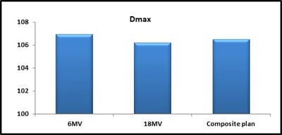
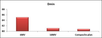
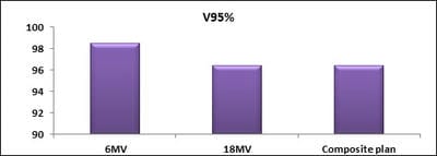
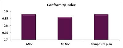
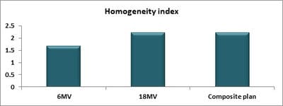
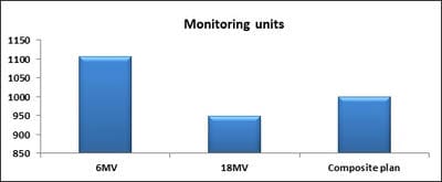
Introduction: Cervical cancer (Ca Cx) is the fourth most frequent cancer in women with an estimated 57000 new cases in 2018 representing 6.6% of all female cancers. Approximately 90% of deaths from cervical cancer occurred in low- and middle-income countries. Material and Methods: A retrospective radiotherapy treatment planning comparative study conducted at the Department of Radiation Physics, Kidwai Memorial Institute of Oncology, Bangalore during June 2018- March 2019. Result: All the plans were normalized to 100 % at Target mean to achieve a similar target dose for quantitative comparison of DVHs. The results for target coverage, OAR sparing, integral dose, and monitoring units. Conclusions: The tradeoff of using 6 MV and 18 MV for cervix patients depends on many parameters. Since the same PTV coverage was forced for both energies by having the same optimization constraints, there was little difference in target coverage and conformity index for both energies.

Keywords: Cervical cancer, Intensity-modulated radiation therapy, Dosimetric plan

Corresponding Author How to Cite this Article To Browse
Ibrahim Khaleel, Assistant Professor, Department of Radiation Physics, Kidwai Memorial Institute of Oncology, Bangalore, Karnataka, India.
Email:
Palaniappan SM, Khaleel I, Varatharaj C, Ganesh K M, Ravikumar M. Dosimetric evaluation of 6 MV and 18 MV intensity-modulated radiotherapy plans for treatment of carcinoma of the cervix. Int J Med Res Rev. 2020;8(4):307-314.
Available From
https://ijmrr.medresearch.in/index.php/ijmrr/article/view/1142

Introduction

Cervical cancer (Ca Cx) is the fourth most frequent cancer in women with an estimated 57000 new cases in 2018 representing 6.6%

of all female cancers. Approximately 90% of deaths from cervical cancer occurred in low- and middle-income countries [GLOBOCON 2018]. Treatment depends on disease extent at diagnosis and locally available resources


Manuscript Received Review Round 1 Review Round 2 Review Round 3 Accepted
2020-07-01 2020-07-15 2020-07-24 2020-08-25
Conflict of Interest Funding Ethical Approval Plagiarism X-checker Note
No Nil Yes 8%

© 2020 by Senthil Manikandan Palaniappan, Ibrahim Khaleel, Varatharaj C., Ganesh K M, Ravikumar M. and Published by Siddharth Health Research and Social Welfare Society. This is an Open Access article licensed under a Creative Commons Attribution 4.0 International License https://creativecommons.org/licenses/by/4.0/ unported [CC BY 4.0].

and might involve radical hysterectomy or chemoradiation or combination of both. Three-dimensional conformal radiation therapy (3DCRT) for Ca Cx is most commonly delivered with high energy photons typically in the range of 6-18 MV with four filed box technique until the intensity-modulated radiotherapy (IMRT) came into practice. Advances in radiotherapy technology, such as intensity-modulated radiotherapy, have resulted in less treatment-related toxicity for women with locally advanced disease. IMRT involves multiple beams from different directions having non-uniform fluences is known to improve target coverage and provide better organ-at-risk (OAR) sparing in comparison with 3DCRT [1.2].

Since high-energy photons provide greater penetration depth and skin-sparing effect, the conventional principle has been that the deeper the target, the higher energy should be used. As known from the literature, energies >10 MV are to be preferred for deep-seated pelvic /abdominal lesions, particularly for larger target volumes or larger size patients due to decrease in the integral dose ratio [3] In addition, it has been shown that dose deposition near and distant from the target is different for different energies.

Low energy produces tighter dose distributions around the target than higher energies but also deposits a higher dose in the surface region near the beam (3-5). In contrast, one of the implied tenets of intensity-modulated radiation therapy (IMRT) has been that energy does not matter or is less important [5,6].

With the advent of Intensity Modulated Radiation Therapy (IMRT), an increase in the number of monitor units (MU) relative to 3DCRT has to lead to a concern about the patient's total body neutron burden incurred during treatment with these high energy photon beams. As a result, the majority of IMRT cervix treatments delivered today are with lower energy (6-10 MV) photons where neutron production in a linear accelerator treatment head is negligible [7-10].

Another investigation [11,12] demonstrated that target dose conformity may be similar for a range of megavoltage photon energies for deep-seated tumors while dose in regions far from the target may receive higher doses for lower energies.

This study aims to find optimal energy that could offer better target coverage, target conformity,

homogeneity, and normal tissue sparing for cervix IMRT. Dosimetric plan evaluations were carried out based on planning target volume (PTV), Organ at risk (OAR) as well as integral dose. To ensure that differences among plans are due only to energy selection, the beam arrangement, number of beams, and dose constraints were kept constant for all plans.

Materials and Methods

Study Setting: Department of Radiation Physics, Kidwai Memorial Institute of Oncology, Bangalore

Duration of Study: June 2018- March 2019

Type of Study: A retrospective radiotherapy treatment planning comparative study

Sampling methods: Retrospective data of all patients of Carcinoma of the cervix treated between in 2018

Sample size calculation Twenty cases of carcinoma of cervix of different stages (II to IIIB) who had been treated with IMRT at our Institution.

Inclusion Criteria: Ca cervix patients of different stages (II to IIIB) whose pelvis separation more than 35 cm

Exclusion Criteria: Ca cervix patients of different stages (II to IIIB) whose pelvis separation less than 35 cm

Data Collection Procedure: Computed tomography simulation was done for all patients in the supine position immobilized with the thermoplastic cast. CT scans were obtained at 2.0 mm slice thickness intervals. Planning target volume (PTV) dose of 50 Gy in 25 fractions such that the PTV received at least 95% of the prescription dose.

The IMRT planning was carried out with the Eclipse treatment planning system (Varian Palo alto CA). The IMRT treatment delivered to the patient using seven co-planner beams of equal angles (0, 51, 102, 153, 204, 255, and 306) with 6 MV energy. The treatment machine is an isocentric linear accelerator (Clinac 2100-DHX, Varian) incorporating a 120 leaf MLC.

A competing IMRT plan with the same beam geometry, contouring, and dose constraints as the clinically delivered plan was developed using 18 MV photons for all 20 patients. Another plan was created with a combination of 6 MV and 18 MV beams with 51, 102, 255, 306 angled beams with


18 MV energy and rest beams (0, 153, 204 angles) with 6 MV energy (Composite plan).

The 51, 102, 255, and 305 angled beams have more depth compared to other angles, hence 18 MV beams were used. All three IMRT planning were calculated using an inverse planning algorithm (Analytic Anisotropic Algorithm AAA) with a dose calculation grid set at 2.5 mm.

The OARs (Organ at Risk) considered in this study include Bladder, Rectum, Bowel, Right, and Left Femoral head. The dosimetric outcomes of IMRT plan with 6 MV, 18 MV, and composite (6 MV and 18 MV) plans were compared qualitatively and quantitatively using standardized dose-volume indices in terms of Dmax, Dmin, target volume coverage (V95%), dose homogeneity, dose conformity, OARs sparing and integral doses (IDs).

Target volume coverage and dose homogeneity were assessed as the volume of the PTV receiving at least 95% (V95%) of the prescribed dose. The conformation of therapeutic dose volume to the target volume was estimated using the conformity index (CI) as defined by [12].

CI = (TV RI/ TV) X (TV RI / V RI)

TVRI is the target volume covered by the reference Isodose, TV is the tumor volume and VRI is the volume of the reference Isodose. The conformity of the target dose is superior if the CI value comes closer to one. Dose homogeneity was evaluated quantitatively using the dose homogeneity index (S‘ index), defined as the standard deviation of the differential dose-volume histogram of PTV by following formula, [13]

S- Index = DSD = √ [Σ (Di – Dmean) 2 x vi/V]

Where DSD represents the standard deviation of the dose, vi is the ith volume element receiving a dose of at least (Di) and V is the total volume. Dmean is the mean dose. If the S index value is near to zero, then the PTV has superior dose homogeneity within the target.

The integral dose (ID) has been defined as the sum of the product of a given dose (Di) and the volume of tissue receiving that dose (Vi) and the density of that tissue volume (Ï?i), as represented by the equation [14].

ID = Σi Di x Vi x Ï?

The integral dose was calculated for non-target normal tissues (NTT) which were actually created by subtracting all targets from the body defined. In addition to the integral dose, the volume of 10% isodose (V10%) and volume of 50% isodose (V50%) coverage lines also investigated.

Any Scoring System: Dosimetric parameters were evaluated and the treatment plans were compared as per the results

Statistical Analysis: Analyses were performed by using a paired two-tailed Student t-test to determine if there was a significant difference in any of the parameters examined. Differences were considered statistically significant at p ≤ 0.01.

Results

All the plans were normalized to 100 % at Target mean to achieve a similar target dose for quantitative comparison of DVHs. The results for target coverage, OAR sparing, integral dose and monitoring units are presented in Tables 1-4. In the interest of clarity, the data are presented as average values over the results calculated for all of the 20 patients with standard deviation values.

In Table 1, the maximum dose (Dmax) was comparable in all three plans studied: 106.9 % for 6 MV plan, 106.2 for 18 MV plan, and 106.5 for the composite plan in PTV (Figure 1).

Table-1: Target coverage for 6 MV, 18 MV, and composite plans.

Dosimetric Parameters 6 MV 18 MV Composite Plan
Mean SD Mean SD P Mean SD p
Dmax (%) 106.9 2.2 106.2 1.3 0.11 106.5 1.8 0.38
Dmin (%) 85.09 1.8 81.1 3.1 0.004 80.8 2.4 <0.001
V95 (%) 98.5 0.4 96.4 1.3 0.001 96.4 1.1 <0.001
Conformity index 0.88 0.03 0.86 0.07 0.18 0.88 0.03 0.92
Homogeneity index 1.69 0.15 2.23 0.3 0.003 2.23 0.2 <0.001

ijmrr_1142_01.jpg

Fig-1: Comparison of Dmax in all three plans.


ijmrr_1142_02.jpg

Fig-2: Comparison of Dmin in all three plans.

ijmrr_1142_03.jpg

Fig-3: Comparison of V95 % in all three plans.

ijmrr_1142_04.jpg

Fig-4: Comparison of conformity index in all three plans.

ijmrr_1142_05.jpg

Fig-5: Comparison of Homogeneity index in all three plans.

But the minimum dose (Dmin) in the target was significantly varied among all three plans. It was 85.09% for 6 MV, 81.1 % (p = 0.004) for 18 MV and 80.8% (p=0.001) for composite plan (Figure 2).

When compared to 6 MV plans, the target coverage and homogeneity index were affected significantly in 18 MV and composite plans. In spite of all plans achieved better target coverage (more than 95%) reduced the target coverage was observed with 18 MV photons when compared to 6 MV plans (Figure 3).

The V95 observed was 98.5% for 6 MV and 96.4% for 18 MV and composite plan. The mean conformity index was 0.88, 0.86, and 0.88 for 6 MV, 18 MV, and

composite plan respectively. These small differences indicate that all plans had good conformity of dose to the target. No significant difference was observed in the conformity index in all three plans (Figure 4). Figure 5 shows that the homogeneity index was better with a 6MV (1.69) plan than 18 MV (2.23) and a Composite plan (2.23).

Table-2: Normal tissue dose values for 6 MV, 18 MV, and composite plans.

Normal Tissues 6 MV Plan 18 MV Plan Composite Plan
Bladder Mean SD Mean SD p Mean SD p
Dmean(%) 80.1 3.1 78.6 3.0 <0.01 78.6 2.9 <0.01
D33% (%) 93.7 2.3 92.7 2.5 0.002 92.7 2.3 0.002
D66% (%) 70.8 5.0 68.7 5.1 <0.01 68.6 5.0 <0.01
Rectum
Dmean (%) 88.43 6.5 88.52 6.4 0.77 89.66 6.6 <0.01
D33% (%) 94.6 4.6 95.0 4.7 0.18 96.0 4.7 <0.01
D66% (%) 85.8 9.5 85.8 9.3 0.86 86.9 9.5 <0.01
Bowel
Dmean (%) 51.1 16.3 50.1 16.4 0.003 50.1 16.4 0.004
D33% (%) 66.3 18.3 64.8 18.7 0.001 64.3 18.4 <0.001
D66% (%) 30.5 25.4 29.6 25.1 0.06 29.8 24.8 0.12
Rfemoral head
Dmean (%) 52.9 15.1 52.6 15.4 0.38 52.6 15.0 0.17
D33% (%) 60.0 14.4 59.6 14.9 0.23 59.5 14.4 0.03
D66% (%) 43.9 17.0 43.8 17.3 0.55 43.8 17.0 0.53
Lfemoral head
Dmean (%) 53.1 13.5 52.9 13.7 0.52 52.9 13.4 0.23
D33% (%) 59.9 12.4 59.7 12.8 0.55 59.2 13.0 0.12
D66% (%) 44.0 8.17 45.0 4.18 0.46 44.7 16.9 0.68

Normal tissue values were shown in Table 2. In the case of bladder, the mean dose, D33%, and D66% were drastically reduced with 18 MV and composite plan compared to 6 MV plan.

It was 80.1% for 6 MV and 78.6% (p = < 0.01) for both 18 MV and composite plans (Figure 7).

There was no much benefit achieved with 18 MV beams for rectum compared to 6 MV plans (Figure 8).

The rectum means dose was increased with the use of composite plans compared to 6 MV and 18 MV beam plans. No much difference was observed between 6 MV and18 MV plans (Table 2).

The mean dose of rectum in composite plan was 89.66 % (p = <0.001) compared to 88.4% and 88.52% for 6 MV and 18 MV plans respectively.

In case of bowel, the mean dose, D33% and D66% were reduced significantly with 18 MV and composi


-te plans. Mean dose was 51.1% (p = 0.003) for 6 MV and 50.1 % (p = 0.004) for 18 MV and composite plan respectively.

Dose to 33% of volume was 66.3% for 6 MV beam and 64.8% (p = 0.001) and 64.3% (p = <0.001) for 18MV and composite plan respectively. There was no much difference observed in 6 MV and 18 MV beams for the bowel.

In the case of Right and Left femoral heads, no such variations in mean, D33%, and D66% dose observed in all three plans.

For the right femoral head, mean dose, D33% and D66% were 52.9%, 52.6%, and 52.6% respectively. D33% was 60% for 6MV and 59.6% and 59.5% for 18MV and composite plans respectively. Dose to 66% was 43.9% for 6 MV and 43.8% was observed for 18 MV and composite plans.

ijmrr_1142_06.jpg

Fig-6: PTV DVHs for 6MV, 18MV, and Composite plans.

ijmrr_1142_07.jpg

Fig-7: Bladder DVHs for 6MV, 18MV, and Composite plans.

ijmrr_1142_08.jpg

Fig-8: Rectum DVHs for 6MV, 18MV, and Composite plans.

Table-3: Integral dose of OARs for 6MV, 18MV, and composite plans.

OAR Integral Dose
6MV 18MV Composite plan
Mean P Mean p
Bladder (J) 15180 14877 0.02 14875 0.02
Rectum (J) 3019 3022 0.80 3061 0.01
Body_PTV (J) 236365 222108 <0.001 226308 <0.001
V10 (cc) 11798 12025 <0.001 12058 <0.001
V50 (cc) 4405 4047 <0.001 4154 <0.001

To account for low dose volumes in the Body_PTV volume, the volume received 50% isodose and 10% isodose were investigated for all three plans. The volume irradiated by a 10% isodose curve was decreased with 6 MV beams compared to 18 MV and composite beams.

For 6 MV plan, it was 11798 J and 12025 J and 12058 J for 18 MV and composite respectively. The volume irradiated by 50% isodose curve was more in 6 MV plan compared to 18 MV and composite plans (Figure 9).

The total integral dose for bladder was drastically reduced with 18 MV plan and composite plan compared to 6 MV beams. It was 15180 J for 6 MV, 14877 J (p = 0.02) for 18 MV and 14875 J (p = 0.02) for composite plans (Figure 10). For the rectum, there were no significant variations observed in all three plans carried out.

It was 3019 J, 3022 J, and 3061 J for 6 MV, 18 MV, and composite respectively (Figure 11). For the Body_PTV volume, the 18 MV plan was superior compared to the 6 MV plan. It was 236365 J for 6 MV, 222108 J (p = <0.001) for 18 MV and 226308 J (p = <0.001) for composite plans (Figure 12).

As expected the 6 MV beam yielded more monitoring units (1107) compared to 18 MV beam (949 (-14.3%) and (999 (-9.75%) for composite beams.

ijmrr_1142_09.jpg

Fig-9.1: 50% isodose distribution for 6 MV plan.


ijmrr_1142_10.jpg

Fig-9.2 50% isodose distributions for 18MV plan.

ijmrr_1142_11.jpg

Fig-9.3 50% isodose distribution for the composite plan.

ijmrr_1142_12.jpg

Fig-10: Comparison of bladder integral dose in all three plans.

ijmrr_1142_13.jpg

Fig-11: Comparison of rectum integral dose in all three plans.

ijmrr_1142_14.jpg

Fig-12. Comparison of the integral dose of Body_PTV.

ijmrr_1142_15.jpg

Fig-13: Monitoring unit values for 6 MV, 18 MV, and Composite plan.

Table-4: Monitoring unit values for 6MV, 18MV, and Composite plans

Monitoring units 6 MV 18 MV Composite plan
1107 949(-14.3%) 999(-9.75%)

Discussion

For PTV, Dmax and conformity index was comparable in 6MV, 18MV, and composite plan. In the case of target coverage and minimum dose, there was no significant benefit observed in using higher energies (18 MV) observed in all three plans. The minimum dose in the target was decreased with 18 MV and composite plans compared to 6 MV plan. This may be due to lack of target coverage in the posterior of the patient where most of the cervix target extends posteriorly. For the same reason, the dose homogeneity in the target also decreased with higher energies (18 MV).

The OAR sparing was better achieved with 18 MV beams only for Bladder and Bowel. It was observed from these results that the larger volume of OAR was spared with higher energy beams compared to the one which has a smaller volume. The rectal dose was slightly increased with 18 MV and composite plan than the 6 MV plan. This may be due to a higher entrance dose with 6 MV photons from the posterior beam. Also, dose modulation is the key to successful IMRT and this modulation is heavily dependent on the lateral fall off provided by the leaves of the multi-leaf collimator. The ability to modulate is impaired at higher energy because the lateral range of electrons widens the lateral fall off. The lateral range increase is the result of the same fundamental physics that produces a deeper depth of maximum dose for high energy photons. The electrons are being set in motion at higher energies, but not so high that they do not scatter. A typical initial kinetic energy of an electron set in motion by 18 MV photons is about 4 or 5 MeV. At this initial kinetic energy, an electron travels about 2 or 3 cm and scatters considerably, even if it is originally set


in motion in the same direction as the photon. This leads to blurring of the lateral boundary and inherently limits the modulation that can be achieved. So high modulation is required when avoidance of the rectum has been assigned high priority in the optimizer. Such high priority demands a very steep gradient between the cervix and rectum. This gradient becomes obviously less steep if a high energy beam is used with its wider penumbra. So irrespective of energy, the dose to the rectum was not much varied between all three plans.

There were no tight dose constraints given for the right and left femoral heads since it is relatively away from the PTV. Since it has no much role in optimization, the dose to femoral heads was not varied much between all three plans irrespective of energy. The total integral dose of the bladder was better achieved with higher energy beams. The integral dose of Body_PTV was also given good results in 18 MV and composite plans compared to lower energies.

In spite of an increase in 10% isodose volume in 18 MV and composite plans, the total integral dose was reduced with 18 MV and composite beams. The volume received by 50% isodose volume was better achieved with 18 MV and composite beams compared to 6 MV beam.

The main disadvantage of using an 18 MV beam is the secondary neutron production in the linac head assembly itself. Neutron is of considerable importance in radiation safety because for any given absorbed dose, neutron irradiation typically yields a much higher biologically effective dose (BED) than photons for practically any biological endpoint.

Since low energy photons (6 MV beam) are normally below the threshold for neutron generation, such concerns are avoided. With conventional radiation therapy or 3 D conformal radiotherapy, the time during which the linac beam is on (i.e. monitoring units) is relatively brief, and therefore, regardless of photon energy, significant amounts of neutron contamination are not likely.

These increased monitoring units required with IMRT greatly increase the odds of neutron generation when high energy photons are used (4). Howell et al [8] measured neutron doses from the delivery of 18 MV conventional and IMRT treatment plans. They found that the IMRT treatment resulted in a higher neutron fluence and higher dose

equivalent. These increases were approximately the ration of the monitoring units used. Chbani et al [7] found that the dose equivalent from photo neutrons at 50 cm off-axis distance produces up to a 2.0% likelihood of fatal secondary cancer for a 70 Gy treatment delivered by a Varian 18 MV beam. Kry et al [10] also found that neutrons were a significant contributor to the out of field dose equivalent for beam energies > 15 MV.

The estimated risks of fatal secondary malignancy associated with IMRT and conventional external beam approaches for prostate cancer and a maximum risk of fatal second malignancy of 1.7 % for conventional radiation therapy and up to 5.1 % for IMRT using 18 MV photons. So the dose produced by neutron with higher energies will have a higher risk of secondary malignancies compared to the more integral dose produced by 6 MV beams.

Limitations

The sample size and specific to one treatment site are limitations of this study.

Conclusion

The tradeoff of using 6 MV and 18 MV for cervix patients depends on many parameters. Since the same PTV coverage was forced for both energies by having the same optimization constraints, there was little difference in target coverage and conformity index for both energies. DVHs for the critical structures showed a little difference between 6 MV, 18 MV, and composite beams. The 6 MV IMRT integral dose was higher than the 18 MV and composite plan. Although integral dose is generally assumed to be improved by the use of higher energy photons, there is concern that the higher neutron contamination with 18 MV beams increases the chance of secondary malignancies.

What does the study add to the existing knowledge?

These results from the study indicate that there is no clinical benefit with respect to target coverage and normal tissue sparing when comparing 6 MV IMRT, 18 MV IMRT, and composite IMRT plans. It is also demonstrated that the ability to generate acceptable IMRT plans is independent of the energies in the 6-18 MV. The higher total body dose in 18 MV IMRT plans due to neutrons should be considered by the clinician when choosing the


optimal treatment course for a patient.

Author’s contribution

Dr. P. Senthil Manikandan: Study Design, Data Collection, and Manuscript Writing

Dr. Ibrahim Khaleel: Treatment Planning, Sample Selection

Dr. C. Varatharaj: Treatment Planning, Data Collection

Dr. K. |M. Ganesh: Manuscript review and data analysis

Dr. M. Ravikumar: Methodology and Manuscript review

Reference

  1. Pirzkall A, Carol M, Pickett B, Xia P, Roach M, Verhey L. The Effect of beam energy and number of fields on photon based IMRT for deep seated targets. Int J Radiat Oncol Biol Phys. 2002;53(2)434-441.
  2. Soderstrom S, Eklof A, Brahme A. Aspects on the optimal photon energy for radiation therapy. Acta Oncologica. 1999;38(2)179-187.
  3. Laughlin JS, Mohan R, Kutcher KJ. Choice of optimum megavoltage for accelerators for photon beam treatment. Int J Radiat Oncol Biol Phys. 1986;12(9)1551-1557.
  4. Boer SF, Kumek Y, Jaggernauth W, Podgarsak MB. The effect of beam energy on the quality of IMRT plans for prostate conformal radiotherapy. Technol Cancer Res Treat. 2007;6(2)139-146.
  5. Garrison H, Anderson J, Laughlin JS. Comparison of dose distributions in patients treated with X ray beams of widely different energies. Radiology. 1952;58(3)361-368.
  6. Sternick ES, Bleier AR, Carol MP, Curran BH, Holmes TW, Kania AA, Lalond R, Larson LS. Intensity modulated radiation therapy- what photon energy is best. In Presentation at The XIIth International Conference on the Use of Computers in Radiation Therapy (ICCR). 1997 May.
  1. Chibani O, Ma CM. Photonuclear dose calculations for high energy photon beams from Siemens and Varian Linacs. Med Phys. 2003;30(8)1990-2000.
  2. Howell RM, Ferenci MS, Hertel NE, Fullerton GD. Investigation of secondary neutron dose for 18 MV dynamic MLC IMRT delivery. Med Phys. 2005;32(3)786-793.
  3. Howell RM, Ferenci MS, Hertel NE. Measurements of secondary neutron dose from 15 MV and 18 MV IMRT. Radiat Prot Dosimetry. 2005;115(1-4)508-512.
  4. Kry SF, Howell RM, Salehpour M, Followil DS. Neutron spectra and dose equivalents calculated in tissue for high-energy radiation therapy. Med Phys. 2009;36(4)1244-1250.
  5. Khan FM. The Physics of Radiation therapy. 3rd Edition; Lippincott, Williams & Wilkins Publishers. (2003).
  6. Feuvret L, Noel G, Mazeron J, Bey P. Conformity Index:-A new homogeneity index based on statistical analysis of the dose-volume histogram. J App Clin Med Physics. 2007;8(2)9-17.
  7. Chan MF, Schupak K, Burman C, Chui CS, Ling CC. Comparison of intensity-modulated radiotherapy with three-dimensional conformal radiation therapy planning for glioblastoma multiforme. Med Dosim. 2003;28(4)261-265.