E-ISSN:2320-8686
P-ISSN:2321-127X

Research Article

Evaluation

International Journal of Medical Research and Review

2019 Volume 7 Number 4 July-August
Publisherwww.medresearch.in

Evaluation of trans thoracic invasive diagnostic procedures in 50 adult patients of mediastinal diseases

Biswas S.1, Saha R.2, Deb J.3*, Dey P.4
DOI: https://doi.org/10.17511/ijmrr.2019.i04.11

1 Siddhartha Biswas, Department of Respiratory Medicine, N.R.S Medical College, Kolkata, West Bengal, India.

2 Rama Saha, Associate Professor, Department of Pathology, Institute of Post Graduate Medical Education and Research, Kolkata, West Bengal, India.

3* Jaydip Deb, Professor, Department of Respiratory Medicine, N.R.S Medical College, Kolkata, West Bengal, India.

4 Prothoma Dey, Department of Respiratory Medicine, N.R.S Medical College, Kolkata, West Bengal, India.

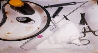
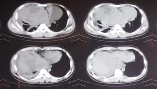
Introduction: Mediastinum is a “Pandora’s box” with many neoplastic and nonneoplastic lesions. Clinico-radiological pattern of mediastinal diseases depends on the size, location and etiology. Hence, non-invasive approach to these cases sometimes leads to diagnostic dilemma. Aims: A prospective study was performed over a 1-year period with the objective of evaluation of diagnostic yields and risk of trans thoracic ultra sound (TTUS) and computed tomography (CT) guided fine‑needle aspiration cytology (FNAC) and Tru-cut biopsy along with comparison of cost-effectiveness among mediastinal diseases where clinical and non-invasive imaging could not conclude the diagnosis. Materials and Methods: A prospective study of mediastinal diseases of the adult population without having any diagnosis admitted in a tertiary care hospital in Eastern India was performed after clearance of the ethical committee of the institute. Fifty cases of mediastinal diseases were seen during the study period. One patient sometimes had undergone more than one procedure. Results: Among 50 patients diagnostic yield of TTUS guided FNA and Tru-cut biopsy were 60% and 63.6% respectively. Diagnostic yield of Thoracic CT guided FNA and Tru-cut biopsy were 85% and 92.3% respectively. As a whole TTUS guided invasive procedure and Thoracic CT guided invasive procedure had a diagnostic yield of 61.5% and 87.9% respectively. Conclusion: Tru-cut biopsy if applicable is much superior to FNAC for a definite diagnosis of the mediastinal diseases. TTUS guided invasive procedures are very much cost-effective and have added advantage of real time guidance and is comparable with CT guided invasive procedures in respect to risk and diagnostic yields.

Keywords: Computed tomography guided invasive procedure, Fine needle aspiration, Mediastinal diseases, transthoracic ultrasound, Tru-cut biopsy, Ultrasound guided invasive procedure

Corresponding Author How to Cite this Article To Browse
Jaydip Deb, Professor, Department of Respiratory Medicine, N.R.S Medical College, Kolkata, West Bengal, India.
Email:
Biswas S, Saha R, Deb J, Dey P. Evaluation of trans thoracic invasive diagnostic procedures in 50 adult patients of mediastinal diseases. Int J Med Res Rev. 2019;7(4):324-331.
Available From
https://ijmrr.medresearch.in/index.php/ijmrr/article/view/1076

Manuscript Received Review Round 1 Review Round 2 Review Round 3 Accepted
2019-07-20 2019-07-30 2019-08-08 2019-08-13
Conflict of Interest Funding Ethical Approval Plagiarism X-checker Note
No Nil Yes 6%

© 2019 by Siddhartha Biswas, Rama Saha, Jaydip Deb, Prothoma Dey and Published by Siddharth Health Research and Social Welfare Society. This is an Open Access article licensed under a Creative Commons Attribution 4.0 International License https://creativecommons.org/licenses/by/4.0/ unported [CC BY 4.0].

Introduction

The thoracic mediastinum is commonly defined as the region between the two pleural sacs, bounded anteriorly by the sternum and posteriorly by the thoracic vertebral column and extending vertically from the thoracic inlet to the diaphragm [1]. The mediastinum is separated into superior and inferior compartments and the inferior compartment can further be divided into anterior (pre-vascular), middle (cardiovascular) and posterior (post vascular) compartments [2]. The most frequent lesions encountered in the mediastinum are thymoma, neurogenic tumors, and benign cysts, altogether representing 60% of patients with mediastinal masses. Neurogenic tumors, germ cell neoplasms, and foregut cysts represent 80% of childhood lesions, whereas primary thymic neoplasms, thyroid masses, and lymphomas are the most common lesions in adults [3].

Anterior mediastinal tumors account for 50% of all mediastinal masses, including thymoma, teratoma, thyroid disease, and lymphoma [4]. Neurogenic tumours, infective lesions and oesophageal pathology were seen most commonly in the posterior and mediastinum. Vascular lesions were mostly seen in middle mediastinum. The nonspecific clinical manifes-tations of most of these disorders and it`s relative inaccessibility for diagnostic examination result in considerable challenges to the clinician for evaluation of mediastinal diseases [5]. The diagnostic evaluation of the mediastinal disorders can be divided into two phases- non-invasive investigations like imaging and invasive procedures to obtain tissue samples.

Among the invasive procedures trans thoracic ultra sound (TTUS) guided and computed tomography (CT) guided fine needle aspiration cytology (FNAC), and Tru‑cut biopsy are not new [6]. TTUS guided procedures are helpful for superior and anterior mediastinal disease evaluation. The advantages of US guidance include the real-time, continuous monitoring of the needle during advancement and sampling; the availability of oblique needle paths; and the ability to perform the biopsy at the bedside in critically ill patients or in patients with dyspnea who cannot tolerate a supine position in semi-sitting positions during the biopsy [7]. Against this background, the present study was conducted to compare diagnostic yields, complication rates and costeffectiveness of those invasive procedures.

Materials and Methods

Study design- This prospective study was performed on patients with having mediastinal mass lesions detected by chest radiograph and/or CT scan and it was conducted in the department of pulmonary medicine in a tertiary care teaching hospital in Eastern India during the period from May 2017 to April 2018. All patients having mediastinal mass lesions were included in thestudy. After exclusion of six patients remaining 50 patients were taken for study.

Patient selection

Inclusion criteria

  1. Patients of any type of mediastinal diseases detected clinically and or radio logically (mediastinal mass lesion >2cm in diameter).
  2. Normal bronchoscopy or unyielding bronchoscopic and other samples.

Exclusion criteria

  1. Any suspected mediastinal diseases proved later to be of chest & pleural origin on investigations
  2. Patients with vascular lesions.
  3. Patients with bleeding tendency
  4. very poor general condition
  5. Patients who were unwilling to give consent.

Study protocol- Detailed previous radiological evaluations were studied in all patients fulfilling the patient selection criteria. All patients were prepared preoperatively by checking their coagulation profile. Ultra sound (USG) and CT guided FNA were maximally employed in the study considering low risk and low cost of the procedure. Thin caliber (18–20 gauge) LP needles were used to obtain specimens for cytological evaluation. Mediastinal lesions, which were touching or near the chest wall were also selected for Tru‑cut biopsy in the same occasion considering detailed and definite result. In addition, cases with inconclusive results from FNAC were all put for Tru‑cut biopsy. Under image guidance after determination of the specific area of interest Tru‑cut biopsy were done. Two types of Tru‑cut biopsy needles were used, one is manually operated Shoney Cut Biopsy needle (16 g × 4.5 cannula) with 20 mm specimen notch and other one is automated Tru‑cut biopsy needle named as automated spring-loaded biopsy gun machine.


After Tru‑cut biopsy impression smears were prepared first from obtained material and the specimen was collected for histopathology and immunohistochemistry if necessary. During the consideration of modes of image guidance, TTUS is preferred first due to its low cost wherever it is helpful considering different topographical distribution of mediastinal lesions. For anterior mediastinal lesions anterior parasternal and suprasternal approaches, for posterior mediastinal lesions paravertebral approach and for middle mediastinal lesions right and left anterior parasternal approaches were applied.Few large masses were approached in TTUS through intercostals spaces. Inconclusive and unapproachable cases by TTUS guided invasive methods were kept for CT guided invasive procedures. Only conclusive results and concordant reports to more sensitive investigations were considered during the evaluation of diagnostic yield of a test in this regard. Post-procedure observation was done in all cases keeping resuscitation kit and the emergency service ready at hand.

ijmrr_1076_01.jpg

Figure-1: Tools of FNA and Tru-cut biopsy procedure

Conclusive results: A definitive diagnosis is made; hence proper therapeutic management can be made.

Inconclusive results: A definitive diagnosis is not possible-

  • Due to poor qualitative and or quantitative sample
  • Nonspecific cytological or histopathological features leading to in conclusion to draw a diagnosis.

Concordant results: High proportion of agreement is present between results of initial and subsequent test where initial result is hinting correctly the result of the subsequent test.

Statistical analysis: All the available information was recorded meticulously, and a database was created. A grand chart was prepared, and statistical analysis was performed.

Results

A total of 50 patients fulfilling case definition were studied in the study period. Many of the patients had undergone more than one invasive procedure. In some cases, one procedure gave the evidence of presence of disease without any confirmation of specific disease. In those situations, other procedures were implicated for knowing histopathological details and immunohistochemistry of the lesion as needed. During the calculation of diagnostic yield of a particular procedure, conclusive results and concordant results to higher procedures were considered.

Diagnostic yield: Of 50 patients undergone invasive procedures [Table 1], TTUS guided FNAs were conducted among 15 patients with conclusive and or concordant results coming from 9 patients of them. So diagnostic yield of TTUS guided FNAC was 60%. Similarly, CT guided FNA was performed among 20 patients with high (85%) diagnostic yield. 11 cases undergone TTUS guided Tru‑cut biopsy of mediastinal lesion and 7 cases out of them were conclusively diagnosed making diagnostic yield of 63.6%. CT guided Tru‑cut biopsy was performed among 13 cases resulting a very high diagnostic yield (92.3%).

Complication rate: Among the procedures [Table 2] TTUS guided FNAC was done in 15 cases, and complication occurred only one occasions (6.6%). Similarly, in 11 cases TTUS guided Tru‑cut biopsy was done and complication occurred only two, making 18.2% complication rate of the procedure. Complications occurred once in 20 occasions of CT guided FNAC making complication rate of 5%, which is the least among all complication rates of the invasive procedures done in the study. CT guided Tru‑cut biopsy were done 13 times, and complication occurred 2 times making the complication rate of 15.4%.

Cost, yield and complication comparison of different invasive procedures- Transthoracic ultrasound guided procedures were less costly than CT guided procedures [Table 3]. TTUS guided FNAC and Tru‑cut biopsy procedures were totally free of cost.


Cost of CT guided FNAC and Tru‑cut biopsy were Rs.600. Tru-cut biopsy is much superior to FNAC for a definite diagnosis of the mediastinal diseases. Complication is less in CT guided invasive procedures (9.1%) compared to Transthoracic USG guided invasive procedures (11.5%) and complication is more common in Tru-Cut biopsy (16.7%) than fine needle aspiration (5.7%).

Table-1: Diagnostic yield of different procedure.

Image‑guided invasive diagnostic procedures diagnostic Patient undergone this procedure Total number of conclusive/ concordant results Percentage (diagnostic yield)
TTUS guided FNAC 15 9 60
TTUS guided Tru-cut biopsy 11 7 63.7
CT guided FNAC 20 17 85
CT guided Tru-cut biopsy 13 12 92.3

TTUS: Trans thoracic ultra sound, FNAC: Fine needle aspiration cytology, CT: Computed tomography


Table-2: Complication rates of different procedures.

Different invasive procedures Pneumothorax Severe chest pain Hemoptysis Total Percentage
TTUS guided FNAC (n=15) 1 Nil Nil 1 6.6
TTUS guided Tru-cut biopsy (n=11) 1 1 Nil 2 18.2
CT guided FNAC (n=20) Nil Nil 1 1 5
CT guided Tru-cut biopsy (n=13) Nil 1 1 2 15.4

TTUS: Trans thoracic ultra sound, FNA: Fine needle aspiration, CT: Computed tomography

Table-3: Cost, yield and complication comparison of different invasive procedures

Parameters TTUS guided FNAC TTUS guided Tru‑cut biopsy CT guided FNAC CT guided Tru‑cut biopsy
Average cost (rupees) Nil Nil 600 600
Diagnostic yield (%) 60٪ 63.7٪ 85٪ 92.3٪
Complication rate (%) 6.6٪ 18.2٪ 15.4٪

TTUS: Trans thoracic ultra sound, CT: Computed tomography, FNAC: Fine needle aspiration cytology

ijmrr_1076_02.jpg

Figure-2: CT Thorax shows Yolk Sac tumour in a 26 years old patients

ijmrr_1076_03.jpg

Figure-3: CT thorax shows Non-Hodgkin’s Lymphomawith pleural effusion

Discussion

Study of cell or tissue obtained from many kinds of procedures is very much important for exact diagnosis of mediastinal masses [8]. Hence, it is obvious that invasive procedures are very necessary for diagnosis of mediastinal diseases. Invasive procedures were needed for 43 cases of the present study (86%) and only 7 cases comprising 14% of total cases were diagnosed by non-invasive investigations. Among the invasive procedures TTUS and Thoracic CT guided FNAC and Tru‑cut biopsy were studied thoroughly in the present study.

Only conclusive results and concordant reports to more sensitive investigations were considered during the evaluation of diagnostic yield of a test. Diagnostic yield of TTUS guided FNAC was 60% in the present study.


This finding has been supported by Hsu et al[9] (diagnostic rate of UG‑FNA was 52%) and Rubens et al [10] (US guided FNAC was 77% sensitive). Another study [11] in Springfield, USA on diagnostic accuracy of image‑guided percutaneous FNA biopsy (FNAB) of the mediastinum also showed a high proportion of agreement (78%) between FNAB and subsequent histological diagnoses for a wide variety of mediastinal lesions. Diagnostic yield of TTUS guided Tru‑cut biopsy was 63.7% in the present study.

A better diagnostic yield was found in the study of Annessi et al. [6] (USG guided biopsy of anterior mediastinal masses among 42 patients showed accurate diagnosis in 100% cases) as that study concentrated only in anterior mediastinal cases, which are easy to approach by TTUS. In the present study, diagnostic yield of thoracic CT guided FNAC, and Tru‑cut biopsy were 85% and 92.3% respectively. Similarly, Deb et al [12] in their study got a very high result before done on trans thoracic cutting needle biopsy where all the cases of intrathoracic mass lesion touching the chest wall were selected for Tru‑cut biopsy and diagnostic yield was 97.95%.

The percutaneous transthoracic FNAC and/or biopsy of the mediastinal lesion under local anesthesia are minimally invasive, cost-effective, easy to perform, and associated with reduced complication rates when performed under ultrasound or CT scan guidance[13]. The accuracy of transthoracic biopsy in the diagnosis of mediastinal lesions ranges from 75% to 90% [14,15,16].

The diagnostic accuracy of mediastinal lesions approached in the present study was 89.9% that is similar to the diagnostic yield in studies by Shaheen et al [17] and Güllüoglu et al, [18] superior compare to studies by Morrissey et al [19] (77%), Assaad et al [20] (82%), Rosenberger and Adler [21] (83%), Adler et al [22] (79%), Pedersen et al [23] (81%), Dubashi et al [24] (50%), and Neyaz et al [25] (74%) and inferior to the studies by Nasit et al [26] (97%) and Annessi et al [6] (100%).

Core biopsy of the lesions provide better yield with more precise diagnosis compared to FNAC[26,27] and the same was observed in the present study. From the present study and different other studies, it is clear that image‑guided procedures are quite helpful to produce good diagnostic yield of FNA and Tru‑cut biopsy of mediastinal masses.

In the present study, diagnostic yield of TTUS guided procedure was a bit low (comparable with study of Hsu et al [8] due to a small number of TTUS guided procedures. TTUS guided FNAC and Tru‑cut biopsies were seen to be more successful among the antero‑superior mediastinal lesions and large mediastinal masses reaching peripheral areas of thorax.

In the present study, neoplastic nature of disease was found in 72%. Malignant nature was seen in 54% of neoplastic cases and in 36 of total cases. Most common etiologies were Germ cell tumours (18%) followed by Lymphomas (16%), Thymic tumours (12%), Infective etiology (10%) and Neurogenic tumours (8%).

Esophageal lesions and vascular lesion were seen among 6% and 4% cases. 33.3% of Thymic tumours were malignant and 77.8% germ cell tumours were malignant. Among the Neurogenic tumours, benign were 75% and alignant were 25% in nature. The study was conducted by Shrivastava et al in Indian scenario among 106 patients shows germ cell tumours were quite high in incidence.

However, Thoracic CT guided procedure showed a very high diagnostic yield. As a whole TTUS guided invasive procedure, and thoracic CT guided invasive procedure had a diagnostic yield of 61.5% and 87.9% respectively. The incidence of complications in this series were very small and compares favorably with previously published series [6,28]. Pneumothorax is the most frequent complication resulting from tru-cut biopsy in thoracic lesion [6,28].

Present study showed complication is less in CT guided invasive procedures (9.1%) compare to Transthoracic USG guided invasive procedures (11.5%) and complication is more common in Tru-Cut biopsy (16.7%) than fine needle aspiration (5.7%). Cost, yield and complication comparison in the present study revealed that in a low cot set up TTUS guided invasive procedures are very much cost‑effective and as comparable as costly CT guided invasive procedures.

Conclusion

  • Transthoracic USG is very much helpful for initial assessment of vascular and cystic mediastinal diseases. However, contrast enhanced CT scan of Thorax is able to describe almost all mediastinal pathologies in details.

  • Neoplastic nature is mostly seen among symptomatic patients. Lymphoma, Thymic tumours, Germ cell tumours are the leading mediastinal diseases.
  • Tru‑cut biopsy procedure if applicable is much superior to FNAC procedure for definite diagnosis of the mediastinal diseases.
  • USG guided procedures are helpful for superior and anterior mediastinal disease evaluation. The advantages of USG guidance include the real-time, continuous monitoring of the needle during advancement and sampling; the availability of oblique needle paths; and the ability to perform the biopsy at the bedside in critically ill patients or in patients with dyspnoea who cannot tolerate a supine position in semi-sitting positions during the biopsy
  • USG guided invasive procedures are very much comparable with CT guided procedures in respect to risk and diagnostic yields, when performed by radiologist experienced in thoracic USG.

What the study adds to the existing knowledge

Realtime USG guide Tru-cut biopsy in the non vascular intra thoracic lesion is likely to replaceFNAC in near future due to its low cost and high yield.Tru‑cut biopsy is helpful among the patients having Mediastinal lesions, which were touching ornear the chest wall.

Author’s contribution

Dr. Siddhartha Biswas: Preparation of cases, collection data,procedure and preparing the write up. Dr. Rama Saha: Pathological evaluation. Prof. Jaydip Deb: Preparation of cases, procedure and collection of data. Dr. Prothoma Dey: Preparation of cases, procedure and preparing the write up.

Reference

  1. Standring Susan, Gray’s Anatomy. The Anatomical Basis of Clinical Practice, Expert Consult. Churchill Livingstone. 40th ed, pp-939.
  2. Banister LH. The respiratory system, In Williams PL (ed)- Gray’s Anatomy. New York- Churchil Livingstone. 1995;1627-82.
  1. Laurent F, Latrabe V, Lecesne R, Zennaro H, Airaud JY, Rauturier JF, et al. Mediastinal masses: diagnostic approach. Eur Radiol. 1998;8(7)1148-59.
  2. Tomiyama N, Honda O, Tsubamoto M, Inoue A, Sumikawa H, Kuriyama K, et al. Anterior mediastinal tumors- diagnostic accuracy of CT and MRI. Eur J Radiol. 2009;69(2)280-8.
  3. Broaddus VC, Martin TR, King TE, Schraufnagel DE. Murray and Nadel's textbook of respiratory medicine, Mason RJ, editor. Elsevier Saunders. 2010;4th ed, Vol-II;1814-1835.
  4. Annessi V, Paci M, Ferrari G, Sgarbi G. Ultra-sonically guided biopsy of anterior mediastinal masses. Interact Cardiovascul Thorac Surg. 2003;2(3)319-21.
  5. Gupta S, Seaberg K, Wallace MJ, Madoff DC, Morello FA Jr, Ahrar K, et al. Imaging-guided percutaneous biopsy of mediastinal lesions- different approaches and anatomic considerations. Radiograph. 2005;25(3)763-86.
  6. Tscheikuna J, Suttinont P. Is cytology necessary in diagnosis of mediastinal mass?. J Med Assoc Thai. 2009;92(2)S24-S29.
  7. Hsu WH, Chiang CD, Hsu JY, Kwan PC, Chen CL, Chen CY. Ultrasonically guided needle biopsy of anterior mediastinal masses- Comparison of carcinomatous and non‐carcinomatous masses. J Clinic Ultrasound. 1995;23(6)349-356.
  8. Rubens DJ, Strang JG, Fultz PJ, Gottlieb RH. Sonographic guidance of mediastinal biopsy- an effective alternative to CT guidance. AJR- Am J Roentgenol. 1997;169(6)1605-1610.
  9. Assaad MW, Pantanowitz L, Otis CN. Diagnostic accuracy of image-guided percutaneous fine needle aspiration biopsy of the mediastinum. Diagn Cytopathol. 2007;35(11)705-709.

  1. Deb J, Saha R, Chaudhury T, Chaudhury L, Ghosh I. Transthoracic cutting needle biopsy- a valuable diagnostic procedure. J Indian Med Associat. 2008;106(4)243-244.
  2. Zafar N, Moinuddin S. Mediastinal needle biopsy- A 15‐year experience with 139 cases. Cancer. 1995;76(6)1065-1068.
  3. Protopapas Z, Westcott JL. Transthoracic hilar and mediastinal biopsy. Radiol Clin North Am. 2000;38(2)281–291.
  4. Wiersema MJ, Vazquez-Sequeiros E, Wiersema LM, et al. Evaluation of mediastinal lymphadenopathy with endoscopic US-guided fine-needle aspiration biopsy. Radiol. 2001;219(1)252–257.
  5. Gupta S, Wallace MJ, Morello FA Jr, Ahrar K, Hicks ME. CT-guided percutaneous needle biopsy of intrathoracic lesions by using the transsternal approach- experience in 37 patients. Radiol. 2002;222(1)57-62.
  6. Shaheen MZ, Sardar K, Murtaza HG, Safdar SS, Hafeez A, Mushtaq MA, et al. CT guided trans-thoracic fine needle aspiration/biopsy of mediastinal and hilar mass lesions- An experience of pulmonology department at a tertiary care teaching hospital. Pak J Chest Med. 2008;12;26-38.
  7. Petranovic M, Gilman MD, Muniappan A, Hasserjian RP, Digumarthy SR, Muse VV, et al. Diagnostic Yield of CT-Guided Percutaneous Trans-thoracic Needle Biopsy for Diagnosis of Anterior Mediastinal Masses. AJR Am J Roentgenol. 2015;205(4)774-779.
  8. Morrissey B, Adams H, Gibbs AR, Crane MD. Percutaneous needle biopsy of the mediastinum- review of 94 procedures. Thorax. 1993;48(6)632-637.
  1. Assaad MW, Pantanowitz L, Otis CN. Diagnostic accuracy of image‐guided percutaneous fine needle aspiration biopsy of the mediastinum. Diagnostic Cytopathol. 2007;35(11)705-9.
  2. Rosenberger A, Adler OB. Fine-needle aspiration biopsy of mediastinal masses- A new approach to diagnosis. Isr J Med Sci. 1981;17(1)49-52.
  3. Adler OB, Rosenberger A, Peleg H. Fine-needle aspiration biopsy of mediastinal masses- evaluation of 136 experiences. AJR Am J Roentgenol. 1983;140(5)893-896.
  4. Pedersen OM, Aasen TB, Gulsvik A. Fine needle aspiration biopsy of mediastinal and peripheral pulmonary masses guided by real-time sonography. Chest. 1986;89(4)504-508.
  5. Dubashi B, Cyriac S, Tenali SG. Clinico-pathological analysis and outcome of primary mediastinal malignancies—a report of 91 cases from a single institute. Annal Thorac Med. 2009;4(3)140-142.
  6. Neyaz Z, Lal H, Thakral A, Nath A, Rao RN, Verma R. Percutaneous computed tomography-guided aspiration and biopsy of intrathoracic lesions- Results of 265 procedures. Lung India- official organ of Indian Chest Society. 2016;33(6)620.
  7. Nasit JG, Patel M, Parikh B, Shah M, Davara K. Anterior mediastinal masses- A study of 50 cases by fine needle aspiration cytology and core needle biopsy as a diagnostic procedure. South Asian J Cancer. 2013;2(1)7-13.
  8. Saha D, Deb J. Diagnostic role of ultra sound and computed tomography guided fine-needle aspiration cytology and Trucut biopsy experienced in 50 adult patients of mediastinal diseases. J Associat Chest Physicians. 2015;3(2)48-52.

  1. Tscheikuna J, Suttinont P. Is cytology necessary in diagnosis of mediastinal mass?. J Med Assoc Thai. 2009;92(2)S24-S29.